Intrahepatic Pancreatic Pseudocyst Complicated by Pancreatitis: A Case Report
Hee Joon Kim, Chung Hwan Jun1, Chang Hwan Park1 and Chol Kyoon Cho
Department of Surgery and Division of Gastroenterology, Department of Internal Medicine1, Chonnam National University Medical School, Gwangju, Korea
Pancreatic pseudocyst is a common complication of acute pancreatitis. Pseudocysts are commonly observed in the lesser sac and retroperitoneum; they are rarely seen in the liver. Herein, we report a case of intrahepatic pseudocyst, complicated by asymptomatic groove pancreatitis, that has successfully been treated with hepatic resection. A 70-year-old woman was referred to our hospital with severe upper abdominal pain. Abdominal computed tomography scan showed 11x10 cm sized cystic lesion in the left lateral section of the liver. Appearance of the pancreas was relatively normal. Endoscopic aspiration revealed a high level of amylase in the cystic fluid. After endoscopy, signs of peritonitis were observed; then, a left hemihepatectomy was performed. Pathologic examination re- vealed an intrahepatic pancreatic pseudocyst. The presence of intrahepatic cystic lesion in patients with suspected pancreatitis should raise the suspicion of intrahepatic pseudocyst. Intrahepatic pancreatic pseudocysts may be the only clinical manifestation even without an episode of acute pancreatitis. (Korean J Gastroenterol 2017;70:202-207)
Key Words: Pancreatitis; Intrahepatic pseudocyst; Hepatectomy
Received August 1, 2017. Revised September 10, 2017. Accepted September 27, 2017.
CC This is an open access article distributed under the terms of the Creative Commons Attribution Non-Commercial License (http://creativecommons.org/licenses/
by-nc/4.0) which permits unrestricted non-commercial use, distribution, and reproduction in any medium, provided the original work is properly cited.
Copyright © 2017. Korean Society of Gastroenterology.
교신저자: 조철균, 61469, 광주시 동구 백서로 160, 전남대학교 의과대학 외과학교실
Correspondence to: Chol Kyoon Cho, Department of Surgery, Chonnam National University Medical School, 160 Baekseo-ro, Dong-gu, Gwangju 61469, Korea.
Tel: +82-61-379-7649, Fax: +82-61-379-7661, E-mail: [email protected] Financial support: None. Conflict of interest: None.
INTRODUCTION
Pancreatic pseudocyst is defined as a collection of pancre- atic juice enclosed by a wall of non-epithelialized granulation tissue or fibrotic capsule. Pancreatic pseudocyst is a com- mon complication of acute pancreatitis that can occur any- where in the abdomen. Most commonly, they occur around the pancreas, lesser sac, and retroperitoneum. However, they have been reported to be found distant from the pan- creas, such as in the mediastinum or scrotum.1,2 Intrahepatic location of the pancreatic pseudocyst is a very rare event. To date, only about 50 cases of intrahepatic pancreatic pseudo-
cysts (IHPPs) have been reported.3 Herein, we report a case of IHPP in the left hepatic lobe that has successfully been treated with left hemihepatectomy.
CASE REPORT
A 70-year-old woman had been hospitalized in another hospital for suspected acute pancreatitis. She visited the hospital with upper abdominal pain that last for 2 days. The initial levels of serum amylase and lipase were 669 U/L and 840 U/L, respectively. Abdominal computed tomography (CT) at the other hospital revealed a 12×10×10 cm sized cyst-
A B
C
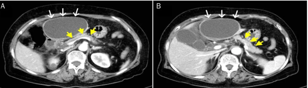
Fig. 2. (A) Follow-up abdominal computed tomography scan demonstrated a huge cystic mass arising from left lateral section of the liver.
(B) Porto-mesenteric junction collapsed by the mass effect of cystic tumor (yellow arrows). The border between the pancreas and cystic mass was clearly demarcated (yellow arrow heads). Soft tissue stranded around the duodenum was seen, suggesting groove pancreatitis (white arrows). (C) Prominent periportal edema in the umbilical fissure was observed (white arrows).
ic tumor between the liver and pancreatic body; however, the appearance of the pancreas was normal (Fig. 1). On the fol- lowing day, she was referred to our hospital due to severely aggravated upper abdominal pain, despite medical treatment.
She had no past history of pancreatitis or alcohol consumption.
A physical examination showed mild epigastric tenderness without signs of peritoneal irritation. Laboratory studies re-
vealed white blood cell count of 12,000/mm3, hemoglobin level of 13.3 g/dL, and platelet count of 250,000/mm3. The level of serum amylase and lipase were elevated to 484 U/L and 215 U/L, respectively. The serum levels of aspartate ami- notransferase (31 U/L), alanine aminotransferase (26 U/L), alkaline phosphatase (48 U/L), and total bilirubin (0.92 mg/dL) were within normal limits. Abdominal CT scan showed a
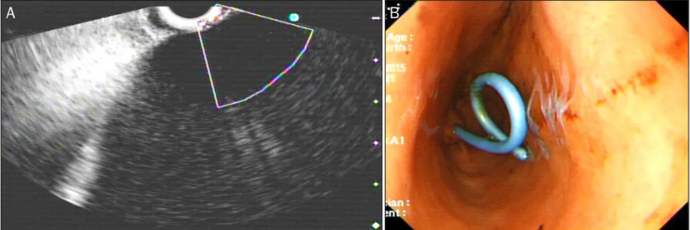
Fig. 3. (A) Endoscopic ultrasonography revealed a huge hypoechoic lesion with internal echogenicity without any solid components or mural nodes. (B) A 7-Fr bipigtailed plastic stent was deployed.
A B
Fig. 4. (A) A huge cystic mass arising from the left lateral section of the liver was seen. (B) The falciform ligament and Glisson pedicle were swollen remarkably and filled with necrotic fatty tissues (arrows).
10.3×6.7×11.7 cm sized cystic mass in the left lateral sec- tion of the liver (Fig. 2A). Internal sepation or irregular papil- lary growth was not observed within the cystic lesion. The por- to-mesenteric junction was collapsed by mass effect of the cystic lesion (Fig 2B). The cystic lesion was not communi- cated with the pancreas. The boundary between the pancre- atic body and cystic mass was clearly delineated (Fig. 2B).
Appearance of the pancreas on the CT scan was relatively normal; however, soft tissue straining was observed around
the duodenum, which resulted in the suspicion of groove pan- creatitis (Fig 2B). The Glisson pedicle in the umbilical fissure was swollen remarkably (Fig. 2C). Endoscopic ultrasonography (EUS)-guided transgatric internal drainage was carried out due to the suspicion of infected hepatic cyst or a pancreatic pseudocyst. An EUS demonstrated a huge hypoechoic lesion with internal echogenicity (Fig. 3A); however, no solid compo- nent or mural nodule was observed. The cystic lesion was punctured with a 19-gauge needle; then, dark green colored
C D
Fig. 5. (A) Gross features: an unilocular cyst containing a ragged inner surface and thick fibrous wall. (B) The cyst is devoid of an epithelial lining (H&E stain, ×40). (C) The lining of the cyst is composed of granulation tissue, inflammatory cells, and fibrous tissue (H&E stain, ×100).
(D) Marked necrosis, fat necrosis, and acute inflammatory cells are observed in the falciform ligament (H&E stain, ×100).
turbid fluid was aspirated. The tract was dilated with 6-8 mm sized CRE™ balloon dilatation catheters (Boston scientific corporation, Boston, MA, USA) and a 7-Fr bipigtailed plastic stent was deployed (Fig. 3B). Cystic fluid analysis revealed high levels of amylase (21,200 U/L), lipase (1,392 U/L), and total bi- lirubin (5.18 mg/dL). The carcinoembryonic antigen level of the cystic fluid was 70.68 ng/mL. Unfortunately, after the en- doscopic procedure, abdominal pain was aggravated. On physical examination, severe epigastric tenderness, re- bound tenderness, and rigidity were observed. The rupture of the cystic lesion was suspected; therefore, an emergency op- eration was performed. According to operative findings, a huge cystic mass was located in the left lateral section of the liver. Cystic mass contained brown turbid fluid and necrotic tissues, and cystic fluid spilled into the lesser sac. There was no communication between the cystic tumor and pancreas.
Porta hepatis, Glisson pedicle in the umbilical portion, and
falciform ligament were swollen remarkably and filled with ne- crotic fatty tissues (Fig. 4). A left hemihepatectomy was performed. Grossly, an intrahepatic unilocular cyst contain- ing a ragged inner surface and thick fibrous wall was ob- served (Fig. 5A). Pathological examination revealed a pseu- docystic nature. The cystic lesion was devoid of an epithelial lining (Fig. 5B). The lining of the pseudocyst was composed of granulation tissue, inflammatory cells, and fibrotic tissue (Fig. 5C). The falciform ligament was composed of marked necrosis with fat necrosis and acute inflammatory cells (Fig. 5D).
They seem to be common findings in acute pancreatitis.
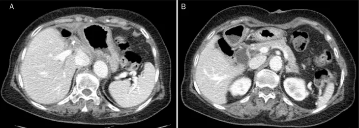
Inclusively, the final diagnosis confirmed intrahepatic pan- creatic pseudocyst. The patient was discharged without any complication on postoperative days 11. No complication was observed during the follow-up period. Twenty-eight months after the operation, the patient was disease-free (Fig. 6).
Fig. 6. Abdominal computed tomography after 28 months of operation. There were no abnormal findings of the liver (A) and pancreas (B).
DISCUSSION
Only about 50 cases of IHPP following pancreatitis have been described in the literature.3 In our case, the patient had no history or symptom of acute pancreatitis, and there was no obvious evidence of acute pancreatitis on an image study, except slightly elevated serum amylase and lipase levels.
Despite normal appearance of the pancreas, soft tissue of the porta hepatis and faliciform ligament was markedly nec- rotized; and there was an occurrence of huge IHPP. IHPP was the only clinical manifestation without any obvious symptom of preceding acute pancreatitis. Patient’s symptom ―upper abdominal pain― was related to IHPP. It is presumed that sub- clinical groove pancreatitis have already existed. Demeusy et al. have reported that of the 54 cases of IHPP since the 1970s, the most common location of IHPP was the left hep- atic lobe (48%).3 CT is generally considered as a modality of choice for the diagnosis of the IHPP. However, diagnosis of IHPP can be difficult sometimes due to its rarity, especially in cases without any history of pancreatitis or obvious findings of pancreatitis. IHPPs should be differentiated from other conditions, such as intrahepatic biliary dilatation, peribiliary cyst, complicated hepatic cyst, liver abscess, biloma, echino- coccal cyst, mucinous cystic neoplasm, and intraductal muci- nous neoplasm of the bile duct. Cystic fluid analysis, which demonstrated a high amylase content, can be a definitive diag- nosis of IHPPs.3 In previous reports, almost all cases were treat- ed with percutaneous drainage or endoscopic procedure.4-8 In our case, there was no obvious evidence of acute pan- creatitis on abdominal CT; instead the scan showed normal
appearance of the pancreas, huge intrahepatic cystic tumor, and enlarged lymph nodes of porta hepatis. Hence, differ- ential diagnosis was required, including complicated heaptic cystic tumors, such as infected hepatic cyst, mucinous cystic neoplasm, or intraductal papillary neoplasm of the bile duct.
Cystic fluid analysis has limited sensitivity in differentiating the hepatic cystic lesion. Surgical resection is ultimately nec- essary to confirm the diagnosis.9 Cystic fluid analysis re- vealed a high level of amylase content, suggesting IHPP.
EUS-guided transgastric internal drainage could be a defini- tive treatment modality. However, after endoscopic proce- dure, signs of peritonitis and clinical deterioration were observed. Therefore, we decided to perform the operation.
There are two mechanisms proposed to explain the patho- physiology of IHPP.4,10-13 The first mechanism is consisted of pancreatic enzyme released following acute pancreatitis into the lesser sac spreading along the lesser omentum and gas- trohepatic ligaments. This mechanism explains why IHPPs are frequently observed in the left lobe. The second mecha- nism is the spreading of pancreatic enzyme from the pancre- atic head into the hepatoduodenal ligament and porta hep- atis, along the portal vein and its branches. In our case, marked swelling and necrosis were observed in hep- atoduodenal ligament, umbilical portion, and falciform ligament. However, there was no obvious finding of acute pancreatitis at the pancreatic body and tail or communica- tion between the cystic mass and the pancreas. Therefore, the second mechanism is more suitable for explaining the pathophysiology in this case.
In conclusion, IHPPs should be considered when a huge in-
10:146-150.
2. Skouras C, Skouras T, Pai M, Zacharakis E, Spalding D. Inguinoscrotal extension of a pancreatic collection: a rare complication of pan- creatitis-case report and review of the literature. Updates Surg 2013;65:153-159.
3. Demeusy A, Hosseini M, Sill AM, Cunningham SC. Intrahepatic pancreatic pseudocyst: a review of the world literature. World J Hepatol 2016;8:1576-1583.
4. Bhasin DK, Rana SS, Chandail VS, et al. An intra-hepatic pancre- atic pseudocyst successfully treated endoscopic transpapillary drainage alone. JOP 2005;6:593-597.
5. Bhasin DK, Rana SS, Nanda M, et al. Endoscopic management of pancreatic pseudocysts at atypical locations. Surg Endosc 2010;24:1085-1091.
1328-1347; quiz 1348.
10. Casado D, Sabater L, Calvete J, et al. Multiple intrahepatic pseu- docysts in acute pancreatitis. World J Gastroenterol 2007;13:
4655-4657.
11. Okuda K, Sugita S, Tsukada E, Sakuma Y, Ohkubo K. Pancreatic pseudocyst in the left hepatic lobe: a report of two cases. Hepatology 1991;13:359-363.
12. Siegelman SS, Copeland BE, Saba GP, Cameron JL, Sanders RC, Zerhouni EA. CT of fluid collections associated with pancreatitis.
AJR Am J Roentgenol 1980;134:1121-1132.
13. Ancel D, Lefebvre M, Peyrin-Biroulet L, et al. Pancreatic pseudo- cysts of the right hepatic lobe during acute biliary pancreatitis.
Gastroenterol Clin Biol 2005;29:743-745.