J Korean Ophthalmol Soc 2019;60(10):1010-1014 ISSN 0378-6471 (Print)⋅ISSN 2092-9374 (Online)
https://doi.org/10.3341/jkos.2019.60.10.1010
Case Report
뇌하수체졸중환자에서 발생한 단독 양측 동안신경마비
Pituitary Apoplexy Presenting as Isolated Bilateral Oculomotor Nerve Palsy
조희정1⋅송영진2⋅류원열1
Heejung Cho, MD1, Young Jin Song, MD, PhD2, Won Yeol Ryu, MD, PhD1
동아대학교 의과대학 안과학교실1, 동아대학교 의과대학 신경외과학교실2 Department of Ophthalmology, Dong-A University College of Medicine1, Busan, Korea
Department of Neurosurgery, Dong-A University College of Medicine2, Busan, Korea
Purpose: To report a case of pituitary apoplexy presenting as isolated bilateral oculomotor nerve palsy.
Case summary: A 46-year-old male presented with bilateral ptosis and acute severe headaches for 6 days. He underwent head surgery and bilateral vitrectomy 12 years prior to his visit because of ocular and head trauma. He mentioned that previous visual acuities in both eyes were not good. The initial corrected visual acuity was finger counting in the right eye and 20/500 in the left eye. Ocular motility testing revealed the limitation of adduction, supraduction, and infraduction with complete bilateral ptosis in both eyes, and his left pupil was dilated. He was diagnosed with an isolated bilateral oculomotor nerve palsy. Magnetic reso- nance imaging indicated pituitary gland hemorrhage with a tumor, which was suspicious of pituitary apoplexy. The patient was treated intravenous with 1.0 g methylprednisolone to prevent the corticotropic deficiency. In addition, he underwent surgical de- compression using a navigation-guided transsphenoidal approach and aspiration biopsy. He was confirmed with pituitary ad- enoma using a pathological examination. The patient’s ocular movements began to dramatically improve by the third day postoperatively. At 4 months postoperative follow-up, his ocular movement and double vision were completely recovered.
Conclusions: This was a rare case of pituitary apoplexy with bilateral isolated oculomotor nerve palsy, which was the first report in the Republic of Korea. A full recovery was achieved after early surgical treatment.
J Korean Ophthalmol Soc 2019;60(10):1010-1014
Keywords: Cranial nerve palsy, Oculomotor nerve disease, Oculomotor nerve palsy, Pituitary adenoma, Pituitary apoplexy
■Received: 2019. 3. 21. ■ Revised: 2019. 4. 17.
■Accepted: 2019. 9. 24.
■Address reprint requests to Won Yeol Ryu, MD, PhD Department of Ophthalmology, Dong-A University Hospital,
#26 Daesingongwon-ro, Seo-gu, Busan 49201, Korea Tel: 82-51-240-2737, Fax: 82-51-254-1987
E-mail: [email protected]
*Conflicts of Interest: The authors have no conflicts to disclose.
ⓒ2019 The Korean Ophthalmological Society
This is an Open Access article distributed under the terms of the Creative Commons Attribution Non-Commercial License (http://creativecommons.org/licenses/by-nc/3.0/) which permits unrestricted non-commercial use, distribution, and reproduction in any medium, provided the original work is properly cited.
뇌하수체졸중은 뇌하수체에서의 갑작스러운 혈류 공급 중단 또는 출혈이 원인이 되어, 뇌하수체 조직의 갑작스러 운 확장 때문에 발생하는 임상증후군으로 보통 뇌하수체샘 종이 동반하는 경우가 많다.1 두통, 구토, 오심, 시력저하,
호르몬 기능 이상 등의 증상이 발생할 수 있고 뇌신경마비 가 동반할 경우 눈운동마비에 의한 복시가 나타날 수 있다. 뇌신경마비에 의한 눈운동마비는 단안성으로 발생하는 경 우가 많고 드물게 양측성으로도 발생할 수 있다.2 뇌하수체 졸중으로 발생한 양안 동안신경마비만 발생하는 경우는 매 우 드물고 지금까지 국내에 보고된 적이 없었다. 본 저자들 은 뇌하수체졸중과 동반한 양안 동안신경마비환자를 진단 하고 조기 수술적 치료 후 빠른 호전을 보였던 환자를 경험 하였기에 이를 보고하고자 한다.
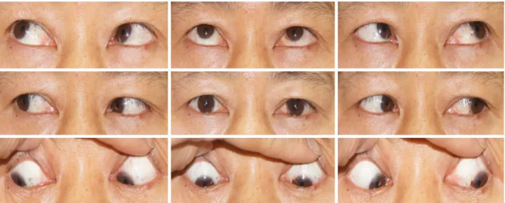
Figure 1. Nine cardinal gaze position at initial visit. Ocular movement was revealed the limitation of adduction, supraduction, and infraduction with complete ptosis in both eyes and a dilated pupil was shown in the left eye.
A B
Figure 2. Magnetic resonance images. Pituitary apoplexy was shown with large cystic lesion on T1 weighted images (arrows) (A, sagittal view, B, axial view).
증례보고
46세 남자가 6일 전부터 발생한 양안 눈꺼풀처짐 및 갑 작스러운 극심한 두통으로 방문하였다. 12년 전에 두부 및 안면부 외상으로 두부 수술, 양안 유리체절제술 및 망막전 막제거술을 받았던 병력이 있어서 양안 시력이 좋지 않았 었다고 하였다. 첫 내원 시 측정한 교정시력은 우안 안전수 지, 좌안 0.05였고 좌안 동공이 산대된 상태였다. 전안부검 사상에서 양안 백내장이 심한 상태였으며, 양안 완전 눈꺼
풀처짐(complete ptosis)도 있었다. 눈운동검사에서 양안의 내전, 상전, 하전 장애가 관찰되고 외하전 방향에서 내회선 이 가능하여 단독 양안 동안신경마비가 의심되었다(Fig. 1).
뇌 자기공명영상에서 뇌하수체에 큰 출혈성 낭성 병변으로 뇌하수체샘종과 동반한 뇌하수체졸중으로 강하게 의심되 었다(Fig. 2). 환자는 즉각 입원하여 메틸프레드니솔론 1.0 g 정맥주사 치료를 3일간 시행하였고, 용량을 서서히 감량하 였다. 수술 전 확인한 호르몬검사에서 갑상샘자극호르몬 (thyroid stimulating hormone)은 0.26 µlU/mL, T3는 0.55 μg/dL
Figure 3. Nine gaze photos at postoperative 4 months. Ocular eye movement and dilated pupil in the left eye were completely recovered.
로 정상 수치보다 다소 낮았고, 항이뇨호르몬(Antidiuretic Hormone)은 13.07 pg/mL로 상승한 것 외에 프로락틴(pro- lactin)이나 코티솔(cortisol) 등 다른 호르몬과 전해질 수치 는 정상이었다. 시야검사에서 양측 모두 전체적인 시야 결 손이 확인되었으나, 반맹을 포함한 뚜렷한 양상의 결손은 아니었다. 안저에서는 망막이나 시신경 이상은 양안 모두 관찰되지 않았고 빛간섭단층촬영(optical coherence tomog- raphy)에서도 특별한 이상 소견은 없었다.
뇌항해기법장치를 이용한 경접형골 접근법(Navigation guided transsphenoidal approach)으로 외과적 감압술(surgical decompression) 및 흡인생검(aspiration biopsy)을 시행하였 다. 병리검사 결과, 괴사성 조직(necrotic tissue)으로 뇌하수 체졸중의 합당한 소견이었다. 수술 치료 2일 후부터 우안 눈꺼풀처짐이 호전되기 시작했고, 술 후 3일째, 우안 안구 운동장애가 모두 회복되었으며 좌안 또한 내전 및 상전장 애가 호전되기 시작하였다. 술 후 2-3개월에 우안 및 좌안 백내장수술을 받은 후 양안 교정시력은 0.6으로 호전되었 다. 술 후 4개월째, 안저검사, 시야검사 및 빛간섭단층촬영 검사에서 시신경 이상 소견은 관찰되지 않았고, 양안 안구 운동 장애 및 복시, 좌측 산대된 동공 모두 완전 회복되었 다(Fig. 3).
고 찰
뇌하수체졸중의 발생 기전에 대해서는 완전하게 밝혀져 있지는 않지만, 뇌하수체 혈류 체계가 뇌하수체샘종으로 인하여 혈류 공급 기능이 저하되는 것과 관계가 있다고 알
려져 있다.1 뇌하수체졸중의 임상 증상은 뇌하수체의 출혈, 괴사, 부종 등 뇌하수체 상태에 따라 매우 다양하게 나타날 수 있다. 그중 시력 및 시야 장애는 출혈성 종양의 갑작스 러운 증가에 의한 시신경 및 시신경 교차의 압박으로 인해 발생하고 환자의 절반 이상에서 나타난다고 알려져 있다.2,3 시야 결손은 다양한 형태로 발생할 수 있고, 양귀쪽반맹이 가장 흔한 걸로 알려 있으나,1 본 환자의 시력저하 및 시야 결손은 뇌하수체졸중 발생 이전에 시행된 망막수술과 그와 연관 있는 심한 백내장 때문에 정확한 시야 결손의 양상을 확인하기 어려웠다.
뇌하수체졸중환자에서 눈운동마비에 의한 복시 또한 비 교적 흔하여 25-78%까지 다양하게 보고되고 있다.2-4 Hage et al2은 235명의 뇌하수체졸중환자에서 눈운동마비는 25%
였고, 안과 검사를 받았던 27명 중에 23명(85.2%)이 단안 눈운동마비였다고 하였다. 동안신경이 침범되어 있는 형태 가 가장 흔한 것으로 알려져 있으나 다른 뇌신경마비와 같 이 동반된 형태가 많고,1 단독 양안 동안신경마비는 비교적 드물어서 몇몇 증례 보고만 있었다.5-7 국내에서도 단독 동 안신경마비에 대한 보고가 있었으나, 모두 단안이었다.4,8 본 증례는 뇌하수체졸중에 의한 단독 양안 동안신경마비의 형태로 나타났다.
본 증례의 환자와 같이 양안 눈꺼풀 처짐을 동반하는 복 시로 나타나는 대표적인 질환으로 눈중증근육무력증, 밀러- 피셔증후군, 기타 해면정맥굴을 침범하는 질환 등이 있고 뇌하수체졸중을 의심할 때 감별해야 할 질환들이다.9 따라 서 양안 눈꺼풀 처짐과 복시를 호소하는 환자에서 위 질환 들을 의심하고 감별하기 위해서는 항GQ1b항체검사 또는
아세틸콜린 수용체 결합 항체검사 등이 필요하다. 본 증례 의 환자는 12년 전 외상으로 인한 두부수술의 병력이 있었 기 때문에, 우선적으로 신경외과적 이상을 감별하기 위한 뇌 영상검사를 시행하여 기타 다른 질환에 대한 혈청검사 없이 조기에 진단되었다. 또한 환자가 호소하였던 극심한 두통은 눈중증근육무력증과 밀러-피셔증후군과 맞지 않는 소견이라고 할 수 있다.
Hage et al2은 뇌하수체졸중환자 110명의 조직병리 소견 을 안구운동장애가 있는 군과 없는 군으로 나누어 비교해 본 결과, 안구운동장애가 있는 군에서 괴사가 있는 경우가 가장 많았다고 하였다. 또한 안구운동장애가 동반된 뇌하수 체졸중환자에서 보다 범뇌하수체저하증(panhypopituitarism) 과 내분비 이상이 나타나는 경우가 많았고, 그것은 이러한 뇌하수체 조직의 높은 괴사 소견과 관련 있기 때문에, 뇌하 수체졸중환자에서 안구운동장애의 동반 자체가 내분비 이 상과 관련한 위험 인자가 될 수 있다고 하였다. 본 환자 또 한 조직병리 소견에서 괴사 소견이 나타났고, 안구운동장 애가 나타났던 증례로 내분비 및 전해질 이상에 대비하여 수술 전 호르몬 및 전해질 검사를 하였으나 다행스럽게 큰 이상 소견을 보이지 않았다.
뇌하수체졸중의 치료 접근 방법에 대하여 대규모 전향적 연구가 없었고, 증상이 매우 다양하기도 하여 정립된 치료 접근 방법은 없다. 스테로이드 결핍증(corticotropic defi- ciency)이 발생할 경우, 생명에 위협을 줄 수 있기 때문에 진단 즉시 고용량 스테로이드 정맥주사 치료가 필수적이 다.1 일부 연구에서는 시력 및 의식 소실이 없는 뇌하수체 졸중환자에서는 보존적인 치료로 호르몬 균형 유지 등의 시도를 우선적으로 시도하자고 주장하고 있으나,10 뇌항해 기법장치 경접형골 접근법을 이용한 외과적 감압술이 확실 한 치료 방법으로 추천된다. 특히 시력 및 시야 이상과 의 식 수준이 감소하기 시작할 경우 외과적 감압술이 우선적 으로 고려되어야 한다. 2011년 영국에서 제시된 뇌하수체 졸중의 치료지침에는 심각한 시력 및 시야 이상, 의식 소실 이 있으면 수술적 치료를 고려하고, 시력 및 시야 이상을 동반하지 않고 단지 제3번, 4번, 6번 뇌신경 마비만 있는 것으로는 수술적 적응이 되지 않으며, 수술이 아주 급한 상 황이 아니라면, 경험 많은 신경외과의에게 증상 발생 후 1주
이내에 수술 받는 것을 추천하였다.11 본 증례는 의식 수준 은 정상이었고 호르몬 및 전해질에 큰 이상은 없었으나 시 력 및 시야 이상은 동반되어 있고, 이미 저시력이었기 때문 에 비교적 빠른 수술적 치료가 필요하는 상황이었다.
본 환자는 뇌하수체졸중으로 발생한 단독 양측 동안신경 마비의 드문 증례로 조기 수술적 치료 후 빠른 호전을 보였 다. 갑작스러운 두통, 시력 및 시야 이상 등을 동반하는 뇌 하수체졸중환자에서도 단독 양안 동안신경마비의 형태로 나타날 수 있고, 호르몬 및 전해질 불균형에 유의하면서 시 력 및 시야 이상 등이 동반한다면, 즉각적인 수술적 치료가 필요할 것으로 생각된다.
REFERENCES
1) Briet C, Salenave S, Bonneville JF, et al. Pituitary apoplexy.
Endocr Rev 2015;36:622-45.
2) Hage R, Eshraghi SR, Oyesiku NM, et al. Third, fourth, and sixth cranial nerve palsies in pituitary apoplexy. World Neurosurg 2016;94:447-52.
3) Bills DC, Meyer FB, Laws ER Jr, et al. A retrospective analysis of pituitary apoplexy. Neurosurgery 1993;33:602-9.
4) Jeon HJ, Kang JH, Cho BM, et al. Clinical manifestations and sur- gical outcome of pituitary apoplexy. J Korean Brain Tumor Soc 2008;7:120-6.
5) Komurcu HF, Ayberk G, Ozveren MF, Anlar O. Pituitary adenoma apoplexy presenting with bilateral third nerve palsy and bilateral proptosis: a case report. Med Princ Pract 2012;21:285-7.
6) Man BL, Fu YP. Pituitary apoplexy presenting with bilateral oculo- motor nerve palsy. BMJ Case Rep 2015;2015. https://casereports.
bmj.com/content/2015/bcr-2015-212049. Accessed Oct 15, 2015.
7) Lau KK, Joshi SM, Ellamushi H, Afshar F. Isolated bilateral oculo- motor nerve palsy in pituitary apoplexy: case report and review. Br J Neurosurg 2007;21:399-402.
8) Lee HG, Noh JS, Rhim JK, Chung BS. Isolated third cranial nerve palsy with ptosis presented in pituitary apoplexy: case report. J Korean Brain Tumor Soc 2011;10:20-1.
9) Yadegari S. Approach to a patient with blepheroptosis. Neuro Sci 2016;37:1589-96.
10) Maccagnan P, Macedo CL, Kayath MJ, et al. Conservative man- agement of pituitary apoplexy: a prospective study. J Clin Endocrinol Metab 1995;80:2190-7.
11) Rajasekaran S, Vanderpump M, Baldeweg S, et al. UK guidelines for the management of pituitary apoplexy. Clin Endocrinol (Oxf) 2011;74:9-20.
= 국문초록 =
뇌하수체졸중환자에서 발생한 단독 양측 동안신경마비
목적: 뇌하수체졸중으로 인해 발생한 양안 단독 동안신경마비 1예를 보고하고자 한다.
증례요약: 46세 남자가 6일 전부터 발생한 양안 눈꺼풀처짐 및 갑작스러운 극심한 두통으로 방문하였다. 12년 전에 외상으로 두부수술 및 양안 유리체절제술을 받았던 병력이 있어서 양안 시력이 좋지 않았던 환자로, 첫 내원 시 교정시력은 우안 안전수지, 좌안 0.05였 다. 좌측 동공이 산대되어 있었고 우측은 정상이었다. 눈운동검사에서 양안 모두 내전, 상전, 하전 장애가 관찰되어 양안 동안신경마비 로 의심되었다. 뇌 자기공명영상에서 뇌하수체졸중으로 진단되어 입원 후 메틸프레드니솔론 1.0 g 전신 정맥주사 치료를 하였고, 뇌항 해기법장치 경접형골 접근법(navigation guided transsphenoidal approach)을 이용한 외과적 감압술(surgical decompression) 및 흡인생검을 시행하였다. 병리검사 결과, 뇌하수체샘종으로 확진되었다. 술 후 3일째에 우안 안구운동장애는 모두 호전되었고, 술 후 4개월째에 양안 눈 운동이 완전히 호전되었다.
결론: 뇌하수체졸중으로 발생한 단독 양안 동안신경마비는 드물고 국내 최초의 보고이며, 수술 치료 후 빠른 완전 회복을 보였기에 보고하고자 한다.
<대한안과학회지 2019;60(10):1010-1014>
조희정 / Heejung Cho
동아대학교 의과대학 안과학교실 Department of Ophthalmology, Dong-A University College of Medicine