DOI : 10.3341/jkos.2009.50.10.1600
= 증례보고 = 접수번호 : 50-10-04-13
우측 7번, 8번 뇌신경 마비를 동반한 선천성 시신경유두결손증
조현경⋅지동현
가톨릭대학교 의과대학 안과 및 시과학교실
목적: 우측의 7번, 8번 뇌신경 마비를 동반한 양안의 선천성 시신경유두결손증을 보고하고자 하였다.
증례요약: 우측 안면마비(7번 뇌신경 마비)와 우측 귓볼저형성증을 동반한 신생아가 미숙아 망막병증 검사를 위해 의뢰되었다. 산동 망막검사에서 양안의 시신경유두결손과 시신경유두 주변부의 맥락막 결손이 발견되었으며, 전안부 소견상 특이사항은 없었다. 추후 시행한 검사에서 후두연화증과 헐거운 후두개(floppy epiglottis)가 발견되었으며, 측두골 컴퓨터단층촬영에서는 좌측의 중이염과 유두염 소견이 관찰되었다. 청성뇌간유발반응검사(Auditory Brain stem Response)에서 우측 전위도가 나오지 않아 우측 8번 뇌신경(Cochlear nerve)마비로 진단되었다. 이후 시행한 염색체 검사와 뇌 자기공명영상에서는 이상이 발견되지 않았다. 심장초음파검사상 심방중격결 손이 있었으며, 상부위장관촬영상 위식도역류증이 있었다.
결론: 선천성유두결손증의 경우 다른 선천성 기형이나 이상을 동반하는 경우가 많으므로 이에 대한 검사가 반드시 필요하다.
<대한안과학회지 2009:50(10):1600-1604>
■ 접 수 일: 2009년 4월 3일 ■ 심사통과일: 2009년 6월 30일
■ 책 임 저 자: 지 동 현
경기도 수원시 팔달구 지동 93-6 가톨릭대학교 성빈센트병원 안과 Tel: 031-249-7343, Fax: 031-251-6225 E-mail: [email protected]
선천성 시신경유두결손증은 시신경에 경계가 명확한 둥근 함몰을 보이는 질환이며, 주로 주변부에 위치하며 아랫쪽이 더 깊은 경향을 보인다. 선천성 시신경유두결손증은 눈술잔 (optic cup)의 양근위부(proximal)의 비정상적인 융합에 기인하는 것으로 알려져 있다.1선천성 시신경유두결손증은 다양한 선천성 이상을 동반하는데 이는 재태 6주에 태아발 달에 이상이 있음을 시사한다.2선천성 시신경유두결손증은 산발적으로 일어나거나 상염색체 우성으로 유전되기도 한 다.3
최근에는 PAX2 유전자의 돌연변이와 연관이 있으며 신장 -시신경유두결손증 증후군(renal-coloboma syndrome)의 일환으로도 받아들여지고 있다.4-7CHARGE 증후군(Colo- boma, Heart defect, Atresia choanae, Retarded growth and development, Genital hypoplasia, Ear anomalies/
deafness)에 동반된 경우에는 CHD7 유전자 돌연변이가 유 발원인의 하나로 생각되고 있다.8,9
선천성 시신경유두결손증의 단안 또는 양안의 발병률은 비슷한 것으로 알려져 있다.10
주변 하측의 망막과 맥락막 결손이 있는 경우에는 명백한 소안구증 역시 동반될 수 있다.11
시력은 다양한 정도로 저하될 수 있는데, 시력 예후에 영
향을 주는 가장 중요한 인자는 시신경 유두결손의 중심와 침범 정도라고 밝혀져 있다.12현저한 굴절이상과 양안 부 등시도 자주 동반되는 것으로 알려져 있다.12 양안의 상염 색체 우성 시신경유두결손 환자에서 진행성의 시신경유두 함몰과 시신경유두테의 감소가 발견되었으나 안압 상승이 나 진행성의 시야결손은 관찰되지 않았다는 보고도 있다.13 국내에서는 선천성 시신경유두결손이 2예14,15 보고되었 으나, 모두 단안의 시신경유두결손이었으며, 동반된 다른 임상양상에 대한 언급은 없었다. 양안의 선천성 시신경유두 결손증에서 우측의 7번, 8번 뇌신경 마비를 동반한 경우는 국내에서 보고된 바가 없어 저자들이 처음으로 보고하는 바이다.
증례보고
우측 안면마비와 우측 귓볼 저형성증을 보이는 재태연령 40+3주의 신생아가 미숙아 망막병증 검사를 위해 본원 안 과로 의뢰되었다. 환아는 재태연령 38+1주에 자연분만으로 출생하여, 체중이 3.12 kg이었으며, 출생 시부터 산소치료의 과거력은 없었다. 가족력에서도 특이사항은 없었다.
산동 망막검사를 시행한 결과 양안의 시신경유두결손과 시신경유두 주변부의 맥락막 결손이 발견되었다(Fig. 1).
전안부 소견상은 특이 사항은 없었다.
우측 안면마비와 우측 귓볼 저형성증 소견이 있어 정밀 검사를 위해 이비인후과 진료를 의뢰하였다. 후두경 검사상 부피가 큰 모뿔연골 형(bulky arytenoids type)의 후두연 화증(laryngomalacia)과 헐거운 후두개(floppy epiglottis)
A B
Figure 1. Fundus photographs showing bilateral optic disc coloboma and peripapillary choroidal defect (A=right;
B=left).
Figure 2. Temporal bone CT scan demonstrating left otitis media (upper arrow) and mastoiditis (lower arrow).
가 발견되었으며, 성대마비 소견은 없었다. 측두골 컴퓨터 단층촬영(Temporal bone CT)(Fig. 2)에서는 좌측 중이염 (otitis media)과 유두염(mastoiditis) 소견이 관찰되었다.
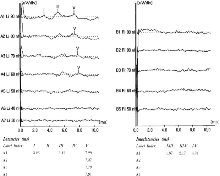
신생아 청력검사의 일환인 청성뇌간유발반응검사(Auditory brain Stem Response)에서 좌측은 정상반응을 보이는 반면 에, 우측은 전위도가 나오지 않아 우측 8번 뇌신경(Cochlear nerve)마비로 진단되었다(Fig. 3).
여러 장기를 침범한 질환 양상을 보여 염색체 검사를 해 보았으나, 특이 소견은 없었다. 뇌 자기공명영상(Brain MRI) 에서도 특이 소견은 보이지 않았다.
심장초음파검사상 심방중격결손(atrial septal defect)가 발견되었고, 상부위장관촬영(Upper Gastrointestinal Series) 상 위식도역류증(Gastroesophageal reflux disease) 소견
이 있었다.
앞서 언급한 헐거운 후두개(floppy epiglottis)로 인한 흡 인을 방지하기 위해 머리를 들고 조금씩 먹이는 방법을 취 하며, 한꺼번에 많이 먹이지 말고, 자주 트림 시킬 것을 교육 하였다. 그러나 환아는 이후 2차례의 기관지염과 폐렴으로 입원 후 항생제 치료를 받았다.
앞서 언급한 헐거운 후두개(floppy epiglottis)로 인한 흡 인을 방지하기 위해 머리를 들고 조금씩 먹이는 방법을 취 하며, 한꺼번에 많이 먹이지 말고, 자주 트림 시킬 것을 교육 하였다. 그러나 환아는 이후 2차례의 기관지염과 폐렴으로 입원 후 항생제 치료를 받았다.
생후 13주째 마지막 외래 경과 관찰시 시신경유두결손은 진행하지 않고 안정적인 상태를 보였다.
고 찰
시신경유두결손은 다양한 질환과 연관되어 있다. Brodsky11 에 의해 기술된 바에 의하면 CHARGE 증후군(Coloboma, Heart defect, Atresia choanae, Retarded growth and development, Genital hypoplasia, Ear anomalies/deaf- ness),8,9Walker-Warburg 증후군,16 Goltz 국소 피부 저 형성증(focal dermal hypoplasia),17Aicardi 증후군,18,19 Goldenhar 증후군(oculoauricularverte-bral dysplasia),20,21 그리고 선상피지모반 증후군(linear sebaceous naevus syn- drome)22들과 관련이 있다고 한다. 최근에는 Dandy Walker 기형(malformation)12및 신장-시신경유두 결손증 증후군 (renal-coloboma syndrome)과의 관련도 보고되고 있다.4-7 1998년에 전문가들이 정의한 CHARGE 증후군의 주진단 기준(Major 4C’s)으로는 시신경유두결손증(Ocular Colo- boma), 후비공폐쇄(Choanal atresia), 뇌신경이상(Cranial nerve anomalies), 귀의 특징적인 이상(Characteristic ear
Latencies (ms)
Label Index I II III IV V
A1 3.25 5.12 7.29
A2 7.37
A3 7.79
A4 7.91
Interlatencies (ms)
Label Index I-III III-V I-V
A1 1.87 2.17 4.04
A2 A3 A4
Figure 3. Auditory brain stem response showing absence of the right electropotential (left), which indicates right cochlear nerve palsy.
anomalies)이 있고, 부진단기준(Minor diagnostic criteria) 으로는 심혈관계 이상(Cardiovascular malformation), 생 식기의 저형성(genital hypoplasia), 구개열/구순열(Cleft palate/lip), 기관식도루(Tracheoesophageal fistula), 특징 적인 CHARGE 얼굴(Distinctive CHARGE faces), 성장부 전(Growth deficiency), 발달지연(Developmental delay) 가 있다.8이 진단 기준 중에 4가지 주진단기준에 모두 해당 하거나 3가지 주진단기준과 3가지 부진단기준에 해당하면 CHARGE 증후군일 가능성이 아주 높다. 이 증례에서는 주 진단기준 중 시신경유두결손증(Ocular Coloboma), 뇌신경 이상(Cranial nerve anomalies), 귀의 특징적인 이상(Cha- racteristic ear anomalies) 세 가지에 해당되며 부진단기준 중에는 심혈관계 이상(Cardio-vascular malformation)이 해당된다. 기관식도루는 발견되지 않았으나 역류성식도염과 기관지연화증이 있어 기관식도루의 변형으로 볼 수 있다.
성장부전이나 발달지연은 뚜렷하게 관찰되지 않았으나 환아
의 나이가 어리므로 명확한 소견은 추후 경과관찰이 필요할 것으로 보인다. 이런 소견에 의해 이 증례의 환아는 CHARGE 증후군의 변형 및 CHARGE 증후군으로 볼 수 있겠다.
이외에 Walker-Warburg 증후군에 동반되는 뇌이랑없 음증(lissencephaly) 및 소뇌 및 뇌간의 이상이나 선천성 근이영양증(congenital muscular dystrophy), Goltz focal dermal hypoplasia에 동반되는 피부의 저형성증 및 유두종 (papilloma), Aicardi 증후군에 동반되는 뇌량무형성증 (callosal agenesis)과 영아 연축(infantile spasms) 소견은 없었다. 또한 Goldenhar 증후군(눈귀척추 형성이상)에 동 반되는 척추이상이나 편측안면소체증(hemifacial micro- somia), 선상피지모반 증후군(linear sebaceous naevus syndrome)에 동반되는 전형적인 정중선 모반(midline nevus)이나 간질 등의 중추신경계 이상은 관찰되지 않았으 며, Dandy Walker 기형(malformation)에 동반되는 수두증 (hydrocephalus) 등 중추신경계의 구조적인 이상, 신장-
시신경유두 결손증 증후군(renal-coloboma syndrome)에 동반되는 신장기능의 이상 및 신장 저형성(renal hypoplasia) 소견도 관찰되지 않았다.
주변 하측의 망막과 맥락막 결손이 있는 경우에는 명백 한 소안구증 역시 동반될 수 있으나11 소안구증 소견은 발 견되지 않았다. 환아는 검사 당시 주시나 따라보기를 할 수 없었으며, 조절마비굴절검사는 시행하지 않았다.
CHARGE 증후군에서는 유발원인의 하나로 CHD7의 유전자 돌연변이가 대두되고 있다. 그러나 Lalani et al23은 임상적으로 CHARGE 증후군으로 진단받은 환자들 중에 58% (64/110)에서만 CHD7 유전자 돌연변이가 검출되었 고, Jongmans et al24도 107명의 코호트를 대상으로 한 연 구에서 유전자형과 표현형 간의 연관성이 명확하지 않다고 보고했다. 신장-시신경유두 결손증 증후군(renal-coloboma syndrome)의 원인으로 대두되고 있는 PAX2 유전자 돌연 변이 역시 Dureau et al25은 50%에서는 검출되지 않았다고 했으며, Cheong et al26도 신장-시신경유두 결손증 증후군 (renal-coloboma syndrome)의 임상양상은 다양하며 유전 자형과 표현형 간의 명확한 연관성은 없다고 하였다. 이 증례 에서 염색체검사는 시행했으나, 유전자 돌연변이에 관련된 검사는 시행하지 않았다. 그러나 위의 연구결과들은 볼 때 유전자 검사가 진단에 필수적인 조건은 아니라고 하겠다.
이 증례에서 양안의 시신경유두결손증이 CHRAGE 증후 군과 연관된 경우를 확인하였으며, 양안의 시신경유두결손증 소견이 있는 경우에는 동반된 전신 이상을 확인하는 것이 필요하겠다.
참고문헌
1) Mann I. Developmental Abnormalities of the Eye, 2nd ed.
Philadelphia, PA: JB Lippincott, 1957;74-91.
2) Duvall J, Miller SL, Cheatle E, Tso MO. Histopathologic study of ocular changes in a syndrome of multiple congenital anomalies.
Am J Ophthalmol 1987;103:701-5.
3) Yamashita T, Kawano K, Ohba N. Autosomal dominantly inherited optic nerve coloboma. Ophthalmic Paediatr Genet 1988;
9:17-24.
4) Schimmenti LA, Manligas GS, Sieving PA. Optic nerve dysplasia and renal insufficiency in a family with a novel PAX2 mutation, Arg115X: further ophthalmologic delineation of the renal- coloboma syndrome. Ophthalmic Genet 2003;24:191-202.
5) Chung GW, Edwards AO, Schimmenti LA, et al. Renal- coloboma syndrome: report of a novel PAX2 gene mutation. Am J Ophthalmol 2001;132:910-4.
6) Salomon R, Tellier AL, Attie-Bitach T, et al. PAX2 mutations in
oligomeganephronia. Kidney Int 2001;59:457-62.
7) Eccles MR, Schimmenti LA. Renal-coloboma syndrome: a multi- system developmental disorder caused by PAX2 mutations. Clin Genet 1999;56:1-9.
8) Blake KD, Prasad C. CHARGE syndrome. Orphanet J Rare Dis 2006;7;1:34.
9) Sanlaville D, Verloes A. CHARGE syndrome: an update. Eur J Hum Genet 2007;15:389-99.
10) Pollock S. The morning glory disc anomaly: contractile move- ment, classification and embryogenesis. Doc Ophthalmol 1987;65 439-60.
11) Brodsky MC. Congenital anomalies of the optic disc. In: Miller NR, Newman NG, eds. Walsh & Hoyt’s Clinical Neuro- ophthalmology, 5th ed. Baltimore, MO: Williams & Wilkins, 1998; chap. 18.
12) Olsen TW, Summers CG, Knobloch WH. Predicting visual acuity in children with colobomas involving the optic nerve. J Pediatr Ophthalmol Strabismus 1996;33:47-51.
13) Moore M, Salles D, Jampol LM. Progressive optic nerve cupping and neural rim decrease in a patient with bilateral autosomal dominant optic nerve colobomas. Am J Ophthalmol 2000;
129:517-20.
14) Hahn KS, Chae BS, Kim JH, Kim SM. A Case of Coloboma of the optic nerve disk. J Korean Opthalmol Soc 1969;10;2:21-2.
15) Lee CY, Kim JH, Shin HH. A Case of Congenital Coloboma of Optic Nerve Head J Korean Opthalmol Soc 1982;23:853-6.
16) Asano Y, Minagawa K, Okuda A, et al. A case of Walker-Warburg syndrome. Brain Dev 2000;22:454-7.
17) Sacoor MF, Motswaledi MH. Three cases of focal dermal hypoplasia (Goltz syndrome). Clin Exp Dermatol 2005;30:735-7.
18) Rosser T. Aicardi syndrome. Arch Neurol 2003;60:1471-3.
19) Aicardi J. Aicardi syndrome. Brain Dev 2005;27:164-71.
20) Strömland K, Miller M, Sjögreen L, et al. Oculo-auriculo- vertebral spectrum: associated anomalies, functional deficits and possible developmental risk factors. Am J Med Genet A 2007;
15:1317-25.
21) Engiz O, Balci S, Unsal M, et al. 31 cases with oculoauriculover- tebral dysplasia (Goldenhar syndrome): clinical, neuroradiologic, audiologic and cytogenetic findings. Genet Couns 2007;18:277-88.
22) Menascu S, Donner EJ. Linear nevus sebaceous syndrome: case reports and review of the literature. Pediatr Neurol 2008;38:
207-10.
23) Lalani SR, Safiullah AM, Fernbach SD, et al. Spectrum of CHD7 mutations in 110 individuals with CHARGE syndrome and genotype-phenotype correlation. Am J Hum Genet 2006;78:
303-14.
24) Jongmans MC, Admiraal RJ, van der Donk KP, et al. CHARGE syndrome: the phenotypic spectrum of mutations in the CHD7 gene. J Med Genet 2006;43:306-14.
25) Dureau P, Attie-Bitach T, Salomon R, et al. Renal coloboma syndrome. Ophthalmology 2001;108:1912-6.
26) Cheong HI, Cho HY, Kim JH, et al. A clinico-genetic study of renal coloboma syndrome in children. Pediatr Nephrol 2007;22:
1283-9.
=ABSTRACT=
Congenital Optic Disc Coloboma Associated With Right Seventh and Eighth Cranial Nerve Palsy
Hyun Kyung Cho, MD, Dong Hyun Jee, MD
Department of Ophthalmology and Visual Science, College of Medicine, The Catholic University of Korea, Suwon, Korea
Purpose: To report a case of bilateral congenital optic disc coloboma associated with the right seventh and eighth cranial nerve palsy.
Case Summary: A female neonate with right facial palsy (seventh cranial nerve palsy) and right earlobe hypoplasia was referred for examination for retinopathy of prematurity (ROP). Bilateral optic disc coloboma and peripapillary choroidal defect was detected on the fundus examination and the anterior segment examination revealed no specific findings. On the otolaryngologic examination, laryngomalacia and floppy epiglottis were observed and left otitis media and mastoiditis were noted on the temporal bone computed tomography (CT). On the auditory brain stem response (ABR), right electro-potential was not detected and right cochlear nerve palsy (eighth cranial nerve palsy) was diagnosed. Further chromosomal analysis and brain magnetic resonance imaging (MRI) revealed no abnormal findings. However, on echocardiography, an atrial septal defect was detected and on upper gastrointestinal series, gastroesophageal reflux disease (GERD) was diagnosed.
Conclusions: Congenital optic disc coloboma is frequently accompanied by other congenital deformities or abnormalities, and therefore, systemic examinations and tests to detect associated findings are required.
J Korean Ophthalmol Soc 2009;50(10):1600-1604
Key Words: Congenital coloboma, Cranial Nerve Palsy, Optic disc hypoplasia
Address reprint requests to Dong Hyun Jee, MD
Department of Ophthalmology and Visual Science, College of Medicine, The Catholic University of Korea
#93-6 ji-dong, Paldal-gu, Suwon 442-723, Korea
Tel: 82-31-249-7343, Fax: 82-31-251-6225, E-mail: [email protected]