척추 측만증의 수술적 치료는 최근 기기 및 수술 방법의 발달로 급속히 발전하였다. 하지만 90° 이상의 고도 척추 측만증의 수술 은 교정각의 급속한 교정으로 인하여 신경학적 합병증을 초래할 수 있으며 과도한 교정력은 척추경 나사못의 이완이나 강봉의 파 손과 같은 기구 실패로 이어질 수 있어 아직까지도 척추외과 의 사에게 과제로 남아 있다.1,2) 또한 고도 척추 측만증은 일반적인 청소년기 특발성 척추 측만증과 달리 다른 질환이 동반되어 있 는 경우가 흔하기 때문에 이에 대한 고려도 필요하다. 이러한 합 병증을 최소화하기 위한 방법 중의 하나로써 halo-skeletal 혹은
halo-gravity 견인 등의 다양한 수술 전 견인법들이 사용되기도 하며 이를 통해 점진적 교정을 통한 측만각의 감소를 얻을 수 있 다.2) 하지만 halo 견인과 관련한 일부 합병증들이 이전 연구들에 서 보고되고 있으며 대표적인 것으로 뇌신경(cranial nerve) 마 비가 발생할 수 있다.3-7)
저자들은 이전에 키아리 I형 기형(Chiari I malformation)에 대한 감압 수술의 병력이 있는 환자의 고도 척추 측만증에서 교 정 수술 전 시행한 halo-pelvic 견인 중 발생한 6번 뇌신경(외전 신경) 마비를 경험한 바, 이에 대해 문헌 고찰과 함께 보고하고자 한다.
증례보고
5개월 전 키아리 I형 기형에 대해 대공 감압술(foramen magna
Copyright © 2020 by The Korean Orthopaedic Association
“This is an Open Access article distributed under the terms of the Creative Commons Attribution Non-Commercial License (http://creativecommons.org/licenses/by-nc/4.0/) which permits unrestricted non-commercial use, distribution, and reproduction in any medium, provided the original work is properly cited.”
The Journal of the Korean Orthopaedic Association Volume 55 Number 6 2020 Received June 5, 2019 Revised October 3, 2019 Accepted November 21, 2019 Correspondence to: Chung-Hwan Kim, M.D., Ph.D.
Department of Orthopedic Surgery, Gangneung Asan Hospital, University of Ulsan College of Medicine, 38 Bangdong-gil, Sacheon-myeon, Gangneung 25440, Korea TEL: +82-33-610-3249 FAX: +82-33-641-8050 E-mail: [email protected] ORCID: https://orcid.org/0000-0002-0630-7930
키아리 1형 기형을 동반한 고도 척추 측만증에서 수술 전 Halo-Pelvic 견인 후 발생한 6번 뇌신경(외전신경) 마비
황재광 • 이춘성* • 최신우 • 김정환
울산대학교 의과대학 강릉아산병원 정형외과학교실, *울산대학교 의과대학 서울아산병원 정형외과학교실
Sixth Cranial Nerve (Abducens Nerve) Palsy after Preoperative Halo-Pelvic Traction for Severe Scoliosis with Chiari I Malformation
Jae-Kwang Hwang, M.D., Ph.D., Choon Sung Lee, M.D., Ph.D.*, Shin Woo Choi, M.D., and Chung-Hwan Kim, M.D., Ph.D.
Department of Orthopedic Surgery, Gangneung Asan Hospital, University of Ulsan College of Medicine, Gangneung, *Department of Orthopedic Surgery, Asan Medical Center, University of Ulsan College of Medicine, Seoul, Korea
The management of severe scoliosis remains a challenge to spine surgeons. The rapid intraoperative correction of severe scoliosis may increase the risk of perioperative complications, such as neurological compromise and implant failure. To minimize these risks, various preoperative traction methods have been employed to achieve partial correction before performing definitive corrective surgery. On the other hand, some studies have shown that one of the complications associated with halo traction could lead to cranial nerve palsy, with the sixth nerve (abducens nerve) being most commonly affected. To reduce the complications, gradual increases in the traction weight and detailed neurological examinations are needed, particularly for patients who have previously undergone brain or cervical surgery. The authors report a case of sixth cranial nerve palsy by preoperative halo-pelvic traction in patients with severe scoliosis who underwent previous decompression surgery for a Chiari I malformation with a review of the relevant literature.
Key words: scoliosis, cranial nerve injury, abducens nerve injury, Arnold-Chiari malformation, halo-pelvic traction
술실에서 국소마취하에 견인 장치를 설치하였으며 24시간 경과 후 신경학적 이상의 유무를 확인하여 이상 없음을 확인한 후에 머리와 골반 쪽으로 각각 1 kg씩 견인을 시작하였다(Fig. 1). 견 인 목표 무게는 체중의 33%에서 50%까지로 계획하였으며 환자 가 견딜 수 있는 정도에 따라서 24시간마다 1–2 kg씩 점진적으 로 증량하였다. 또한 매 8시간마다 견인과 관련한 신경학적 이상 유무를 확인하기 위해 뇌신경 검사 및 상하지의 감각, 운동 이상 을 확인하였다.
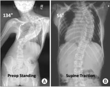
견인 시작 3주 후 견인 무게는 체중의 51.7%인 12 kg까지 증 량되었다. 이때 측정한 관상면 Cobb각은 흉추 3번-흉추 10번 간 134°에서 56°, 흉추 10번-요추5번 간 73°에서 26°로 감소하였으 며 첨단 추체(apical vertebrae)의 전이 거리도 116.4 cm에서 41.6 cm로 감소하여 수술 전 견인에 의한 우수한 교정 정도를 보 였다(Fig. 2). 환자는 무게 증량으로 인한 불편함 호소가 없었으 며, halo-pelvic 견인과 관련한 합병증도 발생하지 않았다. 따라 서 저자들은 2일 후 전신마취하 최종 수술을 계획하였다.
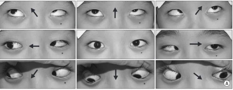
하지만 환자는 다음날(견인 22일째) 갑작스럽게 사물이 겹쳐 보이는 증상을 호소하여 신경학적 검진을 시행하였고 좌측 안구
회복되었으며 안과와 상의하여 6번 뇌신경 마비 발생 3일 후에 측만증에 대한 교정 수술을 진행하였다. 신경생리장치 감시하에 halo 견인 3 kg, 골반 견인 2 kg으로 유지하면서 척추경 나사못 삽입 후 강봉 감염술(rod derotation)에 의한 교정술 및 골유합 술을 시행하였고 수술 직후 시행한 신경학적 검사상 6번 뇌신경 마비 이외에 다른 이상 소견은 없었다.
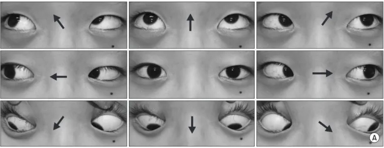
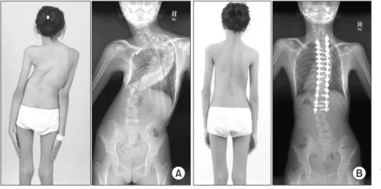
환자는 수술 1주 후 퇴원 시까지 수술과 관련된 다른 합병증은 없었으나 6번 뇌신경 마비에 의한 복시는 유지되었다. 환자 및 보 호자에게 경과 관찰 필요함을 설명하였으며 이후 수술 2개월 추 시관찰상에서 복시 증상 호소 없이 6번 뇌신경 마비의 완전 회복 을 나타냈다(Fig. 4). 환자는 현재 수술 시행 후 5년이 경과하였으 며 매년 경과 관찰 중으로 특별한 합병증 없이 만족스러운 수술 결과를 보였다(Fig. 5).
고 찰
고도 척추 측만증의 교정 수술은 일반적인 척추 측만증의 수술보 다 신경학적 합병증의 발생 가능성이 높으며 그에 따라 보다 신
A B
Figure 2. An eleven-year-old girl had severe scoliosis. (A) Preoperative (preop) standing radiograph demonstrates a 134° major curve. (B) The radiograph three weeks after preoperative halo-pelvic traction demonstrates 56° major curve (58.2% correction).
Figure 1. Halo-pelvic traction is usually started at one to two days after halo application considering pin site pain, adaptation of the halo device, and detection of early complication. Halo and pelvic apparatus were placed on a reverse Trendelenburg position bed with a wood block to avoid going down slides. The initial traction weight was three to five pounds in total, dividing each of the head and pelvic side according to the curve pattern. Traction was increased gradually two to three pounds a day as tolerated.
중한 접근이 필요하다. 이러한 합병증을 줄이기 위한 대안의 하 나로 수술 전 halo 견인을 이용한 점진적 교정이 제시된다.8) 하지 만 halo 견인이 비교적 안전한 방법임에도 불구하고 견인 중 발 생할 수 있는 신경학적 합병증의 가능성이 있기 때문에 이에 대 한 주의 깊은 관찰과 즉각적인 대처가 필요할 수 있다.
뇌신경 마비는 halo 견인과 관련하여 발생할 수 있는 가장 흔 한 신경학적 합병증 중의 하나이다. 이전 연구에 따르면 halo 견 인 중 6번 뇌신경(외전신경), 9번 뇌신경(설인신경), 10번 뇌신경 (미주신경), 12번 뇌신경(설하신경) 등에서의 발생이 보고되었으 며 그 중 6번 뇌신경의 마비를 가장 흔하게 보고하였다.3,4) 본 증
례 환자는 점진적 견인 시행 후 3주에 58.2%의 양호한 주만곡 Cobb각 교정 정도를 보였으나 이후 갑작스럽게 발현한 6번 뇌 신경 마비에 의한 양안 복시를 나타낸 경우이다.
아직까지 halo 견인 후 발생한 뇌신경 마비에 대한 병태생리학 적 원인은 명확하게 밝혀지지 않았지만 생리적 신경차단(neuro- praxia) 혹은 골성 협착에 의한 압박이나 부종에 의한 2차적 허 혈성 변화에 의한 가능성들이 제시되고 있다.5,6) 일반적으로 견인 력이 작용할 경우에 경추부가 흉추부보다 더 많은 견인력을 받게 되며 경추 신경근이 손상에 보다 취약해지게 된다. 특히, 6번 뇌 신경은 두개저에서 구부러진 사행성 경로(tortuous route)를 가
Green glass in front of left eye Green glass in front of right eye
Left Right
A
B
Figure 3. (A) She presented with the complaint of binocular horizontal diplopia worse in the left gaze at 22 days after halo-pelvic traction. (B) Hess chart. The constricted left eye field demonstrates lateral rectus underaction. There was contralateral medial rectus overaction. Very mild right lateral rectus underaction and contralateral overaction were present. The central point was shifted in both eyes, indicating diplopia in the primary gaze.
지기 때문에 더욱 견인력에 취약한 특징을 가지게 된다.6) MacE- wen 등7)은 이러한 뇌신경 마비의 주 원인으로 견인 무게의 급격 한 증가를 제시하였다. 본 환자의 경우, 키아리 1형 기형에 대한 감압술 후 급격히 진행한 고도 척추 측만증의 수술을 위해 3주 동안 비교적 서서히 견인 무게를 체중의 51.7%까지 증가하였음 에도 불구하고 6번 뇌신경 마비가 발생하였다. 견인 무게의 점진 적 증량에도 불구하고 뇌신경 마비가 발생한 원인에 대해, 이는 입원 5개월전 키아리 I 변형에 대한 대공 감압술과 경추 1번 후궁 의 부분절제술로 인한 후방 골성 구조물의 제거 및 후방 근육과 인대의 이완으로 인하여 일반적인 증량 무게에서의 견인력이 과
도하게 6번 뇌신경에 작용했기 때문일 것으로 저자들은 추정하 고 있다.9)
대부분의 뇌신경 마비는 일시적이거나 6–10주 이내에 회복되 는 경우가 많지만 일부 연구에서는 영구적인 장애를 보고하기도 한다.10) 본 환자의 경우는 뇌신경 마비 발생 다음날에 50%의 회 복을 보였으며 안과와의 협진 후 측만증에 대한 교정술을 진행하 기로 한 증례이다. 또한 수술 전후 환자의 신경학적 검진을 추가 적으로 시행하여 수반되는 합병증에 대처하고자 하였다. 따라서 고도 척추 측만증의 halo 견인 시에는 뇌신경 마비의 발생 가능 성을 주지하고 대처하는 것이 필요하겠다. 본 증례와 같이 이전
Green glass in front of left eye Green glass in front of right eye
Left Right
A
B
Figure 4. (A, B) At the two-month follow-up after corrective surgery of severe scoliosis, the binocular diplopia by sixth cranial nerve palsy had completely resolved by simple observation, and she was free of symptoms.
에 뇌수술이나 경추부 수술의 병력이 있는 경우에는 급격한 견인 보다는 점진적으로 견인 무게를 증가시키는 것이 필요할 것으로 생각되며 경추부 측면 방사선 사진을 촬영하여 과도한 견인을 방 지하는 것이 필요하겠다. 또한 환자 및 보호자에게 halo 견인 중 신경학적 손상의 발생 가능성이 있음을 미리 설명하고 신경학적 검진을 주기적으로 시행하여 뇌신경 마비 발생 초기에 대처하는 것이 환자의 예후에 중요할 것으로 생각된다.
CONFLICTS OF INTEREST
The authors have nothing to disclose.
ORCID
Jae-Kwang Hwang, https://orcid.org/0000-0003-4635-953X Choon Sung Lee, https://orcid.org/0000-0002-3263-9410 Shin Woo Choi, https://orcid.org/0000-0002-1091-7694 Chung-Hwan Kim, https://orcid.org/0000-0002-0630-7930
REFERENCES
1. Koller H, Zenner J, Gajic V, Meier O, Ferraris L, Hitzl W. The impact of halo-gravity traction on curve rigidity and pulmo- nary function in the treatment of severe and rigid scoliosis and kyphoscoliosis: a clinical study and narrative review of the literature. Eur Spine J. 2012;21:514-29.
2. Sponseller PD, Takenaga RK, Newton P, et al. The use of trac-
tion in the treatment of severe spinal deformity. Spine (Phila Pa 1976). 2008;33:2305-9.
3. Barsoum WK, Mayerson J, Bell GR. Cranial nerve palsy as a complication of operative traction. Spine (Phila Pa 1976).
1999;24:585-6.
4. Ginsburg GM, Bassett GS. Hypoglossal nerve injury caused by halo-suspension traction. a case report. Spine (Phila Pa 1976). 1998;23:1490-3.
5. Rozario RA, Stein BM. Complications of halo-pelvic traction.
Case report. J Neurosurg. 1976;45:716-8.
6. Limpaphayom N, Skaggs DL, McComb G, Krieger M, Tolo VT. Complications of halo use in children. Spine (Phila Pa 1976). 2009;34:779-84.
7. MacEwen GD, Bunnell WP, Sriram K. Acute neurologi- cal complications in the treatment of scoliosis. A report of the Scoliosis Research Society. J Bone Joint Surg Am.
1975;57:404-8.
8. Rinella A, Lenke L, Whitaker C, et al. Perioperative ha- lo-gravity traction in the treatment of severe scoliosis and kyphosis. Spine (Phila Pa 1976). 2005;30:475-82.
9. Tanaka M, Sugimoto Y, Arataki S, Takigawa T, Ozaki T. A rare course of scoliosis associated with Chiari malformation and syringomyelia. Acta Med Okayama. 2014;68:303-6.
10. Nischal K, Chumas P, Sparrow O. Prolonged survival after atlanto-occipital dislocation: two case reports and review. Br J Neurosurg. 1993;7:677-82.
Preoperative Postperative
A B
Figure 5. Preoperative (A) and postoperative (B) clinical photographs and radio graphs of the same patient showing cosmetic improvement of the deformity after defini- tive corrective surgery.
고도 척추 측만증의 치료는 척추외과 의사에게 어려운 과제로 남아있다. 고도 척추 측만증의 수술 시 급격한 교정은 신경학적 손 상이나 기구 실패 등의 수술 중 합병증의 위험을 증가시킬 수 있다. 이러한 합병증을 최소화하기 위해 최종 수술을 시행하기에 앞 서 부분 교정을 얻기 위한 다양한 수술 전 견인법들이 사용되고 있다. 하지만 이전 연구에 의하면 halo 견인과 관련한 합병증의 하 나로 뇌신경 마비가 발생할 수 있으며 대표적으로 6번 뇌신경(외전신경)의 마비가 가장 흔하게 나타난다. 이러한 합병증을 줄이기 위해 견인 무게의 점진적 증량이나 세심한 신경학적 검진이 필요하며 특히 이전에 뇌수술이나 경추부 수술을 시행한 경우에는 더 욱 주의가 필요할 수 있다. 저자들은 이전에 키아리 1형 기형과 관련하여 감압술을 시행했던 고도 척추 측만증에서 수술 전 halo- pelvic 견인에 의한 6번 뇌신경 마비의 증례를 경험하였기에 문헌 고찰과 함께 보고하고자 한다.
색인단어: 척추 측만증, 뇌신경 손상, 외전신경 손상, 아놀드-키아리 변형, halo-pelvic 견인
접수일 2019년 6월 5일 수정일 2019년 10월 3일 게재확정일 2019년 11월 21일 책임저자 김정환
25440, 강원도 강릉시 사천면 방동길 38, 울산대학교 의과대학 강릉아산병원 정형외과학교실
TEL 033-610-3249, FAX 033-641-8050, E-mail [email protected], ORCID https://orcid.org/0000-0002-0630-7930
Copyright © 2020 by The Korean Orthopaedic Association
“This is an Open Access article distributed under the terms of the Creative Commons Attribution Non-Commercial License (http://creativecommons.org/licenses/by-nc/4.0/) which permits unrestricted non-commercial use, distribution, and reproduction in any medium, provided the original work is properly cited.”