김민석1⋅최 진1⋅정재호2
인제대학교 의과대학 상계백병원 안과학교실1, 부산대학교 의학전문대학원 안과학교실2
목적: 소아 환자에서 엡스타인-바 바이러스(Epstein-Barr virus, EBV) 뇌염과 동반된 양안 외향신경마비 증례를 보고하고자 한다.
증례요약: 증례 1. 특이병력 없는 14개월 남아가 5일전부터 시작된 발열과 45PD 좌안 내사시를 주소로 내원하였다. 양안 외전장애 외에 다른 신경학적 검사상 특이소견은 발견되지 않았으나, 혈청검사와 뇌척수액검사에서 EBV 양성소견을 보여 항바이러스 치료 및 가림 치료를 시행하였고 7개월 후 잔류 내사시에 대해 좌안 내직근 후전술 6.5 mm, 외직근 절제술 6 mm를 시행하였다. 수술 1주일 후 환아는 정위를 보였고 신경학적 후유증은 없었다. 증례 2. 13세 남아가 5일전부터 시작된 두통과 발열, 2일전부터 시작된 의식저하와 발작을 주소로 내원하였고 혈청검사에서 EBV 양성소견, 뇌척수액검사에서 바이러스 감염소견이 있어 항바이러스 치료를 시행하였다.
환아는 2주 뒤 의식회복 후 복시를 호소하였고 30PD 좌안 내사시와 양안 외전장애가 관찰되었다. 4개월간 가림치료 후 환아는 정위를 보였고 외전장애도 관찰되지 않았으나 발작조절을 위해 항경련제를 지속적으로 복용하였다.
결론: 소아에서 엡스타인-바 바이러스 뇌염은 양안 외향신경마비와 동반될 수 있고 이로 인한 잔류 내사시와 신경학적 후유증이 지속 될 수 있다.
<대한안과학회지 2013;54(8):1303-1308>
■Received: 2013. 3. 4. ■ Revised: 2013. 4. 11.
■Accepted: 2013. 6. 27.
■Address reprint requests to Jin Choi, MD
Department of Ophthalmology, Inje University Sanggye Paik Hospital, #1342 Dongil-ro, Nowon-gu, Seoul 139-707, Korea Tel: 82-2-950-1096, Fax: 82-2-935-6904
E-mail: [email protected]
* This study was presented as a e-poster at the 106th Annual Meeting of the Korean Ophthalmology Society 2011.
후천성 외향신경마비는 소아에서 종종 볼 수 있는 증상 으로 그 원인으로는 종양, 외상, 선천 이상, 염증, 감염, 뇌 압상승 등 여러 가지가 있으며, 연구에 따라 차이는 있으나 종양이나 외상이 가장 흔한 원인으로 보고되고 있다. 수막 뇌염, 소뇌염, 뇌농양과 같은 염증, 감염질환에서도 후천성 외향신경마비가 가능하며 소아 외향신경마비의 6-13%가 이런 원인에 의한 것으로 보고되었다.1-4
엡스타인-바 바이러스(Epstein-Barr virus, EBV)는 이 중가닥으로 된 헤르페스 바이러스로 B림프구와 상피세포에 침투하며 전염성 단핵구증의 원인이 된다. 대부분의 EBV 감염은 증상은 없거나 비특이적인 것으로 알려졌으나5 탈 수초성 질환, 급성 뇌염, 뇌수막염, 급성 소뇌 실조증과 같 은 중추신경계 합병증을 유발한다는 보고도 있다.6EBV 뇌 염의 급성 신경학적 증상은 공격적 행동, 발작, 두통, 국소 신경학적 이상소견 등이다.7 이렇게 EBV 뇌염의 임상양상
은 다양하지만 안구운동을 침범하는 경우는 드물다. 우리는 소아환자에서 EBV 뇌염과 동반된 양측의 외향신경마비환 자 2예를 경험하였기에 이를 보고하고자 한다.
증례보고
증례 1
특이병력 없는 14개월된 환아가 5일전부터 시작된 발열 과 좌안의 내사시로 내원하였다(Fig. 1A). 크림스키검사에 서 좌안의 45 프리즘 디옵터(PD)의 내사시를 보였고 한눈 운동/동향운동 검사에서 양안 -4 정도의 외전장애가 관찰 되었다. 안저검사에서 특이소견은 관찰되지 않았고 조절마 비굴절검사상 우안 +2.25 Dsph=-2.00 Dcyl ×90°, 좌안 +1.75 Dsph=-1.75 Dcyl ×90°로 측정되었다. 신경학적 검사상 다른 특이소견은 관찰되지 않았다. 뇌자기공명영상 에서 뇌교, 연수에 T1강조영상에서 저신호강도, T2강조영 상에서 고신호강도를 보이는 병변이 관찰되었다(Fig. 2).
혈청검사에서 EBV 핵항원(EBV Nuclear Antigen, EBNA), 바이러스캡시드항원(Viral Capsid Antigen, VCA) IgG와 IgM이 양성이었고 뇌척수액 중합효소 연쇄 반응 검사에서 EBV 양성소견을 보였다. 뇌척수액검사에서는 30 cells/mm3 (lymphocyte 100%), glucose level 60 mg/dL, protein level
Figure 1. In Case 1, he showed esodeviation of the left eye at
primary gaze position (A). Four months later, there still was esodeviation in the left eye (B). One week after strabismus sur- gery, he showed orthotropia (C).Figure 2. Brain MRI of case 1. T1 weighted sagittal images
show low signal intensity (arrows, A) and T2 weighted axial images show high signal intensity (arrows, B) without en- hancement at anterior portion of pons and medulla.31 mg/dL로바이러스감염소견을 나타내었다. 환아에게 acy- clovir230 mg/day와 prednisolone 12 mg/day을 7일간 정 맥 투여를 하였고 하루2시간의 우안 가림치료를 시행하였 다. 내원 4개월 후 우안의 외전장애는 완전히 호전되었으나 좌안은 -2 정도의 외전장애와 25PD의 내사시가 남아있었 다(Fig. 1B). 하지만 내원 6개월 후 좌안 내사시는 다시 45 PD로 증가하였고 내원 7개월째에 좌안 내직근 후전술 6.5 mm, 외직근 절제술 6.0 mm를 시행하였다. 수술 1주일 후 환아의 눈 위치는 정위를 보였고(Fig. 1C) 수술 1년 후 경 과관찰에서도 유지되었으며 신경학적 후유증은 관찰되지 않았다.
증례 2
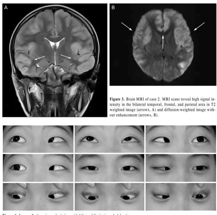
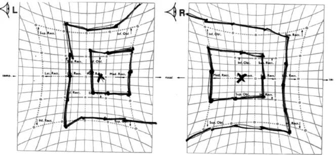
특이병력 없는 13세 남자 환아가 5일전부터 시작된 두통 과 발열, 2일전부터 시작된 의식저하와 발작(seizure)을 주 소로 내원하였다. 환아는 중환자실에 입원하였고 뇌자기공 명영상의 T2강조영상, 확산강조영상에서 양측의 측두엽, 전두엽, 두정엽 부분에 고신호강도를 보였다(Fig. 3). 혈청 검사에서 초기항원(anti-EBV diffuse and restricted early antigen, EADR) IgM이 양성이었고 뇌척수액 검사에서 바 이러스 감염 소견을 나타내어(115 cells/mm3(lymphocyte 83%, monocyte 17%), glucose level 63 mg/dL, protein level 38 mg/dL) acyclovir 정맥 투여를 시작하였다. 2주 후 환자는 의식을 회복하였고 복시를 호소하였다. 환아의 굴절검사상 양안 -3.00 Dsph의 근시가 있었고 교정시력은 양안 20/20 이었다. 정면 주시시 좌안의 30PD 내사시를 보 였고 한눈운동/동향운동 검사에서 양안 -3 정도의 외전장 애가 관찰되었다(Fig. 4). 전안부와 안저검사상 특이소견은 관찰되지 않았고 헤스스크린검사에서 양안의 외전장애가 관찰되었다(Fig. 5). 환아는 종일가림치료를 격일로 양안에 번갈아 시행하였다. 4개월 후 환아의 눈위치는 정위로 관찰
A B
C
A
B
Figure 3. Brain MRI of case 2. MRI scans reveal high signal in-
tensity in the bilateral temporal, frontal, and parietal area in T2 weighted image (arrows, A) and diffusion-weighted image with- out enhancement (arrows, B).Figure 4. In case 2, there is esodeviation with bilateral limitation of abduction.
되었고 외전장애도 소실되었으며 복시증상도 없었다. 하지 만 발작조절을 위한 항경련제 복용이 지속적으로 필요하였 다.
고 찰
EBV 감염에 의해 흔히 나타나는 증상은 발열, 인두염, 림프절병(lymphadenopathy)이지만8폭력적 행동, 발작, 두 통, 국소 신경학정 징후 같은 신경학적 증상들이 개별적으 로 혹은 동시에 일어날 수 있다.7그러나 EBV 뇌염에 의한 후천성 양안 외향신경마비는 드물게 발생하며 단독으로 발
생하는 경우는 더욱 드물다.9,10 우리는 소아환자에서 EBV 뇌염에 의한 양안 외향신경마비 환자 2예를 경험하였고 임 상양상과 경과가 서로 달랐기에 이를 보고하고자 한다.
소아에서 후천성 외향신경마비가 있을 경우 먼저 감염, 외상, 종양 등에 초점을 맞춰 과거력, 현병력에 대해 자세히 청취하고 발열, 두통, 이명, 일시적 시력저하와 같은 동반증 상과 임상경과를 잘 살펴보는 것이 감별에 도움이 된다. 그 리고 안과적, 신경학적 검사가 필요한데 유두부종 감별을 위한 안저검사도 반드시 시행하여야 한다. 영상검사는 종양 의 가능성 때문에 환자의 신경학적 징후가 없더라도 모든 소아환자에서 이른 시기에 고려해야 하며 의심이 된다면
Figure 5. Hess screen test revealed esotropia with bilateral abduction limitation in case 2.
감염에 대한 혈청검사도 시행하여야 한다.1
EBV 뇌염의 특징적인 뇌자기공명영상 소견으로 T2 강 조영상에서 양측의 두정후두에 고신호강도의 병변이 나타 난다는 보고가 있다.7 또 다른 연구에서는 EBV 뇌염의 침 범 부위는 다양하고 그에 따라 예후가 달라진다고 하였다.
즉 대뇌 피질, 수질을 침범한 경우 좋은 예후를 보이고 시 상을 침범하는 경우 후유증을 남길 수 있으며 뇌줄기를 침 범하는 경우 사망률이 가장 높다고 하였다.11 하지만 EBV 뇌염의 영상소견은 환자마다 다양하기 때문에 진단이나 예 후결정에 확정적인 근거가 되지는 못한다. 증례 1의 뇌자기 공명영상에서는 뇌줄기에 이상소견이 보였으나 심각한 합 병증이 나타나지 않았고, 증례 2에서는 대뇌에 이상소견이 있었으나 신경학적 합병증을 동반하였다. 반면 바이러스에 대한 혈청검사는 진단에 있어 중요한데 EBV Viral Capsid Antigen (VCA) IgG와 IgM, EBV Nuclear Antigen (EBNA), anti-EBV diffuse and restricted early antigen (EADR) IgM이 EBV의 중요한 표지이다. 뇌척수액에서 중합효소 연 쇄 반응을 통해 바이러스를 검출하는 것도 EBV 뇌염을 진 단하는데 결정적인 단서가 된다.6
일반적으로 소아환자에서 EBV 뇌염은 특별한 치료 없이 도 호전되는 것으로 알려졌으나 10%에서 신경학적 합병증 이나 사망에 이른다는 보고도 있다.9 Yamashita et al12은 EBV 뇌염 후 발생한 급성 소뇌성 실조증을 보고하였고 Caruso et al13은 5명의 소아환자에서 EBV 뇌염 후 실어증, 실행증, 이상행동, 인지와 판단기능의 저하, 발작, 강박적인 행동과 같은 신경학적 이상이 지속되는 증례를 보고하였다.
Bray et al14은 EBV 감염 후 신경학적 합병증을 보인 5예를 보고하였는데 4명은 다발 경화증, 1명은 급성 파종성 경화
증으로 진단되었고 EBV 감염과 신경학적 합병증의 관련성 은 의심할 수 있으나 EBV가 그 원인이 되는 증거는 될 수 없다고 하였다. 한편 Fujimoto el al6은 EBV가 중추신경계 에 감염된 환자 10예에서 3년간 합병증이 발생하지 않았다 고 보고하였다. 본 증례보고에서는 증례 1에서 7개월 경과 관찰동안 외전장애 및 내사시가 지속적으로 있어 사시수술 을 진행하였고 증례 2에서는 지속적인 발작이 있어 항경련 제를 복용하였다.
EBV 감염 후의 외향신경마비에서 특별한 치료 없이 예 후가 양호했던 보고가 있었다.15 외향신경마비가 자연적으 로 호전되는 데는 원인, 침범부위(단안, 양안), 외전장애의 정도와 같은 몇 가지 요소가 관여할 것으로 생각한다.4 증 례 1에서 환아는 양안에 -4정도의 외전장애가 있었고 7개 월 경과관찰에서 좌안은 호전되지 않았으나 우안은 호전되 는 양상을 보여 한 환자 내에서도 양안의 예후가 다른 것을 볼 수 있었다. 증례 2에서는 양안 -3 정도의 외전장애가 있 었고 가림치료 후 4개월 경과관찰에서 완전히 호전되는 경 과를 보였다. 따라서 첫 증상의 정도가 환자의 예후에 중요 한 요소중의 하나라고 생각한다.
생명에 위협이 되는 EBV 감염에서 acyclovir의 항바이러 스 효과 또는 임상증상 개선에 대한 객관적인 증거는 아직 밝혀지지 않았다.16몇몇 논문에서는 acyclovir와 cortico- steroid가 치료로 추천되고 있고6,10 앞으로 EBV 뇌염에서 항바이러스치료에 대한 대조군 임상연구가 진행되어야 할 것이다. 한편 소아는 시각성숙에 민감한 시기이기 때문에 EBV 뇌염에 의한 외향신경마비에서 약시를 예방하기 위해 우성안에 대한 가림치료도 고려해야 한다.
결론적으로 EBV 뇌염 소아환자에서 후천성 양안 외향신
반해야 한다.
REFERENCES
1) Lee MS, Galetta SL, Volpe NJ, Liu GT. Sixth nerve palsies in children. Pediatr Neurol 1999;20:49-52.
2) Kodsi SR, Younge BR. Acquired oculomotor, trochlear, and abdu- cent cranial nerve palsies in pediatric patients. Am J Ophthalmol 1992;114:568-74.
3) Holmes JM, Mutyala S, Maus TL, et al. Pediatric third, fourth, and sixth nerve palsies: A population-based study. Am J Ophthalmol 1999;127:388-92.
4) Merino P, Gómez de Liaño P, Villalobo JM, et al. Etiology and treatment of pediatric sixth nerve palsy. J AAPOS 2010;14:502-5.
5) Patil AK, Azad ZR, Mathew V, Alexander M. Chronic meningitis and central nervous system vasculopathy related to Epstein Barr virus. Ann Indian Acad Neurol 2012;15:303-6.
6) Fujimoto H, Asaoka K, Imiazumi T, et al. Epstein-Barr virus in- fections of the central nervous system. Intern Med 2003;42:33-40.
7) Kalita J, Maurya PK, Kumar B, Misra UK. Epstein Barr virus ence- phalitis: clinical diversity and radiological similarity. Neurol India
phalitis in children. Pediatr Infect Dis J 1996;15:871-5.
11) Abul-Kasim K, Palm L, Maly P, Sundgren PC. The neuroanatomic localization of Epstein-Barr virus encephalitis may be a predictive factor for its clinical outcome: a case report and review of 100 cas- es in 28 reports. J Child Neurol 2009;24:720-6.
12) Yamashita S, Murakami C, Izumi Y, et al. Severe chronic active Epstein-Barr virus infection accompanied by virus-associated he- mophagocytic syndrome, cerebellar ataxia and encephalitis. Psychiatry Clin Neurosci 1998;52:449-52.
13) Caruso JM, Tung GA, Gascon GG, et al. Persistent preceding focal neurologic deficits in children with chronic Epstein-Barr virus encephalitis. J Child Neurol 2000;15:791-6.
14) Bray PF, Culp KW, McFarlin DE, et al. Demyelinating disease af- ter neurologically complicated primary Epstein-Barr virus infection.
Neurology 1992;42:278-82.
15) Straussberg R, Cohen AH, Amir J, Varsano I. Benign abducens pal- sy associated with EBV infection. J Pediatr Ophthalmol Strabismus 1993;30:60.
16) Sullivan JL, Byron KS, Brewster FE, et al. Treatment of life threat- ening Epstein-Barr virus infection with acyclovir. Am J Med 1982;73(1A):262-6.
=ABSTRACT=
Bilateral Abducens Nerve Palsy in Pediatric Patients with Epstein-Barr Virus Encephalitis
Kim Min Seok, MD1, Jin Choi, MD1, Jae Ho Jung, MD, PhD2
Department of Ophthalmology, Inje University Sanggye Paik Hospital, Inje University College of Medicine1, Seoul, Korea Department of Ophthalmology, Pusan National University School of Medicine2, Busan, Korea
Purpose: To report 2 cases that presented with bilateral abducens nerve palsy associated with Epstein-Barr virus (EBV) encephalitis in children.
Case summary: Case 1. A 14-month-old boy presented with fever and esodeviation of the left eye that started 5 days earlier. On the ophthalmic examination, 45-PD esotropia of the left eye and limitation of abduction in both eyes were observed. On neurological examination, there were no abnormalities. Serologic test and polymerase chain reaction (PCR) from cerebrospinal fluid (CSF) were positive for EBV. The patient was treated with systemic acyclovir and prednisolone.
Part-time occlusion therapy of the right eye for 2 hours/day was also prescribed. The patient underwent a 6.5-millimeter re- cession of the medial rectus and a 6-millimeter resection of the lateral rectus on the left eye 7 months after the presentation. The patient showed orthotropia 1 week after the surgery without neurologic sequelae. Case 2. A 13-year-old boy presented with headaches and fever that started 5 days before and altered consciousness with seizures 2 days previously. Serological test for viral infection was normal, except for EBV, and CSF examination showed viral infection.
After the patient recovered consciousness, he complained of diplopia. A 30-PD esotropia of his left eye with bilateral limi- tation of abduction was present. Alternating full-time occlusion of both eyes was prescribed. At 4 months after pre- sentation, diplopia disappeared and the patient showed orthotropia without abduction limitation; however, anticonvulsants were prescribed to control seizures.
Conclusions: In children, EBV encephalitis can be accompanied by acquired bilateral abducens nerve palsy. Residual nerve palsy and other neurologic sequelae can remain after several months.
J Korean Ophthalmol Soc 2013;54(8):1303-1308
Key Words: Acquired abducens nerve palsy, Encephalitis, Epstein-Barr virus, Neurologic sequelae
Address reprint requests to Jin Choi, MD
Department of Ophthalmology, Inje University Sanggye Paik Hospital
#1342 Dongil-ro, Nowon-gu, Seoul 139-707, Korea
Tel: 82-2-950-1096, Fax: 82-2-935-6904, E-mail: [email protected]