• 검색 결과가 없습니다.

Temporal Expression of Interleukin 1 β in the Dorsal Root Ganglion in a Rat Model of Lumbar Disc Herniation

N/A
N/A
Protected

Academic year: 2021

Share "Temporal Expression of Interleukin 1 β in the Dorsal Root Ganglion in a Rat Model of Lumbar Disc Herniation"

Copied!
6
0
0

로드 중.... (전체 텍스트 보기)

전체 글

(1)

Su-Jeong Kim, PhD; Hee-Jin Gu, MS

1

; Yun-Woo Cho, MD, PhD

2

; Hea-Woon Park, MD, PhD

3

; Joon-Ha Lee, PhD

4

; Se-Jin Hwang, MD, PhD

5

; Sang-Ho Ahn, MD, PhD

2

Institute of Medical Science, Yeungnam University;

1

Clinical Trial Center for Medical Devices of Yeungnam University Hospital;

2

Department of Rehabilitation Medicine, College of Medicine, Yeungnam University;

3

Department of Rehabilitation Medicine, School of Medicine, Catholic University of Deagu;

4

Department of Biochemistry and Molecular Biology, Yeungnam University;

5

Department of Anatomy and Cell Biology, College of Medicine, Hanyang University

Purpose: To investigate temporal changes in IL-1β mRNA expression in spinal dorsal horn (DH) and dorsal root ganglion (DRG) in a rat lumbar disc herniation (LDH) model.

Methods: Autologous nucleus pulposus, harvested from the tail disc between the second and third coccygeal vertebrae (Co2-3), was implanted next to the left L5 nerve root just proximal to the DRG after partial laminectomy. IL-1β mRNA expression was investigated in DRG and DH in our LDH model. Real-time PCR assays were done using a 7500 Real Time PCR system (Applied Biosystems, USA).

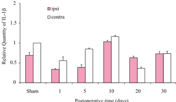
Results: Expression of IL-1β in DRG and DH was observed for 30 days postoperatively. Expression of IL-1β mRNA in the ipsilateral DRG of the LDH group gradually increased from 5 to 30 days after surgery. The amount of IL-1β in the contralateral DRG peaked 10 days after surgery and then gradually decreased. However, there was no difference in IL-1β mRNA expression in spinal DH between the LDH group and the sham-operated group.

Conclusion: Long-term expression of IL-1β in the LDH model may worsen the chronic pain state. Future studies on inhibition of IL-1β expression in the LDH model will be needed to develop selective treatment strategies for patients with LDH.

Keywords: Interleukin-1β, Lumbar disc herniation, pain, Dorsal root ganglion Received: May 13, 2010

Revised: June 10, 2010 Accepted: June 14, 2010

Corresponding author: Se-Jin Hwang, [email protected]; Sang-Ho Ahn, [email protected]

Ganglion in a Rat Model of Lumbar Disc Herniation

The Journal Korean Society of Physical Therapy

I. Introduction

Local application of autologous nucleus pulposus may induce spinal nerve damage and causes radicular pain characterized by hyperalgesia and allodynia. It has been suggested that nucleus pulposus in the epidural space induces pain, not only by mecha- nical but also by chemical mechanisms. The mechanism of the pain, however, has not been fully elucidated. Many of reports suggested that inflammatory reactions rather than mechanical compression may play a major role in the radicular pain after lumbar disc herniation (LDH).

1-3

The nucleus pulposus has been known to be inflammatory in nature and may sensitize the spinal nerves directly or indirectly through release of inflamma-

tory mediators. Proinflammatory cytokines have been shown to play key roles in such chemically induced nerve root injury.

Cytokines are generally defined as any polypeptide that affects

the function of other cells. Cytokines such as interleukin-1α

(IL-1α),

2

interleukin-1β (IL-1β),

2,4-6

inteleukin-6 (IL-6)

4-6

and

tumor necrosis factor-α (TNF-α),

6,7

and prostaglandin E2 (PGE

(2))

4,8

have been reported to be strongly related to the effects of

nucleus pulposus on nerve roots. The intraplantar injection of

TNF-α in normal animal had been shown both to upregulate

IL-1β and to induces hyperalgesia.

9

IL-1β released from the facet

joint in the degenerative spinal disorder was reported to be

associated with leg pain.

10

IL-1β was suggested to serve as a

mediator to sensitize nociceptors in chronic inflammation and

(2)

possibly in hyperalgesia through long-term changes in neuronal plasticity.

11

The expression of IL-1β has been reported in glial cells and neurons in the central nervous system (CNS),

12,13

and sensory neurons in dorsal root ganglion (DRG) of the peripheral nervous system (PNS).

4,14,15

However, the temporal expression of IL-1β in the spinal dorsal horn (DH) and DRG was rarely reported. The purpose of present study was to investigate the temporal changes of mRNA expression of IL-1β in spinal DH and DRG in rat LDH models.

II. Materials and Methods

1. Animals

The experimental protocol was reviewed and approved by the Institutional Animal Care and Use Committee at the Yeungnam University, South Korea. Fifty-four male Sprague-Dawley rats (200∼250 g) were used in this study (n = 9 for real-time PCR for each time course). Rats were housed in an animal room on a 12-hour light/12-hour dark cycle with free access to water and food.

2. Lumbar disc herniation

Rats were anesthetized by Zoletil (Virbac, 50 mg/kg, i.p.) additional doses were used as required to maintain anesthesia throughout the experiment. A midline dorsal longitudinal incision was made over the lumbar spine, and the multifidus muscles were removed along the spinous processes from L4 to S1. The left L5 nerve roots and DRG were exposed after partial laminectomies. Nucleus pulposus, harvested from the tail disc between the second and third coccygeal vertebrae (Co2-3), was implanted next to the left L5 nerve root just proximal to the DRG.

16

Surgery in control rats was identical, except for the implantation of nucleus pul- posus. The dura mater was left intact in all procedures.

3. Real-time PCR

Total RNA was isolated from lumbar spinal cord and DRG tissue, corresponding to L5 root at 1, 5, 10, 20 and 30 days after surgery. Total RNA was isolated from each sample with Trizol reagent and purity was checked with spectrophotometer. Reverse transcription of 1 μg aliquots of total mRNA was carried out at 45℃ using a cDNA Reverse Transcription Kit (Applied Bio- systems, USA). Real-time PCR assays were performed using a

7500 Real Time PCR system (Applied Biosystems, USA). Primers and the TaqMan probe were designed using ProbeFinder software (Universal Probe Library (UPL), Roche, Switzerland).

To amplify IL-1β and hypoxanthine guanine phosphoribosyl- transferase (HPRT) transcripts the following primers were used:

sense primer 5′- TGT GAT GAA AGA CGG CAC AC -3′ and antisense primer 5′- CTT CTT CTT TGG GTA TTG TTT GG -3′ for IL-1β (GenBank accession number : NM031512) and sense primer 5′-GGT CCA TTC CTA TGA CTG TAG ATT TT-3′ and antisense primer 5′-CAA TCA AGA CGT TCT TTC CAG TT-3′ for HPRT (GenBank accession number:

NM012583). The HPRT gene was used as an internal control to adjust for differences between samples. The mastermix consisted of 250 nM of UPL probe, 700 nM of each primer (sense and antisense), 10 μl of 2X TaqMan master and 2 μl of cDNA. All PCR reactions were run in duplicate. After pre-incubation at 95℃

for 10 minutes, PCR was performed using 50 amplification cycles of denaturation at 95℃ for 15 seconds and annealing at 60℃ for 60 seconds.

4. Statistical analysis

Data were analyzed with the Wilcoxon signed rank test and Mann Whitney U test using SPSS/PC v. 15.0 and expressed as mean±standard deviation. Significance was set at p<0.05.

III. Results

1. Expression of mRNA for IL-1β in the DRG In previous our study, mechanical allodynia was developed at 1 day after surgery and prolonged to 30 days after surgery in a rat LDH model.

16

Real-time PCR analysis for the expression of IL-1β mRNA revealed that the amount of IL-1β was increased in the LDH group compared to the sham-operated group. The expression amount of mRNA for IL-1β in the ipsilateral DRG of LDH group was gradually increased from 5 days to 30 days after surgery (p<0.05). But, the amount of IL-1β in the contralateral DRG was peaked at 10days after surgery and gradually decreased (Figure 1).

2. Expression of mRNA for in the spinal DH

The amount of mRNA of IL-1β at 10 days after surgery was

(3)

Figure 1. The mRNA expression of IL-1β in the ipsilateral (ipsi) and contralateral (contra) sides of DRG after application of nucleus pulposus to the nerve root for 30 days. *p<0.05,

p

<0.01 compared to contralateral of sham value. The expre- ssion amount of mRNA for IL-1β in the ipsitateral DRG of lumbar disc herniation group was gradually increased from 5 days to 30 days after surgery (p<0.05). But, the amount of IL-1β in the contralateral DRG was peaked at 10 days after surgery and gradually decreased.

Figure 2. The mRNA expression of IL-1β in the ipsilateral (ipsi) and contralateral (contra) sides of DH after application of nucleus pulposus to the nerve root for 30 days. There was no statistically significant difference of the mRNA expression of IL-1β between the lumbar disc herniation group and the sham- operated group.

expressed relatively high in DH of the LDH group compare to the sham-operated group. However, there was no statistically significant difference of the mRNA expression of IL-1β between the LDH group and the sham-operated group (Figure 2).

IV. Discussion

In this study, we found that the mRNA expression of IL-1β in DRG of a rat LDH model was increased according to time course after surgery. The amount of mRNA for IL-1β in the ipsilateral DRG of LDH group was gradually increased during the experimental period. However, the expression of IL-1β in the contralateral DRG was peaked at 10 days after surgery and gradually decreased. We also observed that there was no significant difference in the mRNA expression in spinal DH between the LDH group and the sham-operated group.

The expression of IL-1β has been reported to be produced in disc tissues of patients with disc herniation.

1,4,17

In a patient with the protrusion type orthe extrusion type herniation, cells pro- ducing IL-1β was detected by enzyme-linked immunosorbent assay in their harvested tissues. From histological analysis of many other samples from patients, almost all of cells in the

harvested tissues are classified as histiocytes, fibroblasts, or endothelial cells.

1

Interleukin-1β is suggested to an early mediator in the inflammatory response

17,18

and may play an important role in the generation and persistence of the inflammatory response observed in the extruded disc. It has been suggested that if the nucleus pulposus extruded into the spinal canal, a repair process may occur through the normal inflammatory response. At this stage, the immunological system can contact with the nucleus pulposus, which is known to contain antigenic components, resulting in an autoimmune response to the extruded disc.

17

Among the proinflammatory cytokines, IL-1β is a particulary

known to modulate pain sensitivity. Peripheral or spinal admini-

stration of IL-1β usually produces both mechanical allodynia

and thermal hyperalgesia.

19,20

According to the previous study

on the expression of IL-1β mRNA in the nerve root and DRG of

a LDH rat model, the mRNA expression increased at 1 week

postoperatively compared with control group. This increase

correlated with the increase in mechanical hyperalgesia only at 1

week postoperatively.

4

In our previous behavioral study on the

LDH model, the mechanical allodynia was developed at 1 day

after surgery and prolonged to 30 days after surgery.

16,21

We have

found increased mRNA expression of IL-1β in DRG of a LDH

rat model. The expression of IL-1β gradually increased from 5

days to 30 days after surgery. Related to our result, several reports

also suggested chronic expression of IL-1β correlated with chronic

pain.

22-24

IL-1 receptor was spontaneously secreted by cells

(4)

isolated from herniated lumbar discal tissue after discectomy.

24

IL-1β mRNA and protein were expressed for 4 weeks in cutaneous tissue of a rat tibia fracture model and the fracture-caused mechanical allodynia reduced by IL-1 receptor antagonist.

22

In addition, IL-1β blockade was reported to improve the pain at least 75% in patients with chronic active gout.

23

Many mechanisms have been proposed to account for IL-1β and neuropathic pain. Firstly, local inflammation by IL-1β may be important in disc tissue pathophysiology, possibly also in discogenic pain mechanisms.

25,26

Recruited macrophage by nerve injury release IL-1β and other proinflammatory cytokines, and the recruitment of macrophage increase neuropathic pain.

27

In an animal herniation model, cells consisting mainly of macro- phages with some neutrophils appeared three days after herniation.

28

Actually, macrophage depletion after partial ligation of the sciatic nerve could alleviate thermal hyper- algesia.

25

Second, activation of spinal cord glia, including both microglia and astrocytes following nerve injury is another possible neurophathic pain cause.

29

Intrathecal administration of IL-1β increased wind-up activity in the spinal cords of both normal and monoarthritic rats without propentofylline (glial inhibitor), but resulted in decreased wind-up activity in normal and monoarthritic propentofylline-treated animals. Thus, glial inactivation reverted to inhibition of the excitatory effect of IL-1 β on the spinal cord wind-up of the normal or monoarthritic condition of rats.

30

The glial cells are not only activated, but they may also create and maintain pathological pain states by locally releasing proinflammatory cytokines.

31

While the expression of IL-1β in DRG of LDH has been often reported,

4,6,32

the expression of IL-1β in spinal DH of LDH rarely reported. However, many studies reported that the expression of IL-1β in spinal DH of various pain models strongly involved in neuropathic pain.

33-36

In our study, we could observe expression of IL-1β in spinal DH but we did not find the significant difference of the mRNA expression of IL-1β in spinal DH between the LDH group and the sham-operated group.

The present study showed that the time course of IL-1β expression in LDH model and the expression of IL-1β may contribute to the chronic painful and inflammatory reaction in LDH. The blockade of IL-1β might be one of useful treatments for LDH patients.

V. Conclusion

We have found the temporal expression of IL-1β in DRG of a rat LDH model. Long-term expression of IL-1β in LDH models may influence to chronic pain state. Future studies on inhibition of IL-1β in LDH model will be needed to develop the selective treatment strategy for patients with LDH.

Author Contributions Research design: Kim SJ

Acquisition of data: Kim SJ, Gu HJ

Analysis and interpretation of data: Kim SJ, Park HW, Cho YW, Hwang SJ, Ahn SH

Drafting of the manuscript: Kim SJ

Administrative, technical, and material support: Lee JH, Cho YW, Gu HJ

Research supervision: Ahn SH, Hwang SJ

Acknowledgement

This work was supported by the Korea Research Foundation Grant funded by the Korean Government (MOEHRD, Basic Research Promotion Fund) (KRF- 2008-532-E00019).

References

1. Takahashi H, Suguro T, Okazima Y et al. Inflammatory cyto- kines in the herniated disc of the lumbar spine. Spine (Phila Pa 1976). 1996;21(2):218-24.

2. Le Maitre CL, Freemont AJ, Hoyland JA. The role of inter- leukin-1 in the pathogenesis of human intervertebral disc degeneration. Arthritis Res Ther. 2005;7(4):R732-45.

3. Ahn SH, Cho YW, Ahn MW et al. mRNA expression of cytokines and chemokines in herniated lumbar intervertebral discs. Spine (Phila Pa 1976). 2002;27(9):911-7.

4. Kawakami M, Matsumoto T, Kuribayashi K et al. mRNA expression of interleukins, phospholipase A2, and nitric oxide synthase in the nerve root and dorsal root ganglion induced by autologous nucleus pulposus in the rat. J Orthop Res.

1999;17(6):941-6.

5. Ozaktay AC, Kallakuri S, Takebayashi T et al. Effects of interleukin-1 beta, interleukin-6, and tumor necrosis factor on sensitivity of dorsal root ganglion and peripheral receptive fields in rats. Eur Spine J. 2006;15(10):1529-37.

6. Ozaktay AC, Cavanaugh JM, Asik I et al. Dorsal root

(5)

sensitivity to interleukin-1 beta, interleukin-6 and tumor necrosis factor in rats. Eur Spine J. 2002;11(5):467-75.

7. Aoki Y, Rydevik B, Kikuchi S et al. Local application of disc-related cytokines on spinal nerve roots. Spine (Phila Pa 1976). 2002;27(15):1614-7.

8. Kawakami M, Tamaki T, Hashizume H et al. The role of phospholipase A2 and nitric oxide in pain-related behavior produced by an allograft of intervertebral disc material to the sciatic nerve of the rat. Spine (Phila Pa 1976). 1997;22(10):

1074-9.

9. Woolf CJ, Allchorne A, Safieh-Garabedian B et al. Cytokines, nerve growth factor and inflammatory hyperalgesia: the contribution of tumour necrosis factor alpha. Br J Pharmacol.

1997;121(3):417-24.

10. Igarashi A, Kikuchi S, Konno S. Correlation between in- flammatory cytokines released from the lumbar facet joint tissue and symptoms in degenerative lumbar spinal disorders.

J Orthop Sci. 2007;12(2):154-60.

11. Jeanjean AP, Moussaoui SM, Maloteaux JM et al. Inter- leukin-1 beta induces long-term increase of axonally trans- ported opiate receptors and substance P. Neuroscience. 1995;

68(1):151-7.

12. Molenaar GJ, Berkenbosch F, van Dam AM et al. Distri- bution of interleukin 1 beta immunoreactivity within the porcine hypothalamus. Brain Res. 1993;608(1):169-74.

13. Watt JA, Hobbs NK. Interleukin-1beta immunoreactivity in identified neurons of the rat magnocellular neurosecretory system: evidence for activity-dependent release. J Neurosci Res. 2000;60(4):478-89.

14. Copray JC, Mantingh I, Brouwer N et al. Expression of interleukin-1 beta in rat dorsal root ganglia. J Neuroimmunol.

2001;118(2):203-11.

15. Eliav E, Benoliel R, Herzberg U et al. The role of IL-6 and IL-1beta in painful perineural inflammatory neuritis. Brain Behav Immun. 2009;23(4):474-84.

16. Cho YW, Park HW, Kim SJ et al. Expression of MCP‐ in spinal dorsal horn in a rat model of lumbar disc herniation. J Kor Soc Phys Ther. 2009;21(3):95-102.

17. Doita M, Kanatani T, Harada T et al. Immunohistologic study of the ruptured intervertebral disc of the lumbar spine.

Spine (Phila Pa 1976). 1996;21(2):235-41.

18. Feghali CA, Wright TM. Cytokines in acute and chronic inflammation. Front Biosci. 1997;2:d12-26.

19. Ferreira SH, Lorenzetti BB, Bristow AF et al. Interleukin-1

beta as a potent hyperalgesic agent antagonized by a tripeptide analogue. Nature. 1988;334(6184):698-700.

20. Reeve AJ, Patel S, Fox A et al. Intrathecally administered endotoxin or cytokines produce allodynia, hyperalgesia and changes in spinal cord neuronal responses to nociceptivestimuli in the rat. Eur J Pain. 2000;4(3):247-57.

21. Kim WR, Ahn SH, Cho YW et al. The long-lasting pattern of the neuropathic radicular pain in an autologous nucleus pulposus model of rat. J Korean Acad Rehabil Med. 2009;

33(4):477-82.

22. Li WW, Sabsovich I, Guo TZ et al. The role of enhanced cutaneous IL-1beta signaling in a rat tibia fracture model of complex regional pain syndrome. Pain. 2009;144(3):303-13.

23. Terkeltaub R, Sundy JS, Schumacher HR et al. The inter- leukin 1 inhibitor rilonacept in treatment of chronic gouty arthritis: results of a placebo-controlled, monosequence cross- over, non-randomised, single-blind pilot study. Ann Rheum Dis. 2009;68(10):1613-7.

24. Koch H, Reinecke JA, Meijer H et al. Spontaneous secretion of interleukin 1 receptorantagonist (IL-1ra) by cells isolated from herniated lumbar discal tissue after discectomy. Cytokine.

1998;10(9):703-5.

25. Liu T, van Rooijen N, Tracey DJ. Depletion of macrophages reduces axonal degeneration and hyperalgesia following nerve injury. Pain. 2000;86(1-2):25-32.

26. Myers RR, Campana WM, Shubayev VI. The role of neuro- inflammation in neuropathic pain: mechanisms and thera- peutic targets. Drug Discov Today. 2006;11(1-2):8-20.

27. Kiguchi N, Maeda T, Kobayashi Y et al. Macrophage in- flammatory protein-1alpha mediates the development of neuropathic pain following peripheral nerve injury through interleukin-1beta up-regulation. Pain. 149(2):305-15.

28. Yoshida M, Nakamura T, Sei A et al. Intervertebral disc cells produce tumor necrosis factor alpha, interleukin-1beta, and monocyte chemoattractant protein-1 immediately after her- niation: an experimental study using a new hernia model.

Spine (Phila Pa 1976). 2005;30(1):55-61.

29. Watkins LR, Maier SF, Goehler LE. Cytokine-to-brain communication: a review & analysis of alternative mecha- nisms. Life Sci. 1995;57(11):1011-26.

30. Constandil L, Hernandez A, Pelissier T et al. Effect of interleukin-1beta on spinal cord nociceptive transmission of normal and monoarthritic rats after disruption of glial function.

Arthritis Res Ther. 2009;11(4):R105.

(6)

31. Watkins LR, Maier SF. Beyond neurons: evidence that immune and glial cells contribute to pathological pain states. Physiol Rev. 2002;82(4):981-1011.

32. Takebayashi T, Cavanaugh JM, Cuneyt Ozaktay A et al.

Effect of nucleus pulposus on the neural activity of dorsal root ganglion. Spine (Phila Pa 1976). 2001;26(8):940-5.

33. Pineau I, Lacroix S. Proinflammatory cytokine synthesis in the injured mouse spinal cord: multiphasic expression pattern and identification of the cell types involved. J Comp Neurol.

2007;500(2):267-85.

34. Martucci C, Trovato AE, Costa B et al. The purinergic

antagonist PPADS reduces pain related behaviours and interleukin-1 beta, interleukin-6, iNOS and nNOS over- production in central and peripheral nervous system after peripheral neuropathy in mice. Pain. 2008;137(1):81-95.

35. Ledeboer A, Sloane EM, Milligan ED et al. Minocycline attenuates mechanical allodynia and proinflammatory cytokine expression in rat models of pain facilitation. Pain. 2005;

115(1-2):71-83.

36. Fiorentino PM, Tallents RH, Miller JN et al. Spinal inter- leukin-1beta in a mouse model of arthritis and joint pain.

Arthritis Rheum. 2008;58(10):3100-9.

수치

Figure 1. The mRNA expression of IL-1β in the ipsilateral (ipsi)  and contralateral (contra) sides of DRG after application of  nucleus pulposus to the nerve root for 30 days

참조

관련 문서

(((((((((((((((((chronic low back pain) OR nerve root compres- sion) OR lumbosciatic pain) OR radicular pain) OR radicu- litis) OR sciatica) OR disc herniation) AND

This book contains hundreds of complete, working examples illustrating many common Java programming tasks using the core, enterprise, and foun- dation classes APIs.. Java Examples

The index is calculated with the latest 5-year auction data of 400 selected Classic, Modern, and Contemporary Chinese painting artists from major auction houses..

○ 해외 시장에서의 높은 신뢰도와 인지도를 바탕으로 한국 경제의 성장 동력으로 자리잡아가는 국내 유명 화장품 제조업체를 보호하고 , 국내 뿐만

Recognizing that a S&amp;T Co–operation Fund of 10 million USD, contributed for the period 2011–15, has been utilized to implement joint research.. projects in the

The aqueous extract of Rhodiola sachalinensis root enhances the expression of inducible nitric oxide synthase gene in RAW264.7 macrophages.. Camptothecin

- CERAGEM center owners, staffs and customers from Bihar state participated by donation to support daily necessities &amp; living expenses

Rule 6(1)에 따라 원칙적으로 매입 재료 또는 서비스가 면세 재화의 제조 또는 면세 서비스의 공급에 사용될 경우 Cenvat Credit를 이용할 수 없다. 따라서