위장관의 급, 만성 출혈의 원인 질환으로는 종양, 궤양, 염증 성 질환 등이 있지만, 노인에서는 특히 혈관성 병변이 중요한 원인이 되고 있다 (1). 동정맥기형 (AVM, arteriovenous malformations), 혈관이형성증(angiodysplasia)이 흔한 원인이 고, 드문 혈관병변으로는 모세혈관확장증 (telangiectasia), 정맥 확장증(phlebectasia), 그리고 혈관종이 있다.
증례 보고
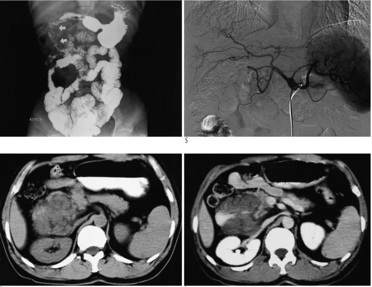
53세 남자환자가 약 4년 전부터 소량씩 지속되어온 혈변을 주소로 내원하였다. 내원 당시 경도의 호흡곤란과 어지러움을 보이고 있었다. 혈액검사상 혈색소(Hgb) 3.8g/dl, 헤마토크리 트(Hct) 13.7%, 그리고 백혈구 3700/μl이었으며, 말초혈액도 말 검사상 저색소성 소적혈구성 빈혈(hypochromic microcytic anemia)을 보였다. 4년 전부터 몇 차례 위내시경, 대장내시경 검사를 받았으나 이상소견이 없었고, 내원 후 시행한 소장 조 영술에서 점막이상이 보이지 않고 소장 통과 시간이 정상으로 이상소견이 없는 것으로 보고되었다(Fig. 1A). 십이지장근처 의 혈관계통의 질환을 의심하여 복강동맥 촬영술을 하였으나 십이지장 주위로 혈관이상도 보이지 않았다(Fig 1B). 이후 시 행한 위내시경상 십이지장 하행부의 바터씨 팽대부(ampulla of Vater) 직하부에 외부에서의 눌림같이 보이는 점막의 융기가 보이고, 백태로 덮인 궤양과 소량출혈이 보였다. 복부 CT 조 영증강전 영상에서 크기 8×7 cm의 소엽상의 경계를 가진 비 교적 균일한 저음영의 연조직 종괴가 십이지장 구부부터 횡행 부(3rd portion)까지 둘러싸고 있었다. 종괴와 접한 췌장 두부 는 넓어지고 바터씨 팽대부의 근위부 총담관을 침범하고 있었 지만 십이지장이나 총담관의 근위부가 늘어나지 않았다. 지연 기 조영후 CT영상에서 종괴는 비교적 균일한 약한 조영증강 을 보였다(Fig. 1C, D).
수술 소견상 십이지장과 췌장사이 장간막에서 생긴 종괴가
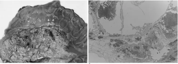
주변 조직으로 파고 들어가는 양상을 보이며, 출혈로 인해 박 리가 쉽지 않아서 위플씨 수술(Whipple’s operation)을 시행 하여 췌장두부와 십이지장까지 종괴와 함께 절제하였다. 육안 소견상 약 10×4 cm 크기의 경계가 불분명한 황갈색의 반고 형 종괴로 단면은 혈관이 풍부한 스펀지형태를 보였다(Fig 2A). 현미경 조직 소견상 늘어난 혈관, 성숙지방세포, 섬유성 간질로 구성된 해면상 혈관종으로 진단되었다. 악성변화는 보 이지 않았고 췌장실질과 십이지장의 점막까지 침범을 보여 혈 관종의 분류 중에서도 혈관종증 (diffuse hemangioma, angiomatosis)로 구분되었다(Fig. 2B). 수술 후 현재까지 환자 는 위장관 출혈의 재발은 없었다.
고 찰
소장을 침범한 장간막 혈관종이 재발성 혈변의 원인으로 몇 증례가 보고 되어 있다(1, 2). 본 증례는 십이지장과 췌장 두 부사이의 장간막에서 발생해서 십이지장 벽의 전 층, 췌장두 부, 횡행결장 장간막을 침범한 경우이다.
일반적으로 혈관종은 선천성 병변으로 출생 시부터 있거나 생후 수개월 내 자라는 경우가 많다. 기형종과 진성 신생물 (true neoplasm)의 중간에 위치하지만 보통 한 장기에 국한되 어 있고 종괴효과를 보이기 때문에 종양으로 구분한다. 장간막 뿐만 아니라 신체 어디에서 발생한 경우라도 경계가 분명한 종 괴부터 비정형, 침윤성 종괴까지 그 범위가 다양하다. 혈관종 은 임상적으로는 양성이므로 주변장기 침범이나 원격전이의 가 능성이 없지만, 장간막에 생긴 경우는 장관과 가깝게 있어서 장관벽이 침범되고 출혈도 일으킨다.
구성하는 혈관의 크기에 따라 모세혈관상(capillary), 해면상 (cavernous)과 혼합형(mixed) 혈관종으로 나눈다. 대부분 단 일 종괴로 나타나는데, 동시에 여러 군데에 나타나거나 주변 장 기 를 침 범 하 면 혈 관 종 증 (diffuse hemangioma, angiomatosis)이라고 한다(3). 보고된 장간막 혈관종은 대부 분이 해면상 혈관종이고 정맥혈관종과 혈관임파종이 각각 1예
─ 321 ─ 대한영상의학회지 2004;51:321-323
위장관 출혈을 일으킨 장간막 혈관종: 증례 보고1
김 기 남
장간막에서 기원한 혈관종은 매우 드문 질환으로 문헌상 간혹 보고된 바가 있다. 저자는 53 세 남자환자에서 십이지장 궤양을 형성하고, 근처의 췌장두부, 횡행결장 장간막을 침범한 장간 막 혈관종을 보고한다. 복부 CT상 약한 조영증강을 보이는 저밀도 음영의 종괴였다.
1동아대학교 의과대학 진단방사선과학교실
이 논문은 2004년 3월 23일 접수하여 2004년 7월 21일에 채택되었음.
씩의 보고가 있다(2, 4). 위치는 소장 장간막이 가장 많고 대 망, 소망, 결장, 충수돌기 장간막의 증례도 보고되어 있다(2, 5, 6). 발견시 대부분 복통이나 만져지는 종괴가 있고 2,4-8), 드 물게 위장관 출혈이 있다(1). Hanatate F 등(2)의 보고에 따 르면 출혈여부에 관계없이 증례의 62%(11/18)에서 빈혈이 있 었고, 혈관종의 크기가 클수록 정도가 심하였다.
현재까지 문헌에 보고된 장간막 혈관종의 영상소견은 조영 증강 CT상 경계가 잘 그려지는 불균질의 저, 고음영이 혼합된 종괴(1, 2)와 비교적 균질한 조영증강이 잘 되는 종괴(6)가 있고, 자기공명영상은 내부에 출혈을 가진 장간막 혈관종이 T2 강조 스핀에코 영상에서 비균질의 저, 고신호강도로 보인 경우 (7)와 소망에 발생한 해면상 혈관종이 조영 증강되는 격막을 가지고 T1, T2강조영상에서 각각 저, 고음영을 보인 경우(5) 가 보고 되어 있다. 드물게 공장 장간막에 내부가 출혈로 채워 진 다방성 낭종으로 나타난 예도 있다(6). 혈관임파종 (mixed
hemangioma and lymphangioma) 1예는 조영증강 CT상 비교 적 균일한 연조직 종괴로 보였다(4). 혈관조영술은 본 증례를 포함해서 종괴를 나타내지 못한 경우가 있었는데 종괴 내부의 심한 혈전증 등이 가능한 원인으로 생각된다(2). 대부분의 증 례에서 위장관 조영술이나 일반촬영은 진단에 도움이 되지 못 하였고, 초음파, CT, 혈관촬영의 확인이 필요하였다. 최근에 영 상진단이 급속히 발전함에도 불구하고 장간막 혈관종의 특정 한 진단은 무척 어렵다.
장간막 혈관종의 치료방법은 종류, 위치, 범위, 증상, 수술가 능성 등에 따라 달라질 수 있는데, 대부분 수술로 완전 제거하 며 수술적 제거가 어렵거나 다병소성인 경우는 소량의 방사선 치료를 하고 있는데 방사선 치료로 인한 악성변화가 보고 되 고 있다(4). 장간막 혈관종의 완전 제거후 재발보고는 없었다.
하지만 종격동 혈관종에서 술 후 재발이 보고된 바 있어서 가 능성을 배제하기는 어렵다(9).
─ 322 ─
김기남: 위장관 출혈을 일으킨 장간막 혈관종
A B
C D
Fig. 1. A. Small bowel follow-through shows widening of the C-loop in 2nd portion of the duodenum (arrows).
B. Celiac angiography shows no area of increased vascularity or site of active bleeding. selective gastroduodenal arteriogram was not obtained, because angiography was done before performing of CT scans or endoscopy.
C, D. Unenhanced CT scan shows a large and heterogeneous mass with lobulated margin, extending from duodenal 2nd portion to the pancreatic head. On contrast-enhanced CT, the mass shows focal area of marked enhancement.
참 고 문 헌
1. Ruiz AR Jr, Ginsberg AL. Giant mesenteric hemangioma with small intestinal involvement: an unusual cause of recurrent gas- trointestinal bleed and review of gastrointestinal hemangiomas.
Dig Dis Sci 1999;44:2545-2551
2. Hanatate F, Mizuno Y, Murakami T. Venous hemangioma of the mesoappendix: report of a case and a brief review of the Japanese literature. Surg Today 1995;25:962-964
3. Juan Rosai. Ackerman’s surgical pathology. 8th Ed. Mosby, 1996:
2060-2064
4. Tai PT, Jewell LD. Case report: mesenteric mixed haemangioma
and lymphangioma; report of a case with 10 year follow-up after radiation treatment. Br J Radiol 1995;68:657-661
5. Chung J, Kim M, Lee JT, Yoo HS. Cavernous hemangioma arising from the lesser omentum: MR findings. Abdom Imaging 2000;
25:542-544
6. McHugh K, Spitz L. Capillary haemangioma of the greater omen- tum. Pediatr Radiol 2002;32:148-149
7. Takamura M, Murakami T, Kurachi H, Kim T, Enomoto T, Narumi Y, et al. MR imaging of mesenteric hemangioma: a case re- port. Radiat Med 2000;18:67-69
8. Rathnaraj S, Aggarwal S, Verghese M. Giant mesenteric heman- gioma. Indian J Gastroenterol 1995;14:113
9. Cohen AJ, Sbaschnig RJ, Hochholzer L, Lough FC, Albus RA.
Mediastinal hemangioma. Ann Thorac Surg 1997;43:656-659
─ 323 ─ 대한영상의학회지 2004;51:321-323
A B
Fig. 2. A. Surgical specimen shows an ill-defined dark red brown colored and semisolid mass with spongy-like consistency in the mesentery and between the duodenum and the pancreas (black arrows), measuring 10×4 cm. Note focal ulceration of duodenal mucosa (white arrows).
B. Micrograph shows that the mesenteric mass is composed of dilated interconnecting, endothelial-line vascular spaces. In some ar- eas, the vascular spaces are nearly coalescent to produce a pattern reminiscent of cavernous hemangioma (H & E, ×20).
J Korean Radiol Soc 2004;51:321-323
Address reprint requests to : Ki Nam Kim, M.D., Department of Diagnostic Radiology, College of Medicine, Dong-A University 1. 3-ga, Dongdaesin-dong, Seo-gu, Pusan 602-103, Korea.
Tel. 82-51-240-5367 Fax. 82-51-253-4931 E-mail: [email protected]
Radiologic Findings of Mesenteric Hemangioma with Gastrointestinal Bleeding: Case Report1
Ki Nam Kim, M.D.
1Department of Diagnostic Radiology, College of Medicine, Dong-A University
Mesenteric hemangioma is a rare disease entity. To our knowledge, only scattered reports about this condi- tion have appeared in the literature. Herein, the author presents a rare case of mesenteric hemangioma with duodenal ulceration and invasion of the adjacent pancreatic head and transverse mesocolon. The tumor ap- peared in the form of a mild contrast enhancement of a low attenuation mass on contrast-enhanced CT.
Index words :Neoplasms
Gastrointestinal tract