© 2012 The Korean Academy of Medical Sciences.
This is an Open Access article distributed under the terms of the Creative Commons Attribution Non-Commercial License (http://creativecommons.org/licenses/by-nc/3.0) which permits unrestricted non-commercial use, distribution, and reproduction in any medium, provided the original work is properly cited.
pISSN 1011-8934 eISSN 1598-6357
Jejunal Variceal Bleeding Successfully Treated with Percutaneous Coil Embolization
A 52-yr-old male with alcoholic liver cirrhosis was hospitalized for hematochezia. He had undergone small-bowel resection due to trauma 15 yr previously.
Esophagogastroduodenoscopy showed grade 1 esophageal varices without bleeding. No bleeding lesion was seen on colonoscopy, but capsule endoscopy showed suspicious bleeding from angiodysplasia in the small bowel. After 2 weeks of conservative treatment, the hematochezia stopped. However, 1 week later, the patient was re-admitted with hematochezia and a hemoglobin level of 5.5 g/dL. Capsule endoscopy was performed again and showed active bleeding in the mid-jejunum. Abdominal computed tomography revealed a varix in the jejunal branch of the superior mesenteric vein. A direct portogram performed via the transhepatic route showed portosystemic collaterals at the distal jejunum. The patient underwent coil embolization of the superior mesenteric vein just above the portosystemic collaterals and was subsequently discharged without re-bleeding.
At 8 months after discharge, his condition has remained stable, without further bleeding episodes.
Key Words: Hematochezia; Capsule Endoscopy; Abdominal Computed Tomography;
Jejunal Varices; Embolization So My Koo1, Soung Won Jeong1,
Jae Young Jang1, Tae Hee Lee1, Seong Ran Jeon1, Hyun Gun Kim1, Jin Oh Kim1, and Yong Jae Kim2 Institute for Digestive Research and Digestive Disease Center, Departments of 1Gastroenterology and 2Radiology, Soonchunhyang University Hospital, Seoul, Korea
Received: 2 May 2011 Accepted: 4 November 2011 Address for Correspondence:
Soung Won Jeong, MD
Institute for Digestive Research and Digestive Disease Center, Soonchunhyang University Hospital, 59 Daesagwan-ro, Yongsan-gu, Seoul 140-743, Korea
Tel: +82.2-709-9863, Fax: +82.2-709-9797 E-mail: [email protected]
http://dx.doi.org/10.3346/jkms.2012.27.3.321 • J Korean Med Sci 2012; 27: 321-324
CASE REPORT
Gastroenterology & Hepatology
INTRODUCTION
Jejunal varices are an uncommon manifestation of portal hy- pertension and are rarely symptomatic (1). Acute bleeding has been reported in 5.5% of patients with portal hypertension and small bowel varices (2). Jejunal varices are associated with por- tal hypertension, which may be due to cirrhosis or extrahepatic portal venous obstruction, chronic alcoholism, portal vein throm- bosis, or intrahepatic arterioportal fistulas (3). Ectopic varices tend to develop at sites of tissue adhesion in patients who have previously undergone abdominal surgery (4) and may occur in combination with jejunal variceal bleeding. Esophagogastric var- ices, in contrast, are caused primarily by portal hypertension.
Traditionally, jejunal variceal bleeding has been treated sur- gically (5, 6). Non-surgical treatment options include porto-ca- val shunt (7, 8), endoscopic sclerotherapy (9), embolization (8, 10-12), and balloon dilatation and stent placement in the portal vein for extrahepatic portal venous obstruction (4, 13).
Here, we report a case of ectopic jejunal variceal bleeding that was treated successfully by percutaneous coil embolization via the superior mesenteric vein.
CASE DESCRIPTION
A 52-yr-old male was admitted for hematochezia on 5 February
2009. Fifteen years previously, he had undergone a laparotomy for the repair of a small bowel perforation due to blunt trauma.
Seven years ago, he was diagnosed with alcoholic liver cirrho- sis. Balloon-occluded retrograde transvenous occlusion (BRTO) was performed for gastric variceal bleeding 3 months prior to admission. Upon admission to our hospital, he underwent esophagogastroduodenoscopy (EGD), which showed a grade 1 esophageal varix without bleeding. Colonoscopy revealed no lesion that explained the hematochezia. Capsule endoscopy showed angiodysplasia in the small bowel, suspected of caus- ing the hematochezia. The patient was treated conservatively for 2 weeks. His melena ceased and he was discharged 5 weeks after admission. However, 1 week after discharge, he was re-ad- mitted for hematochezia, which reportedly occurred 3-4 times a day. He had not consumed any alcohol after the discharge.
His blood pressure was 106/37 mmHg and his heart rate was 91 beats/min. He had a hemoglobin count of 5.5 g/dL, a platelet count of 149,000/μL, a prothrombin time of 15.8 s (international normalized ratio: 1.36), and an albumin concentration of 3.0 g/
dL. There was no encephalopathy but he had mild ascites and a Child-Pugh score of 7. Serologic viral markers showed HBsAg/
Ab (-/+), anti-HCV Ab (-).
Emergency EGD showed non-bleeding, grade 1 esophageal varices. Hematochezia at the sigmoid colon and rectum was noted on colonoscopy, but there was no active bleeding lesion.
Koo SM, et al. • Jejunal Variceal Bleeding Treated by Embolization
322 http://jkms.org http://dx.doi.org/10.3346/jkms.2012.27.3.321
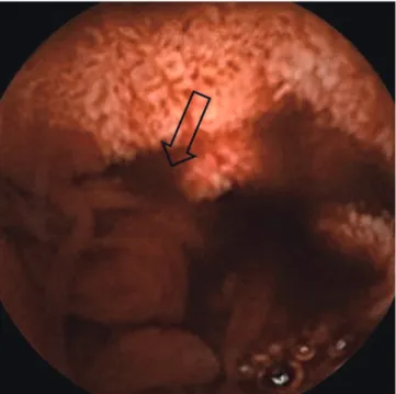
Intestinal segments above the sigmoid colon could not be ex- amined because of poor visualization due to the presence of a blood clot. Capsule endoscopy showed active bleeding at the mid-jejunum and bloody staining of the bowel wall below the mid-jejunal level (Fig. 1). Abdominal enhanced computed to- mography (CT) showed liver cirrhosis, splenomegaly with mild ascites, varices of the jejunal branch of the superior mesenteric
vein, and edematous change at the ascending colon and gall bladder (Fig. 2). Based on these findings, jejunal variceal bleed- ing was suspected.
A direct portogram performed via the transhepatic route showed hepatofugal flow into the superior mesenteric vein and multiple dilated portosystemic shunts from the superior mes- enteric vein to both internal iliac veins (Fig. 3A-D). There was no evidence of active bleeding. Coil embolization was performed at the superior mesenteric vein just above the portosystemic collaterals to decompress the variceal pressure (Fig. 3E).
The patient was discharged without re-bleeding after the pro- cedure. Eight months after discharge he remains in stable con- dition, with no further bleeding.
DISCUSSION
Jejunal variceal bleeding, although rare, can be life threatening without treatment. The three clinical features of small-bowel varices are portal hypertension, a history of abdominal surgery, and hematochezia without hematemesis (5, 14). The case de- scribed here shows that jejunal variceal bleeding can be treated successfully with percutaneous coil embolization via the supe- rior mesenteric vein.
Portal hypertension due to baseline alcoholic liver cirrhosis, a history of abdominal surgery, and recent BRTO likely explained our patient’s jejunal varices. Baseline alcoholic liver cirrhosis is known to induce portal hypertension and, frequently, esopha- geal and jejunal varices. Moreover, the severity of hepatic failure is a risk factor for gastroesophageal varix (15, 16). In contrast, in Fig. 1. Capsule endoscopic findings show blood-stained mucosa below the mid-jeju-
num, with suspected active bleeding (arrow) of a jejunal varix.
Fig. 2. Jejunal varices and main portal vein in multidetector CT; Axial scan and multiplanar reformation in multidetector CT show multiple and dilated jejunal varices (arrow). Note the main portal vein (small arrows) in the multiplanar reformation.
A B
Koo SM, et al. • Jejunal Variceal Bleeding Treated by Embolization
http://jkms.org 323
http://dx.doi.org/10.3346/jkms.2012.27.3.321
patients with portal hypertension, there is less association be- tween other factors, such as age, gender, presence of cirrhosis, gastroesophageal varices, and Child-Pugh class C, and varices occurring in the small bowel (2).
A history of abdominal surgery is also a known predisposing factor for jejunal variceal bleeding (5). Previous abdominal sur- gery can lead to the development of ectopic varices at the ab- dominal surgical site or they may be a consequence of postsur- gical stricture in the small bowel (4). Small-bowel varices due to prior abdominal surgery may result from the mesenteric hy- pertension caused by mesenteric vein stenosis or from portal hypertension. Thus, in a patient with obscure gastrointestinal bleeding, a history of abdominal surgery, and portal hyperten- sion, small-bowel varices at the anastomotic site or the postop- erative stricture site should be considered.
BRTO procedures for the treatment of gastric varices may in- duce a significant elevation in the portal systemic pressure gra- dient (17). In our patient, recent BRTO might have aggravated the portal systemic pressure gradient, thereby inducing jejunal variceal bleeding.
While the patient may have had jejunal varices at first admis- sion, there was no evidence for their presence on a capsule en-
doscopy image and it was assumed that the cause of hemato- chezia was angiodysplasia of the small bowel. Jejunal variceal bleeding is difficult to diagnose and is best confirmed using ab- dominal enhanced CT, capsule endoscopy, abdominal angiog- raphy, or 99mTc-labeled red blood cell scanning (14). In some cases, push enteroscopy and ileoscopy may be useful diagnos- tically (18). Capsule endoscopy is highly sensitive for the detec- tion of fresh blood in the small bowel. However, it is of limited usefulness for the diagnosis of small-bowel varices because the mucosal layer covering the varices can exhibit a mosaic pattern, a shining pattern, or normal features. Blood clots can cover the varices. Therefore, clinical suspicion is key to diagnosis (19).
The treatment of jejunal varices includes surgery (5, 6), tran- sjugular intrahepatic porto-systemic shunt (TIPS) (7, 8), entero- scopic sclerotherapy (9), percutaneous embolization (8, 10-12), and dilatation of a stenosed portal vein followed by stent place- ment (4, 13). Treatment for segmental varices in which there is superior mesenteric vein stricture without portal hypertension involves surgical resection. In the presence of systemic portal hypertension, a TIPS or a decompressive shunting procedure is recommended (18).
For small-bowel varices with extrahepatic portal vein obstruc- Fig. 3. Serial direct portogram and coil embolization; Serial direct portogram shows hepatofugal flow into the superior mesenteric vein and multiple dilated portosystemic shunts from the superior mesenteric vein to both internal iliac veins (A-D). Coil embolization of the superior mesenteric vein was performed just above the portosystemic shunts (E).
A B C
D E
Koo SM, et al. • Jejunal Variceal Bleeding Treated by Embolization
324 http://jkms.org http://dx.doi.org/10.3346/jkms.2012.27.3.321
tion, surgery and TIPS are not only invasive but are relatively difficult to perform. Additionally, inflammation, trauma, and/or severe abdominal adhesion are common postoperatively (10).
Superior mesenteric vein embolization and portal vein angio- plasty with stent insertion are options in patients in whom sur- gery is not possible due to portal hypertension and extrahepatic obstruction (10, 13). These procedures can also be performed successfully in patients with jejunal variceal bleeding related to extrahepatic portal vein obstruction but without portal hyper- tension (20). The Child-Pugh score of our patient at admission was 7, and he had no evidence of hepatic failure. Possible causes of jejunal variceal bleeding were portal hypertension due to stric- ture at the anastomotic site, intra-abdominal adhesion, and ex- trahepatic portal venous obstruction from previous small-bowel surgery, rather than aggravation of alcoholic liver cirrhosis. He had no portal vein obstruction. Intervention angiography was selected as a first-line therapy rather than TIPs, due to the pres- ence of mild ascites and grade 1 esophageal varices, and the risk of hepatic encephalopathy. Coil embolization as a first-line treat- ment resulted in a successful outcome for our cirrhotic patient.
In summary, we report the case of a patient with jejunal vari- ceal bleeding who was treated successfully by percutaneous coil embolization. Further studies are needed to confirm the use of embolization as an interventional first-line therapy for small- bowel variceal bleeding.
REFERENCES
1. Hamlyn AN, Morris JS, Lunzer MR, Puritz H, Dick R. Portal hypertension with varices in unusual sites. Lancet 1974; 2: 1531-4.
2. Figueiredo P, Almeida N, Lérias C, Lopes S, Gouveia H, Leitão MC, Fre- itas D. Effect of portal hypertension in the small bowel: an endoscopic approach. Dig Dis Sci 2008; 53: 2144-50.
3. Filik L, Odemiş B, Köklü S, Tola M, Yurdakul M, Sahin B. Arterioportal fistula causing jejunal variceal hemorrhage. Turk J Gastroenterol 2003;
14: 266-9.
4. Hiraoka K, Kondo S, Ambo Y, Hirano S, Omi M, Okushiba S, Katoh H.
Portal venous dilatation and stenting for bleeding jejunal varices: report of two cases. Surg Today 2001; 31: 1008-11.
5. Yuki N, Kubo M, Noro Y, Kasahara A, Hayashi N, Fusamoto H, Ito T, Kamada T. Jejunal varices as a cause of massive gastrointestinal bleed- ing. Am J Gastroenterol 1992; 87: 514-7.
6. Bhagwat SS, Borwankar SS, Ramadwar RH, Naik AS, Gajaree GI. Isolat- ed jejunal varices. J Postgrad Med 1995; 41: 43-4.
7. Vangeli M, Patch D, Terreni N, Tibballs J, Watkinson A, Davies N, Bur- roughs AK. Bleeding ectopic varices: treatment with transjugular intra- hepatic porto-systemic shunt (TIPS) and embolisation. J Hepatol 2004;
41: 560-6.
8. Robert B, Yzet T, Bartoli E, M’Bayo D, Dupas JL, Nguyen-Khac E. Embo- lization of recurrent bleeding jejunal varices. Gastroenterol Clin Biol 2010;
34: 100-3.
9. Getzlaff S, Benz CA, Schilling D, Riemann JF. Enteroscopic cyanoacrylate sclerotherapy of jejunal and gallbladder varices in a patient with portal hypertension. Endoscopy 2001; 33: 462-4.
10. Sato T, Yasui O, Kurokawa T, Hashimoto M, Asanuma Y, Koyama K. Jejunal varix with extrahepatic portal obstruction treated by embolization using interventional radiology: report of a case. Surg Today 2003; 33: 131-4.
11. Lim LG, Lee YM, Tan L, Chang S, Lim SG. Percutaneous paraumbilical embolization as an unconventional and successful treatment for bleed- ing jejunal varices. World J Gastroenterol 2009; 15: 3823-6.
12. Sasamoto A, Kamiya J, Nimura Y, Nagino M. Successful embolization therapy for bleeding from jejunal varices after choledochojejunostomy:
report of a case. Surg Today 2010; 40: 788-91.
13. Sakai M, Nakao A, Kaneko T, Takeda S, Inoue S, Yagi Y, Okochi O, Ota T, Ito S. Transhepatic portal venous angioplasty with stenting for bleeding jejunal varices. Hepatogastroenterology 2005; 52: 749-52.
14. Joo YE, Kim HS, Choi SK, Rew JS, Kim HR, Kim SJ. Massive gastrointes- tinal bleeding from jejunal varices. J Gastroenterol 2000; 35: 775-8.
15. Burroughs AK. The natural history of varices. J Hepatol 1993; 17: S10-3.
16. Benedeto-Stojanov D, Nagorni A, Mladenović B, Stojanov D, Denić N.
Risk and causes of gastroesophageal bleeding in patients with liver cirrho- sis. Vojnosanit Pregl 2007; 64: 585-9.
17. Tanihata H, Minamiguchi H, Sato M, Kawai N, Sonomura T, Takasaka I, Nakai M, Sahara S, Nakata K, Shirai S. Changes in portal systemic pres- sure gradient after balloon-occluded retrograde transvenous obliteration of gastric varices and aggravation of esophageal varices. Cardiovasc In- tervent Radiol 2009; 32: 1209-16.
18. Tang SJ, Jutabha R, Jensen DM. Push enteroscopy for recurrent gastroin- testinal hemorrhage due to jejunal anastomotic varices: a case report and review of the literature. Endoscopy 2002; 34: 735-7.
19. Tang SJ, Zanati S, Dubcenco E, Cirocco M, Christodoulou D, Kandel G, Haber GB, Kortan P, Marcon NE. Diagnosis of small-bowel varices by capsule endoscopy. Gastrointest Endosc 2004; 60: 129-35.
20. Deshpande A, Sampat P, Bhargavan R, Sharma M. Bleeding isolated jeju- nal varices without portal hypertension. ANZ J Surg 2008; 78: 814-5.