Multidisciplinary Approach to Refractory Upper Gastrointestinal
Bleeding: Case Series of Angiographic Embolization
Although medical and endoscopic hemostasis is now considered as the first-line therapy for nonvariceal upper gastrointestinal (UGI) bleeding, refractory bleeding still occurs in 5%–10% of the patients. In these patients, transcatheter arterial embolization (TAE) or surgery is required, but research on embolization for unmanageable UGI bleeding in Korea is scanty. We reviewed the medical records of 518 patients who underwent endoscopic hemostasis during 4 years. Among these subjects, 8 patients who required embolization due to failure of endoscopic hemostasis were enrolled. Mean patient age was 74.00 ± 8.25 years, and rebleeding occurred in 4 patients within 48 hours after TAE. Three patients with duodenal rebleeding underwent surgery, and the other patient with a gastric ulcer underwent endoscopic hemostasis. Nonvariceal UGI bleeding remains a serious clinical challenge, especially in older patients. A multidisciplinary approach including endoscopists, interventional radiologists, and surgeons may be important for the treatment of
nonvariceal UGI bleeding.
Keywords: Gastrointestinal Hemorrhage; Embolization; Angiography; Endoscopic Hemostasis
Ko Eun Lee,1 Ki-Nam Shim,1
Chung Hyun Tae,2 Min Sun Ryu,1
Sun Young Choi,3 Chang Mo Moon,1
Seong-Eun Kim,1 Hey-Kyung Jung,1
and Sung-Ae Jung1
1Department of Internal Medicine, Ewha Medical
Research Institute, Ewha Womans University School of Medicine, Seoul, Korea; 2Department of Health
Promotion Medicine, Ewha Medical Research Institute, Ewha Womans University School of Medicine, Seoul, Korea; 3Department of Radiology,
Ewha Medical Research Institute, Ewha Womans University School of Medicine, Seoul, Korea Received: 16 October 2015
Accepted: 29 April 2016 Address for Correspondence: Ki-Nam Shim, MD
Department of Internal Medicine, Ewha Medical Research Institute, Ewha Womans University School of Medicine, 1071 Anyangcheon-ro, Yangcheon-gu, Seoul 07985, Korea E-mail: [email protected]
https://doi.org/10.3346/jkms.2017.32.9.1552 • J Korean Med Sci 2017; 32: 1552-1557
INTRODUCTION
Upper gastrointestinal (UGI) bleeding is defined as gastrointes-tinal blood loss proximal to the Treitz ligament (1). The prevalence of acute UGI bleeding is approximately 160 cases per 100,000 people in the United States, which amounts to over 400,000 peo-ple per year (1). About 80%–90% of acute UGI bleeding is due to nonvariceal causes, and the most common cause is gastro-du-odenal peptic ulcer (20%–50%) (2,3). Gastrodugastro-du-odenal erosions, Mallory-Weiss tears, and arterio-venous malformation can also cause acute UGI bleeding (3). The mortality rate associated with nonvariceal bleeding is high. In the United Kingdom, the in-hos-pital mortality rate is reported to be as high as 9.6%, and is es-pecially high in the elderly (2,4). In Korea, the rate of rebleeding after a successful endoscopic hemostasis for peptic ulcer dis-ease is 17.8%, and the 30-day mortality rate is reported as 2.15%, and it is as high as 7.65% in patients older than 80 years old (5-7). In cases of UGI bleeding, early upper endoscopy is recom-mended within 24 hours of presentation for both diagnostic and treatment purposes, and in most cases the bleeding is managed by endoscopic hemostasis such as sclerotherapy, thermocoag-ulation, and hemoclipping (8). However, in some cases, rebleed-ing occurs after endoscopic hemostasis, usually within the first 7 days after the procedure, and the risk varies according to age,
size, depth, concurrent comorbidities, and presentation with shock (9,10). When rebleeding occurs, repeated endoscopic hemostasis, transcatheter arterial embolization (TAE), or sur-gery can be attempted. However, to our knowledge, research on outcome of TAE in Korea was scanty, and there was a study about factors associated with rebleeding after TAE in nonvariceal UGI bleeding recently, such as coagulopathy and number of embo-lization territories (11). Here, we report 8 cases of TAE perform-ed for nonvariceal UGI bleperform-eding.
CASE DESCRIPTION
We performed a retrospective analysis on 518 patients who had received upper endoscopic hemostasis procedures from Janu-ary 2010 to August 2014 in a single tertiJanu-ary hospital. We reviewed clinical data (age, gender, underlying disease, drug causing bleed-ing, patient status, and laboratory findings), endoscopic data (bleeding site, Forrest classification), and angiographic data (site of embolization, procedural outcome, material used for embo-lization). In 128 (24.7%) patients, rebleeding occurred after first endoscopic hemostasis, and in 28 (5.4%) patients, rebleeding occurred after repeated endoscopic hemostasis, which was fol-lowed by additional treatments such as surgery or TAE. Among these patients, TAE was performed in 8 (1.5%) patients (Fig. 1). Gastroenterology & Hepatology
2017-03-16 https://crossmark-cdn.crossref.org/widget/v2.0/logos/CROSSMARK_Color_square.svg
Fig. 1. A flow diagram of patients’ selection is shown. 518 patients with UGI bleeding received upper endoscopic hemostasis from January
2010 to August 2014
128 patients with rebleeding after first endoscopic hemostasis
28 patients with rebleeding after repeated endoscopic hemostasis required additional
treatments
8 patients received transcatheter arterial embolization
Excluded (n = 390): no more bleeding
Excluded (n = 20): received only surgery Excluded (n = 100): controlled with endoscopic hemostasis
Fig. 2. EGD and angiographic findings of case 1. (A) There is a 2.5-cm sized huge ulcer with blood oozing at the posterior wall of midbody. (B) Blood oozing is still observed af-ter sclerotherapy and hemoclipping. (C) Selective angiogram of LGA (arrow) shows no definite extravasation. (D) Afaf-ter embolization at the LGA using absorbable gelatin sponge particles, LGA angiogram shows near complete obstruction of the distal branches of the LGA.
EGD = esophagogastroduodenoscopy, LGA = left gastric artery.
C D
A B
Of the 8 cases analyzed in this study, 4 represented gastric ul-cer bleeding, 3 were duodenal ulul-cer bleeding, and one was du-odenal gastrointestinal stromal tumor (GIST) bleeding identi-fied by surgery. We reviewed one case of gastric ulcer bleeding and GIST bleeding each in detail.
Case 1
A 75-year-old man visited the emergency room after experienc-ing syncope and history of melena on August 7, 2014. He had underlying hypertension, a history of single vessel coronary ar-tery occlusion disease, end-stage renal disease managed with hemodialysis, and was taking aspirin regularly. His vital signs were stable and his hemoglobin level was decreased to 3.9 g/ dL. Emergency esophagogastroduodenoscopy (EGD) was per-formed, and a huge ulcer with blood oozing was found at the posterior wall of the midbody. Sclerotherapy was performed
but the oozing continued, and hemoclipping also failed (Fig. 2A and B). Therefore, emergency TAE was performed on the same day. Celiac artery angiogram showed no extravasation, and em-pirical embolization of the left gastric artery (LGA) was performed (Fig. 2C and D). The patient did not experience more bleeding, and was discharged without any complications.
Case 2
A 74-year-old man visited the emergency room with melena on April 16, 2012. He had underlying hypertension, a history of myocardial infarction and cerebrovascular accident (CVA), and was taking warfarin. He was transferred to our hospital after EGD and hemoclipping for duodenal Dieulafoy’s lesion bleed-ing at another hospital 2 days ago. His initial vital signs were stable, and hemoglobin level was decreased to 8.2 g/dL. Levin tube irrigation was negative, and digital rectal examination
sug-gested melena. The patient was admitted, treated with intrave-nous proton pump inhibitors. One day later, follow-up EGD was performed and hemoclips were observed near the protruded mass at the second portion of the duodenum without active bleeding. Two days later, he experienced syncope and present-ed 1,000 mL of melena with a systolic blood pressure (BP) of 50 mmHg and a pulse rate over 120, and was transferred to the in-tensive care unit. Emergency EGD was performed. A protruded mass with minor blood oozing was observed, and sclerothera-py was performed. However, his hemoglobin level did not re-cover despite transfusion, and follow-up EGD was performed again the next day. Blood spurting was observed at the center of the mass, and hemoclipping was performed. However, blood oozing continued (Fig. 3). The patient was referred to the radi-ology department for TAE. Angiographic findings showed focal nodular hypervascular staining in the second portion of the
du-Fig. 3. EGD findings of case 2. (A) On the next day of admission, about 2 cm sized protruded mass with hemoclipping is observed. (B) On the mass, surface ulceration with mi-nor blood oozing is observed on 3 days after admission. (C) Four days after admission, blood spurting is observed at the center of mass, but (D) blood oozing still remains de-spite application of hemoclips.
EGD = esophagogastroduodenoscopy.
A B
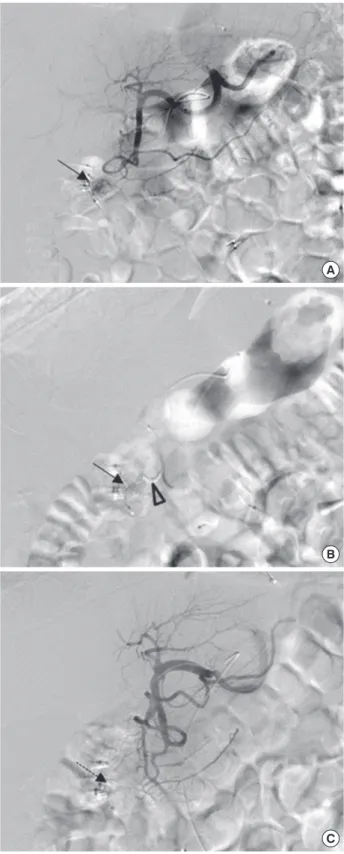
Fig. 4. Angiographic findings of case 2. (A) Celiac artery angiogram shows no definite extravasation, but a focal nodular hypervascular staining (arrow) in the second por-tion of the duodenum suggesting hypervascular tumor staining. (B) After superselec-tion of the feeding artery, the SPDA, with microcatheter tip (arrowhead), embolizasuperselec-tion is performed using absorbable gelatin sponge particles. The arrow indicates the same blood vessel in A. (C) GDA angiogram shows no further focal hypervascular staining (dotted arrow) in the duodenum, and the distal branches of GDA are normal. SPDA = superior pancreaticoduodenal artery, GDA = gastroduodenal artery.
A
B
C
odenum. Embolization of the superior pancreaticoduodenal artery (SPDA) was performed (Fig. 4). His hemoglobin level was still low with hematochezia, and surgery was performed. Mass excision was performed at the second portion of the duodenum, and the pathologic findings revealed a 2.1 × 1.4 cm sized benign GIST. The patient was discharged without complications. DISCUSSION
In cases of UGI bleeding, the rate of rebleeding after early endo-scopic hemostasis is about 10%–30%, and in 5%–10% of cases, endoscopic procedures fail and require TAE or surgery (12,13). In this report, the rate of rebleeding after early endoscopic he-mostasis was 24.7% (128/518) and 5.4% (28/518) of the patients underwent TAE or surgery due to rebleeding after repeated en-doscopic hemostasis, which is similar to the rates reported by previous studies.
We reviewed 8 rare cases in which TAE was performed for nonvariceal UGI bleeding. Among these cases, there were 4 males, the mean age was 74.00 ± 8.25 years, and most of the patients were taking drugs known to cause bleeding, such as aspirin, or warfarin (Table 1). All patients had at least one un-derlying disease. The initial hemoglobin levels of all patients were below 9 g/dL, prothrombin time (PT) was prolonged in 4 patients, and initial systolic BP of 3 patients was under 90 mmHg. Initial endoscopic findings revealed gastric origin bleeding in 4 cases and duodenal origin bleeding in the other 4 cases. Forrest classification Ia with spurting hemorrhage and Ib with oozing hemorrhage were also found in 4 cases each. Hemoclipping or sclerotherapy was performed in initial endoscopic hemostasis. In 3 cases, TAE was performed due to rebleeding despite suc-cessful primary hemostasis, and in 5 cases, TAE was performed after failure of primary endoscopic hemostasis. Regarding angi-ographic findings, extravasation was observed in 2 cases out of 8 cases, and embolization was performed at the arterial bleed-ing site. In the other 6 cases, empirical embolization was per-formed. The major limitation of TAE is that if it is not performed at the time of bleeding, it is difficult to identify the bleeding ves-sel because the injected contrast is not extravasated into the bowel lumen, and in this situation, empirical embolization is performed based on the patient’s clinical signs, endoscopic find-ings, and imaging findings (14). Of the 8 cases of embolization observed in this study, 4 were successful, but the other 4 cases showed rebleeding within 48 hours after the procedure. Among the cases with rebleeding, 1 patient was successfully managed with endoscopic hemostasis, and 3 patients eventually received surgery. TAE procedures showed a 50% success rate, but the other 50% of the cases resulted in rebleeding which required additional endoscopic hemostasis or surgery. Several studies have reported that the rebleeding rate of TAE in nonvariceal UGI bleeding ranges from 9%–47%, and this may be due to the
Table 1.
Baseline and clinical characteristics for each case undergoing
TAE Patient No. Age, yr Sex Underlying disease Causative drugs Hb, g/dL PL T, 10 3/µL PT, INR Systolic BP, mmHg Initial present -ing symptom Location of hemorrhage Forrest classifi -cation Extravasa -tion on an -giography Arter y of embolization Embolization meterial Outcome of embolization (consequent procedure)
1 83 F HTN, DM NSAIDs (ketorolac) 7.1 222 1.23 70 Melena Duodenum, bulb Ia No GDA Gelatin sponge Success 2 75 M HTN, IHD, CKD Antiplatelets (aspirin) 3.9 61 1.06 161 Melena Stomach, body Ib No LGA Gelatin sponge Success 3 74 M HTN, IHD, CV A Anticoagulants (warfarin) 8.2 126 1.29 110 Melena Duodenum, second portion Ia No SPDA Gelatin sponge Failure (surger y) 4 77 F IHD, P AOD Antiplatelets (aspirin, clopidogrel) 8.2 471 1.15 110 Hematochezia Duodenum, second portion Ib Ye s SPDA Gelatin sponge Failure (surger y) 5 73 F HTN NSAIDs (loxoprofen) 6.5 134 1.85 88 Hamatemesis Stomach, body Ia Ye s LGA NBCA
Failure (endoscopic hemostasis)
6 58 M CVA NSAIDs (ketorolac) 3.6 203 1.10 70 Melena Stomach, antrum Ib No GDA, SPDA NBCA Success 7 85 F HTN None 5.5 390 0.98 112 Syncope Duodenum, bulb Ia No SPDA NBCA Failure (surger y) 8 74 M HTN, DM Antiplatelet (sarpogrelate) 8.4 88 1.03 101 Hematemesis Stomach, cardia Ib No LGA Gelatin sponge Success TAE =
transcatheter arterial embolization,
Hb = hemoglobin, PL T = platelets, PT = prothrombin time, BP = blood pressure, F = female, HTN = hypertension, DM = diabetes mellitus, NSAIDs = nonsteroidal antiinflammator y drugs, GDA = gastroduodenal arter y, M = male, IHD =
ischemic heart disease,
CKD
=
chronic kidney disease,
LGA
=
left gastric arter
y, CV A = cerebrovascular accident, SPDA =
superior pancreaticoduodenal arter
y, P AOD = peripheral ar -ter y occlusive disease, NBCA = N-butyl cyanoacr ylate.
varying severity and etiology of the patients’ conditions (15). In this study, the patients who received TAE were old aged with medical comorbidities and drug history known to cause bleed-ing, which may have led to a higher clinical failure rate. In pati-ents who experienced rebleeding after TAE, more patipati-ents showed initial coagulopathy (3/4 vs. 1/4), duodenal origin bleeding (3/4 vs. 1/4), and spurting hemorrhage (3/4 vs. 1/4), compared to the patients who had successful TAE.
TAE is performed in cases of refractory acute UGI bleeding, which is not well controlled by endoscopic procedures, or in cases of massive bleeding or hemodynamic compromise (15). There are no absolute contraindications, but cases with renal insufficiency, contrast allergy or uncorrectable coagulopathy are considered as relatively contraindicated (15). Most of the patients who failed endoscopic hemostasis or had rebleeding after hemostasis were old aged (≥ 70 years old) with comorbid-ities. These patients were at high risk for complications under general anesthesia, and also were taking antiplatelets or antico-agulants. Therefore, bleeding may have occurred at the incision site after surgery. In some studies, TAE is reported to be associ-ated with fewer complications relassoci-ated to underlying conditions compared to surgery, but the rate of rebleeding is known to be higher (16-18). TAE is thought to be relatively safe, but the pos-sibility of complications such as access site hematoma, arterial dissection, contrast nephrotoxicity, and bowel ischemia should also be considered (14). In UK guidelines, it is recommended to repeat endoscopy when rebleeding occurs after endoscopy for initial treatment of UGI bleeding, and to perform TAE or sur-gery when repeated endoscopy also fails (19). Several studies claimed that success rates and complication rates of TAE and surgery are similar, whereas in some studies TAE is reported to be better than surgery in terms of success and complication rates, therefore, more prospective studies are required in the future (16-20).
In conclusion, in cases of refractory UGI bleeding after endo-scopic hemostasis, treatment plans should be decided based on the patient’s condition and the characteristics of the bleed-ing lesions after discussions between the relevant endoscopists, interventional radiologists, and surgeons. TAE may be attempt-ed prior to surgery when a radiology expert is immattempt-ediately avail-able, especially in old aged patients with high comorbidity who have high operation risk.
DISCLOSURE
The authors have no potential conflicts of interest to disclose. AUTHOR CONTRIBUTION
Conceptualization: Shim KN, Tae CH. Data curation: Shim KN, Choi SY, Ryu MS, Jung HK. Investigation: Lee KE, Choi SY, Jung
HK, Kim SE. Writing - original draft: Lee KE, Tae CH, Shim KN. Writing - review & editing: Lee KE, Tae CH, Shim KN, Choi SY, Moon CM, Kim SE, Jung SA.
ORCID
Ko Eun Lee https://orcid.org/0000-0002-1261-5702 Ki-Nam Shim https://orcid.org/0000-0003-4004-6292 Chung Hyun Tae https://orcid.org/0000-0002-0764-7793 Min Sun Ryu https://orcid.org/0000-0003-2613-4501 Sun Young Choi https://orcid.org/0000-0003-2488-1183 Chang Mo Moon https://orcid.org/0000-0003-2550-913X Seong-Eun Kim https://orcid.org/0000-0002-6310-5366 Hey-Kyung Jung https://orcid.org/0000-0002-6653-5214 Sung-Ae Jung https://orcid.org/0000-0001-7224-2867 REFERENCES
1. Gralnek IM, Barkun AN, Bardou M. Management of acute bleeding from a peptic ulcer. N Engl J Med 2008; 359: 928-37.
2. Hearnshaw SA, Logan RF, Lowe D, Travis SP, Murphy MF, Palmer KR. Acute upper gastrointestinal bleeding in the UK: patient characteristics, diagno-ses and outcomes in the 2007 UK audit. Gut 2011; 60: 1327-35.
3. Esrailian E, Gralnek IM. Nonvariceal upper gastrointestinal bleeding: epi-demiology and diagnosis. Gastroenterol Clin North Am 2005; 34: 589-605.
4. Yachimski PS, Friedman LS. Gastrointestinal bleeding in the elderly. Nat Clin Pract Gastroenterol Hepatol 2008; 5: 80-93.
5. Bae S, Kim N, Kang JM, Kim DS, Kim KM, Cho YK, Kim JH, Jung SW, Shim KN. Incidence and 30-day mortality of peptic ulcer bleeding in Korea. Eur J Gastroenterol Hepatol 2012; 24: 675-82.
6. Hong MJ, Lee SY, Kim JH, Sung IK, Park HS, Shim CS, Jin CJ. Rebleeding after initial endoscopic hemostasis in peptic ulcer disease. J Korean Med Sci 2014; 29: 1411-5.
7. Lee YJ, Kim ES, Hah YJ, Park KS, Cho KB, Jang BK, Chung WJ, Hwang JS. Chronic kidney disease, hemodynamic instability, and endoscopic high-risk appearance are associated with 30-day rebleeding in patients with non-variceal upper gastrointestinal bleeding. J Korean Med Sci 2013; 28:
1500-6.
8. Spiegel BM, Vakil NB, Ofman JJ. Endoscopy for acute nonvariceal upper gastrointestinal tract hemorrhage: is sooner better? A systematic review.
Arch Intern Med 2001; 161: 1393-404.
9. Lau JY, Sung J, Hill C, Henderson C, Howden CW, Metz DC. Systematic review of the epidemiology of complicated peptic ulcer disease: incidence, recurrence, risk factors and mortality. Digestion 2011; 84: 102-13.
10. Suk KT, Kim HS, Lee CS, Lee IY, Kim MY, Kim JW, Baik SK, Kwon SO, Lee DK, Ham YL. Clinical outcomes and risk factors of rebleeding following endoscopic therapy for nonvariceal upper gastrointestinal hemorrhage.
Clin Endosc 2011; 44: 93-100.
11. Lee HH, Park JM, Chun HJ, Oh JS, Ahn HJ, Choi MG. Transcatheter arteri-al embolization for endoscopicarteri-ally unmanageable non-varicearteri-al upper gastrointestinal bleeding. Scand J Gastroenterol 2015; 50: 809-15.
12. Loffroy R, Estivalet L, Cherblanc V, Sottier D, Guiu B, Cercueil JP, Krausé D. Transcatheter embolization as the new reference standard for endo-scopically unmanageable upper gastrointestinal bleeding. World J Gas-trointest Surg 2012; 4: 223-7.
13. Guglielmi A, Ruzzenente A, Sandri M, Kind R, Lombardo F, Rodella L, Cat-alano F, de Manzoni G, Cordiano C. Risk assessment and prediction of rebleeding in bleeding gastroduodenal ulcer. Endoscopy 2002; 34:
778-86.
14. Shin JH. Recent update of embolization of upper gastrointestinal tract bleeding. Korean J Radiol 2012; 13 Suppl 1: S31-9.
15. Shin JH. Refractory gastrointestinal bleeding: role of angiographic inter-vention. Clin Endosc 2013; 46: 486-91.
16. Ang D, Teo EK, Tan A, Ibrahim S, Tan PS, Ang TL, Fock KM. A comparison of surgery versus transcatheter angiographic embolization in the treat-ment of nonvariceal upper gastrointestinal bleeding uncontrolled by en-doscopy. Eur J Gastroenterol Hepatol 2012; 24: 929-38.
17. Wong TC, Wong KT, Chiu PW, Teoh AY, Yu SC, Au KW, Lau JY. A compar-ison of angiographic embolization with surgery after failed endoscopic hemostasis to bleeding peptic ulcers. Gastrointest Endosc 2011; 73: 900-8.
18. Venclauskas L, Bratlie SO, Zachrisson K, Maleckas A, Pundzius J, Jönson C. Is transcatheter arterial embolization a safer alternative than surgery when endoscopic therapy fails in bleeding duodenal ulcer? Scand J Gas-troenterol 2010; 45: 299-304.
19. Mirsadraee S, Tirukonda P, Nicholson A, Everett SM, McPherson SJ. Em-bolization for non-variceal upper gastrointestinal tract haemorrhage: a systematic review. Clin Radiol 2011; 66: 500-9.
20. Schenker MP, Duszak R Jr, Soulen MC, Smith KP, Baum RA, Cope C, Frei-man DB, Roberts DA, Shlansky-Goldberg RD. Upper gastrointestinal hem-orrhage and transcatheter embolotherapy: clinical and technical factors impacting success and survival. J Vasc Interv Radiol 2001; 12: 1263-71.