PGHN
Case Report
A Newborn with Gastric Hemangioma Treated Using Propranolol
Huseyin Kaya, Ismail Kursad Gokce, Sukru Gungor*, Hatice Turgut, and Ramazan Ozdemir
Divisions of Neonatology and *Pediatric Gastroenterology, Department of Pediatrics, Turgut Ozal Medical Center, Inonu University School of Medicine, Malatya, Turkey
Gastric hemangiomas are rare benign vascular tumors that can cause severe gastrointestinal system bleeding. We presented the case of a neonate with fresh bleeding and melena from the orogastric tube and detected gastric he- mangioma in esophagogastroduodenoscopic examination. Propranolol is widely used in treatment of cutaneous hemangiomas and non-gastric gastrointestinal system hemangiomas. However, the surgical approach is preferred for treating gastric hemangiomas, and there are few reports of gastric hemangiomas associated with non-surgical treatment. Gastric hemorrhage decreased with antacid and somatostatin treatment. Propranolol treatment was ini- tiated before the surgery decision. After three weeks of treatment, we observed regression in the hemangioma with endoscopic evaluation. During the course of treatment, the patient’s gastrointestinal system bleeding did not recur, and there were no side effects associated with propranolol.
Key Words: Hemangioma, Newborn infant, Propranolol, Stomach
Received:October 28, 2017, Revised:December 21, 2017, Accepted:December 29, 2017
Corresponding author: Huseyin Kaya, Department of Pediatrics, Turgut Ozal Medical Center, Inonu University School of Medicine, Campus Street, Malatya 44280, Turkey. Tel: +90-422-341-0660-5310, Fax: +90-422-341-0736, E-mail: [email protected]
Copyright ⓒ 2018 by The Korean Society of Pediatric Gastroenterology, Hepatology and Nutrition
This is an openaccess article distributed under the terms of the Creative Commons Attribution NonCommercial License (http://creativecommons.org/licenses/by-nc/4.0/) which permits unrestricted noncommercial use, distribution, and reproduction in any medium, provided the original work is properly cited.
INTRODUCTION
Gastrointestinal system (GIS) hemangiomas are benign vascular tumors. Although GIS hemangio- mas may occur anywhere along the gastrointestinal tract, they usually occur in the small intestine fol- lowed by the colon and rectum. GIS hemangiomas in the stomach are rare. So far, 22 cases of gastric he- mangioma have been reported in pediatric patients;
of these, 13 were isolated cases [1-4]. Esophagogas- troduodenoscopy, angiography and computed to-
mography are valuable in diagnosis of gastric hemangiomas. In the case of gastric hemangiomas that can cause life-threatening hemorrhage with le- sion ulceration, surgical involvement (total or partial gastrectomy, wedge excision) is the primary treat- ment option [5,6]. Propranolol is commonly used with corticosteroids to treat non-gastric GIS he- mangiomas [7]. However, pharmacotherapy has of- ten been performed in a small number of hemangio- ma patients with multiple lesions in the GIS, where- in surgical possibilities are limited [2-4,8]. We report
Fig. 1. Corpus and fundus of the stomach hemangioma.
the case of a neonate with an isolated gastric he- mangioma, presented with massive gastrointestinal bleeding, and the hemangioma shrunk with propra- nolol therapy after bleeding was controlled with an- tisecretory drugs. This is the first case of a neonate with gastric hemangioma treated with pharmaco- therapeutic agents alone.
CASE REPORT
A female infant weighting of 2,220 g was born via vaginal route at 33 weeks of gestation. The neonate was admitted to the neonatal clinic at another centre due to a 24-hour history of preterm rupture of mem- branes and respiratory distress. The mother was age
26 years and this was her first pregnancy. On second postnatal day, fresh bleeding from the orogastric catheter and melena was detected and supportive treatment was initiated. Bleeding continued, and patient was administered erithrocyte suspension and referred to our hospital. Physical examination revealed that body weight was 2,200 g (50th-75th percentile), the length 45 cm (50th-75th percentile), and head circumference 32 cm (50th-75th percen- tile). The patient’s axillary body temperature was 36.7oC, blood pressure was 65/35 mmHg, the pulse and respiratory rate 130 beats/minute and 50 beats/mi- nute, respectively. The patient had phenotypic char- acteristics of Down syndrome, and other system ex- aminations were normal. Results of the laboratory
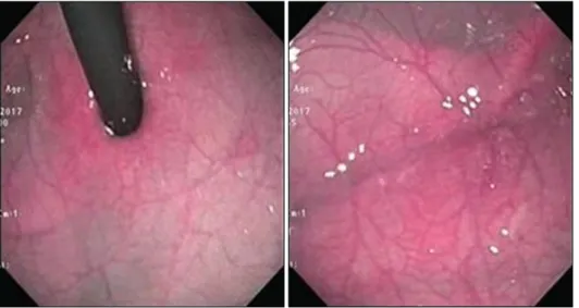
Fig. 2. Control endoscopy images.
investigation were as follows: hemoglobin, 10.5 g/dL; leukocytes, 6.680/mm3; thrombocytes, 242.000/mm3. Bleeding time, coagulation and other biochemical test results were normal. GIS bleeding continued and somatostatin, omeprazol and raniti- din treatments were initiated. Enteral feeding was interrupted and abdominal graphy and portal vein Doppler yielded normal results. In the following days, gastric bleeding regressed with antacid therapy and somatostatin and the patient did not require transfusion. On day eight, the patient underwent esophagogastroduodenoscopy. Hemangioma was detected in the corpus and fundus of the stomach (Fig. 1). Biopsy specimen was not obtained as it may have caused additional bleeding. No cutaneous he- mangioma was present and ultrasonographic ex- amination revealed no hemangioma in the liver.
The patient was diagnosed with gastric hemangio- ma, and treatment with propranolol was initiated with 1 g/kg per day divided into three doses and subsequently increased to 2 g/kg per day the follow- ing days. Enteral feeding was increased. Somatosta- tin treatment was tapered and discontinued on day fourteen. No side effects (hypoglycemia, brady- cardia, or respiratory distress) were observed in asso- ciation with gastric bleeding and propranolol on sub- sequent days. On postnatal day twenty-two, patient was discharged with propranolol and antiacid treat-
ment, and called for endoscopic examination three weeks later. Thecontrol endoscopy revealed marked regression in the size of the hemangioma (Fig. 2).
Treatment was maintained to be completed at three months. Control endoscopy was to be performed af- ter treatment is completed.
DISCUSSION
Hemangiomas may be observed in all organs, es- pecially on the body surface as isolated or multiple lesions. However, visceral hemangiomas occur rarely except in the liver. Childhood GIS hemangiomas most commonly involve the small intestine and pres- ent with lower GIS bleeding. In more than half of these patients, cutaneous lesions are present, and some of these patients are diagnosed with posterior fossa malformations, hemangiomas, arterial anoma- lies, coarctation of the aorta and other cardiac de- fects, and eye abnormalities (PHACE) syndrome [9].
Gastric hemangiomas, that occur more rarely, are frequently observed as isolated lesions. So far, 22 cases of gastric hemangioma have been reported in pediatric patients; of these, 13 were isolated cases [1-4]. Four cases of hemangiomas were reported in the neonatal period, three of which were reported as isolated gastric hemangioma [2,4,5,10].
GIS hemangiomas may occur in association with
Maffucci syndrome, Klippel-Trénaunay syndrome, widespread neonatal hemangiamatosis and blue rubber-bleb nevus syndrome [11]. In our patient, chromosome analysis revealed presence of mozaic type trisomy 21 (46, XX/47, XX, +21), and he- mangioma was not detected in other regions except the stomach. Interestingly, risk of vascular anomaly is lower in patients diagnosed with Down syndrome than in the general population. This is explained by increased expression of angiogenic factors [12].
As in this case, the most common symptom of gas- tric hemangioma is hematemesis, and is frequently accompanied by melena. More rarely, the patient may present with epigastric pain or symptoms of anemia secondary to chronic bleeding. Diagnostic tools that can be used irrespective of age and ana- tomic characteristics of the patients include imaging techniques such as computed tomography, magnetic resonance imaging, abdominal ultrasonography, scintigraphy and angiography. If it is thought that if bleeding originates from a lesion in stomach or duo- denum, esophagogastroduodenescopic examination should be performed; if it originates from a focus in colon or rectum, colonoscopic examination is preferred. However, since most GIS hemangiomas are in the small intestine, esophagogastroduodeno- scopy and colonoscopy may not be sufficient for imaging the lesion. In these patients, capsule endos- copy or scintigraphy are other options that can be performed for diagnosis [13]. In this case, contrast abdominal tomography and portal vein Doppler ex- aminations were normal. A definitive diagnosis was made in detection of hemangioma by esophagogas- troduodenoscopy in corpus and fundus of the stomach.
In treatment of GIS hemangioma, if there is a small, isolated lesion that causes mild symptoms, pa- tients may be administered conservative treatments such as blood transfusion or iron supplementation [6]. First-line agents for symptomatic GIS he- mangiomas are corticosteroids and propranolol, as in other infantile hemangiomas [14,15]. Combined use of these agents may be performed in refractory cases.
In addition, vincristine, interferon 1 alpha and other
antiangiogenic drugs may be used [2,9]. For patients not responding to medical treatment, argon plasma coagulation, laser photocoagulation, sclerotherapy, band ligation or surgical resection are other treat- ment options [4,8,16]. In a case series of 16 patients with gastrointestinal hemangioma, 8 diagnosed in neonatal period, 14 received pharmacological treatment. Of these patients, only two required in- testinal resection as well as pharmacotherapy.
However, none of these 14 patients had gastric he- mangioma [9].
Treatment apporoach for gastric hemangiomas may differ from that for gastrointestinal hemangiomas.
The clinical course of asymptomatic gastric he- mangiomas is not completely known. However, in symptomatic gastric hemangiomas, that may be po- tentially life-threatening due to ulceration of the le- sion, surgical resection is the preferred treatment op- tion [1,5]. For this purpose, depending on the size and location of the lesion, excision of mass, an- trectomy, and subtotal or total gastrectomy may be performed [17].
After gastric hemangioma is diagnosed, appro- priate endoscopic and/or surgical treatment should be performed as soon as possible in to prevent serious complications that may potentially be life threatening.
In this case, after bleeding was controlled with medi- cal treatment, propranolol treatment was instituted before deciding on surgical intervention. In the weeks following propranolol treatment initiation, the patient was closely monitored in the clinic for GIS bleeding. There was no GIS bleeding and in con- trol endoscopic examination performed three weeks later, the lesion had regressed and medical treat- ment continued. In a previous study of 22 gastric he- mangioma cases reported in childhood, surgical in- tervention was performed in all cases except four [1-4]. Presently, oral propranolol may be considered as first-line treatment in infantile hemangiomas [18]. Propranolol is also used commonly with corti- costeroids in treatment of gastrointestinal he- mangiomas [2,7,9]. However, few studies have re- ported treatment of gastric hemangiomas with methods other than surgical intervention, and these
cases are usually with multiple lesions in which sur- gical options are limited. In a previous study, three patients were successfully treated with pharmaco- therapy with additional argon plasma coagulation and electrocoagulation [2,4,8]. Similar to this case, a patient with multiple hemangiomas in the stomach and intestines successfully treated with propranolol was reported [3]. The most serious known adverse e ffects of propranolol are bradycardia, hypoglycemia and bronchospasm [19]. Hypoglycemia is common during propranolol therapy in the newborn and in- fantile period due to insufficient glycogen deposits and high rates of glucose consumption rates [20].
We did not observe side effects that may be asso- ciated with propranolol in our patient.
Unlike cutaneous lesions, it is not possible to di- rectly visualise GIS hemangiomas to evaluate effi- cacy of drug treatment; therefore, endoscopic exami- nation is required for evaluation of treatment. In this case, as regression was determined in control endo- scopic examination, medical treatment was con- tinued to be completed at three months. Repeat en- doscopic examination was planned after completion of treatment.
In conclusion, we report a case of gastric he- mangioma in a newborn. Although it is rare, he- mangioma should be considered during differential diagnosis of cases with GIS bleeding. Unlike gastric hemangiomas, of which the most conservative and preferred treatment is pharmacotherapy, surgical is the preferred approach to treating gastric heman- giomas. We observed regression in hemangioma with propranolol therapy in our patient. We think that propranolol therapy must be performed before surgical intervention in selected cases under GIS bleeding control such as in our case.
REFERENCES
1. Parolini F, Colusso M, Giannotti G, Cheli M, Alberti D.
Gastric hemangiomas in children. Int J Gastroenterol Hepatol Transpl Nutr 2016;1:10-4.
2. Hansen LF, Wewer V, Pedersen SA, Matzen P, Paerregaard A. Severe blue rubber bleb nevus syn-
drome in a neonate. Eur J Pediatr Surg 2009;19:47-9.
3. Akcam M, Pirgon O, Salman H, Kockar C. Multiple gas- trointestinal hemangiomatosis successfully treated with propranolol. J Pediatr Gastroenterol Nutr 2015;60:e16.
4. Lee YA, Chun P, Hwang EH, Lee YJ, Kim CW, Park JH.
Gastric hemangioma treated with argon plasma coagu- lation in a newborn infant. Pediatr Gastroenterol Hepatol Nutr 2017;20:134-7.
5. Holden KR, Alexander F. Diffuse neonatal heman- giomatosis. Pediatrics 1970;46:411-21.
6. Han EC, Kim SH, Kim HY, Jung SE, Park KW.
Gastrointestinal hemangioma in childhood: a rare cause of gastrointestinal bleeding. Korean J Pediatr 2014;57:245-9.
7. Morris GA, Stratchko L, Sabri M. Intestinal hemangio- ma presenting as recurrent hematochezia in a 6-week- old male. J Pediatr Surg Case Rep 2015;3:280-2.
8. Khan K, Weisdorf-Schindele S. Gastric hemangiomas in an infant managed with argon plasma coagulation.
Pediatr Endosurgery Innov Tech 2003;7:185-8.
9. Soukoulis IW, Liang MG, Fox VL, Mulliken JB, Alomari AI, Fishman SJ. Gastrointestinal infantile hemangio- ma: presentation and management. J Pediatr Gastro- enterol Nutr 2015;61:415-20.
10. Nagaya M, Kato J, Niimi N, Tanaka S, Akiyoshi K, Tanaka T. Isolated cavernous hemangioma of the stom- ach in a neonate. J Pediatr Surg 1998;33:653-4.
11. Magnano A, Privitera A, Calogero G, Nanfito' L, Basile G, Sanfilippo G. Solitary hemangioma of the small in- testine: an unusual cause of bleeding diagnosed at cap- sule endoscopy. J Pediatr Surg 2005;40:e25-7.
12. Greene AK, Kim S, Rogers GF, Fishman SJ, Olsen BR, Mulliken JB. Risk of vascular anomalies with Down syndrome. Pediatrics 2008;121:e135-40.
13. Kavin H, Berman J, Martin TL, Feldman A, Forsey- Koukol K. Successful wireless capsule endoscopy for a 2.5-year-old child: obscure gastrointestinal bleeding from mixed, juvenile, capillary hemangioma-angioma- tosis of the jejunum. Pediatrics 2006;117:539-43.
14. Chen TS, Eichenfield LF, Friedlander SF. Infantile he- mangiomas: an update on pathogenesis and therapy.
Pediatrics 2013;131:99-108.
15. Bertrand J, McCuaig C, Dubois J, Hatami A, Ondrejchak S, Powell J. Propranolol versus prednisone in the treatment of infantile hemangiomas: a retro- spective comparative study. Pediatr Dermatol 2011;28:
649-54.
16. Agnese M, Cipolletta L, Bianco MA, Quitadamo P, Miele E, Staiano A. Blue rubber bleb nevus syndrome.
Acta Paediatr 2010;99:632-5.
17. Desa LA, Bridger J, Grace PA, Krausz T, Spencer J.
Primary jejunoileal tumors: a review of 45 cases. World J Surg 1991;15:81-6.
18. Léauté-labrèze C, Hoeger P, Mazereeuw-Hautier J, Guibaud L, Baselga E, Posiunas G, et al. A randomized, controlled trial of oral propranolol in infantile hemangioma. N Engl J Med 2015;372:735-46.
19. Horev A, Haim A, Zvulunov A. Propranolol induced hypoglycemia. Pediatr Endocrinol Rev 2015;12:308-10.
20. de Graaf M, Breur JMPJ, Raphaël MF, Vos M, Breugem CC, Pasmans SGMA. Adverse effects of propranolol when used in the treatment of hemangiomas: a case ser- ies of 28 infants. J Am Acad Dermatol 2011;65:320-7.