283
Copyrights © 2013 The Korean Society of Radiology
INTRODUCTION
Sclerosing hemangioma is relatively rare, the second most common benign pulmonary neoplasm (1), which is initially considered to be of vascular origin. However, recent immuno- histochemical and genetic studies suggest that sclerosing hem- angioma is an epithelial tumor, related to the pulmonary epithe- lium. Typically, this is a solitary, generally asymptomatic, and well-described lesion located in the periphery of the lung.
Therefore, sclerosing hemangioma which contains endobron- chial portion is extremely rare (2). The histological characteris- tics of sclerosing hemangioma have been well known for show- ing solid, papillary, sclerotic and hemorrhagic patterns (1).
However, the specimens obtained by bronchoscopic biopsy may be limited to the papillary or other specific histologic patterns. It
can also be misdiagnosed. Therefore, we report an extremely rare case of central sclerosing hemangioma in 58-year-old wom- an with radiological-pathological findings, which was initially misdiagnosed as papillary adenoma by bronchoscopic biopsy and mimicked central lung malignancy on non-invasive image evaluations.
CASE REPORT
A solitary lung mass with central location in right upper lobe was discovered in 58-year-old female without specific symp- toms. The initial chest PA radiograph showed about 3 cm sized nodular opacity in the right hilar area (Fig. 1A), and the initial chest CT revealed a well-defined centrally located mass of about 3 cm diameter, and heterogeneous contrast enhancement in the
Case Report
pISSN 1738-2637 / eISSN 2288-2928 J Korean Soc Radiol 2013;69(4):283-286 http://dx.doi.org/10.3348/jksr.2013.69.4.283
Received May 28, 2013; Accepted July 26, 2013 Corresponding author: Hyun Ju Seon, MD
Department of Radiology, Chonnam National University Hospital, Chonnam National University Medical School, 160 Baekseo-ro, Dong-gu, Gwangju 501-746, Korea.
Tel. 82-62-220-5888 Fax. 82-62-226-4380 E-mail: [email protected]
This is an Open Access article distributed under the terms of the Creative Commons Attribution Non-Commercial License (http://creativecommons.org/licenses/by-nc/3.0) which permits unrestricted non-commercial use, distri- bution, and reproduction in any medium, provided the original work is properly cited.
Sclerosing hemangioma is relatively rare, the second most common benign pulmonary neoplasm, which usually presents the peripheral location. Central location of this neo- plasm is extremely rare with only a few reports. Herein, we would like to report an ex- tremely rare case of central sclerosing hemangioma with descriptions of radiological characteristics. It was initially misdiagnosed as a papillary adenoma by bronchoscopic biopsy and mimicked central lung malignancies such as carcinoid tumors on non-inva- sive image evaluations. However the patient was finally confirmed with surgery.
Index terms
Sclerosing Hemangioma Pneumocytoma Endobronchial Pulmonary Neoplasm
Radiological-Pathological Findings of Central Sclerosing
Hemangioma Initially Misdiagnosed as Papillary Adenoma by Bronchoscopic Biopsy: A Case Report
1기관내시경 조직검사에서 유두상 선종으로 오진된 중심성 경화혈관종의 영상의학적-병리학적 소견: 증례 보고1
Soo Hyun Kim, MD
1, Hyun Ju Seon, MD
1, Jang-Hyeon Song, MD
1, Seo Yeon Park, MD
1, Yun-Hyeon Kim, MD
1, Yoo-Duk Choi, MD
2, Sang-Yun Song, MD
3Departments of 1Radiology, 2Pathology, 3Thoracic and Cardiovascular Surgery, Chonnam National University Hospital, Chonnam National University Medical School, Gwangju, Korea
Central Sclerosing Hemangioma Initially Misdiagnosed as Papillary Adenoma
284
J Korean Soc Radiol 2013;69(4):283-286 jksronline.orgfindings suggested a slow growing low grade central lung malig- nancy with some hypervascularity such as a carcinoid tumor rather than benign neoplasm. For accurate diagnosis and treat- ment, the patient underwent posterior segmentectomy of the RUL, and the mass was completely removed. The mass showed papillary, hemorrhagic, and sclerotic pattern composed of sur- face cuboidal cells and sheets of round cells (Fig. 1G). There was no necrosis or mitotic figures. On the immunohistochemical stains, surface cuboidal cells were positive for cytokeratin (CK), epithelial membrane antigen (EMA), and thyroid transcription factor-1 (TTF-1), but round cells were positive for EMA and TTF-1, and negative for CK. Therefore, the final histopathologic diagnosis of the specimen was a sclerosing hemangioma.
DISCUSSION
Sclerosing hemangioma is relatively rare, the second most common benign pulmonary neoplasm, and named due to his- tologically prominent features of sclerotization and vasculariza- tion of the tumor (1). Many recent immunohistochemical stud- right upper lobe (RUL) abutting the posterior segmental bron-
chus (Fig. 1B). There was no evidence of infiltration on the sur- rounding lung parenchyma or lymphadenopathy. Bronchoscop- ically, there was an endobronchial lesion obliterating posterior segmental bronchus of RUL with easy touch bleeding, and the bronchoscopic suspicion was a carcinoid tumor (Fig. 1C). The pathologic result of initial bronchoscopic biopsy was a papillary adenoma (Fig. 1D).
However, the serial of follow-up chest CT scans show gradual increases of the mass with small endobronchial portion. The last follow-up CT scan obtained 7 years after the initial exam indi- cated about 1.1 cm interval growth of central mass in the poste- rior segment of RUL with ice-burg sign. The large extraluminal portion with relatively small endobronchial portion is known as a typical radiologic finding of slow growing central carcinoid tu- mor (3), with newly developed distal obstructive pneumonitis and subsegmental atelectasis (Fig. 1E). The follow-up positron emission tomography-CT (PET-CT) scan also revealed high fluorodeoxyglucose (FDG) uptakes of the mass [maximal stan- dardized uptake value (SUVmax): 5.8] (Fig. 1F). These image
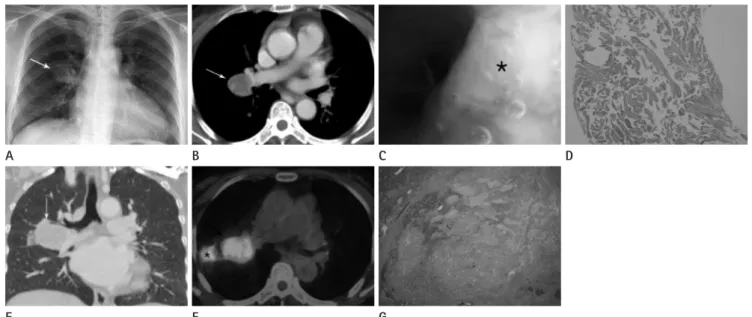
Fig. 1. Centrally located sclerosing hemangioma with endobronchial extension in a 58-year-old female.
A. Initial chest PA radiograph shows about 3 cm sized nodular opacity (arrow) in right hilar area.
B. The initial chest CT image shows about 3 cm sized well-defined central mass (arrow) with heterogeneous enhancement in right upper lobe (RUL).
C. Bronchoscopy shows a protruding endobronchial lesion in posterior segmental bronchus of RUL (*).
D. Only papillary pattern of tumor was obtained on initial biopsy (H&E, × 100).
E. Follow-up CT image after 7 years from initial examination shows gradual increase of a central mass in RUL with endobronchial extension to posterior segmental bronchus (arrow).
F. Follow-up positron emission tomography-CT image shows high fluorodeoxyglucose uptake of the central mass (arrow) and distal obstructive pneumonia and atelectasis (*) of RUL.
G. A mixture of papillary, hemorrhagic, and sclerotic patterns is shown on final surgical specimen (H&E, × 10).
E A
F B
G
C D
Soo Hyun Kim, et al
285
jksronline.org J Korean Soc Radiol 2013;69(4):283-286
diagnosis of sclerosing hemangioma which has four different pathologic components. Nevertheless, a tiny specimen does not show the entire features of sclerosing hemangioma and may re- sult in inaccurate diagnosis. Therefore, surgical methods, in- cluding wedge resection or segmentectomy, can exhibit entire features of sclerosing hemangioma and establish complete re- section of the tumor. Furthermore, sclerosing hemangioma can grow and usually show high FDG-uptakes. Therefore, within such situations, results of limited biopsy specimens should be doubted.
Sclerosing hemangioma is a progressively growing benign tu- mor with high FDG uptakes and may have endobronchial por- tion which is extremely rare. Therefore, it can mimic slow grow- ing central lung malignancy such as carcinoid tumor on imaging findings. Furthermore, this tumor is composed of four patho- logic patterns: the papillary, sclerotic, solid and hemorrhagic components. Thus, the diagnosis by limited specimen through bronchoscopic biopsy may be insufficient. For accurate diagno- sis, a complete surgical resection should be recommended in ra- diological suspicions of central lung malignancy or central scle- rosing hemangioma with endobronchial extensions.
REFERENCES
1. Sugio K, Yokoyama H, Kaneko S, Ishida T, Sugimachi K. Scle- rosing hemangioma of the lung: radiographic and patho- logical study. Ann Thorac Surg 1992;53:295-300
2. Boudaya MS, Falcoz PE, Alifano M, Camilleri-Broet S, Rég- nard JF. Endobronchial sclerosing hemangioma: a rare presentation of a parenchymal tumor. Asian Cardiovasc Thorac Ann 2008;16:57-58
3. Chong S, Lee KS, Chung MJ, Han J, Kwon OJ, Kim TS. Neu- roendocrine tumors of the lung: clinical, pathologic, and imaging findings. Radiographics 2006;26:41-57; discus- sion 57-58
4. Devouassoux-Shisheboran M, Hayashi T, Linnoila RI, Koss MN, Travis WD. A clinicopathologic study of 100 cases of pulmonary sclerosing hemangioma with immunohisto- chemical studies: TTF-1 is expressed in both round and surface cells, suggesting an origin from primitive respira- tory epithelium. Am J Surg Pathol 2000;24:906-916 5. Kim GY, Kim J, Choi YS, Kim HJ, Ahn G, Han J. Sixteen cases ies supported that pulmonary sclerosing hemangioma arises
from epithelial cells, probably the type II pneumocyte (4).
Typically, it occurs in middle-aged adults, with a predilection on females, who are usually asymptomatic until the time of di- agnosis similar to this case (5). The tumor has well-defined mar- gins, but not a definite capsule, and can grow expansively by compressing the adjacent lung parenchyma or bronchus. The CT findings show round or ovoid shape, a smooth margin, ho- mogeneous attenuation, calcification, and strong early enhance- ment (6). In PET-CT scans, the majority of sclerosing hemangi- oma show increased FDG uptakes, the SUVmax of tumors ranged from 0.60 to 4.7 (median 2.30), and tumor ≥ 2 cm can frequent- ly be falsely interpreted as malignancy (7). The tumor in our case shows intense FDG uptakes (SUVmax: 5.8). Single tumor lo- calizations in the pulmonary parenchyma is a major concern.
However, there are exceptional presentations such as endobron- chial localization, which is extremely rare and mimics a slow growing low grade central lung malignancy such as carcinoid tumor. The reported doubling time of carcinoid tumor was 417 days and shorter than that of sclerosing hemangioma, which was reported to be 965 days (8, 9). The presence of sclerosing hem- angioma inside a bronchus may not be a proliferation from the bronchus itself but a proliferation from peribronchial lung pa- renchyma invading adjacent bronchus (2). Histologically, the sclerosing hemangioma is a mixture of solid, papillary, sclerotic or hemorrhagic patterns. The proportions of these four compo- nents in the tumor usually vary, although one of them tends to predominate. Sugio et al. (1) reported that most sclerosing hem- angioma cases exhibit a papillary pattern, while Devouassoux- Shisheboran et al. (10) reported that predominant endobronchi- al component by bronchial biopsy of this tumor was papillary and solid pattern.
The sclerosing hemangioma of our case exhibited endo- bronchical portion in the posterior segmental bronchus of RUL, and this tumor was initially misdiagnosed as a papillary adeno- ma. The initial specimen obtained by bronchoscopic biopsy contained only papillary patterns of the mass. The endobronchi- al mass biopsy obtained by bronchoscopy is widely used for the diagnosis of central lung lesions with endobronchial portion, re- gardless of benignity or malignancy as this method is less inva- sive and simpler than surgical biopsy. However, the broncho- scopic biopsy may not be a sufficient method for the pathologic
Central Sclerosing Hemangioma Initially Misdiagnosed as Papillary Adenoma
286
J Korean Soc Radiol 2013;69(4):283-286 jksronline.orget al. [Surgical treatment for patients with pulmonary sclerosing hemangioma]. J UOEH 2011;33:41-45
9. Greengard O, Head JF, Goldberg SL, Kirschner PA. Pulmonary carcinoid tumors: enzymic discriminants, growth rate, and early age of inception. Cancer Res 1986;46:2600-2605 10. Devouassoux-Shisheboran M, de la Fouchardière A, Thivo-
let-Béjui F, Sourisseau-Millan ML, Guerin JC, Travis WD.
Endobronchial variant of sclerosing hemangioma of the lung: histological and cytological features on endobron- chial material. Mod Pathol 2004;17:252-257
of sclerosing hemangioma of the lung including unusual presentations. J Korean Med Sci 2004;19:352-358
6. Chung MJ, Lee KS, Han J, Sung YM, Chong S, Kwon OJ. Pul- monary sclerosing hemangioma presenting as solitary pul- monary nodule: dynamic CT findings and histopathologic comparisons. AJR Am J Roentgenol 2006;187:430-437 7. Lee E, Park CM, Kang KW, Goo JM, Kim MA, Paeng JC, et
al. 18F-FDG PET/CT features of pulmonary sclerosing hem- angioma. Acta Radiol 2013;54:24-29
8. Oka S, Ono K, Kuwata T, Nagata Y, Baba T, Shigematsu Y,
기관내시경 조직검사에서 유두상 선종으로 오진된 중심성 경화혈관종의 영상의학적-병리학적 소견: 증례 보고1
김수현
1· 선현주
1· 송장현
1· 박서연
1· 김윤현
1· 최유덕
2· 송상윤
3경화혈관종은 상대적으로 드문, 폐의 양성 종양 중 두 번째로 흔한 종양으로서 일반적으로 폐의 주변부에 위치한다. 중심 성 경화혈관종은 극히 드물다고 알려져 있기 때문에 이의 영상의학적 소견에 대한 증례를 보고한다. 이 증례는 최초 기관 지 내시경 생검 결과 유두상 선종으로 진단되었으나 영상의학적 검사에서 유암종 같은 중심성 악성 종양처럼 보였다. 수술 적 절제를 시행하였고 경화혈관종으로 최종 진단되었다.
전남대학교 의과대학 전남대학교병원 1영상의학과, 2병리과, 3흉부외과