Brief Report
Vol. 29, No. 4, 2017 501
Received May 27, 2016, Revised July 20, 2016, Accepted for publication August 7, 2016
Corresponding author: Yu Sung Choi, Department of Dermatology, Ulsan University Hospital, University of Ulsan College of Medicine, 877 Bangeojinsunhwan-doro, Dong-gu, Ulsan 44033, Korea. Tel: 82-52-250- 7090, Fax: 82-52-250-7155, E-mail: [email protected]
This is an Open Access article distributed under the terms of the Creative Commons Attribution Non-Commercial License (http://creativecommons.
org/licenses/by-nc/4.0) which permits unrestricted non-commercial use, distribution, and reproduction in any medium, provided the original work is properly cited.
Copyright © The Korean Dermatological Association and The Korean
Society for Investigative Dermatology Fig. 1. (A, B) Multiple various-sized erythematous plaques and nodules on the face.
https://doi.org/10.5021/ad.2017.29.4.501
Blastic Plasmacytoid Dendritic Cell Neoplasm Presenting as Erythematous Nodules with Gallbladder Involvement
Sook Hyun Kong, Seok Hyun Han, Ho Seok Suh, Yu Sung Choi
Department of Dermatology, Ulsan University Hospital, University of Ulsan College of Medicine, Ulsan, Korea
Dear Editor:
Blastic plasmacytoid dendritic cell neoplasm (BPDCN) is a rare, highly aggressive hematopoietic malignancy which is derived from the precursors of plasmacytoid dendritic cells1. It was recently classified in the 2008 World Health Organization classification2. BPDCN can be characterized by a marked predilection for cutaneous involvement in the initial phase, and later develops leukemic dissem- ination3.
A previously healthy 43-year-old woman presented with 3-week history of nodular eruptions on the face. She had no systemic symptoms. Physical examination did not re- veal any abnormalities, except for the skin lesions. Skin examination revealed multiple various-sized erythematous plaques and nodules on the face (Fig. 1). We obtained in- formed consent for using the photos from the patient.
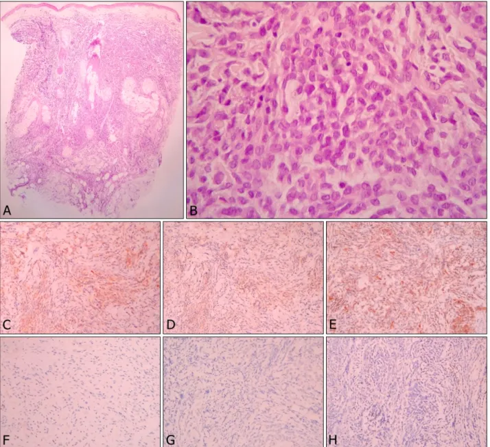
Histopathologic findings showed a diffuse cellular in- filtrate in the entire dermis (Fig. 2A). The cells were mono- morphic medium sized atypical lymphocytes containing irregular round or indented nuclei with pale cytoplasm (Fig. 2B). Immunohistochemical stains were positive for CD4 (Fig. 2C), CD56 (Fig. 2D), and CD123 (Fig. 2E) and negative for CD3 (Fig. 2F), CD20 (Fig. 2G), myeloperox- idase (Fig. 2H), and terminal deoxynucleotidyl transferase.
Microscopic examination of peripheral blood failed to de- tect neoplastic cells. Abdominal computed tomography
scan disclosed wall thickening of gallbladder (GB). After cholecystectomy, GB involvement of BPDCN was identi- fied by biopsy. Bone marrow biopsy with immunohisto- chemical stain also revealed anaplastic cells. Thus, BPDCN with skin, GB and bone marrow involvement was diagnosed. The patient received chemotherapy after lym- phoblastic lymphoma-type induction protocol. She sub- sequently underwent allogeneic bone marrow transplan- tation. There has been no disease progression following 12 months.
BPDCN is a rare hematologic malignancy typically affects older males4. In majority of cases, skin is affected at the in- itial presentation as bruise-like tumefaction or an eryth- ematous nodule1,5. BPDCN is often accompanied by ex- tracutaneous involvement, including bone marrow, lymph node, and peripheral blood but practically any organ can be affected1. Our patient presented with multiple eryth- ematous nodules on the face, which is a rare pattern in BPDCN to the best of our knowledge1,5. After review of the literature, we believe that the GB involvement de- scribed herein has not been documented before.
Pathologic evaluation and immunophenotyping play an important role in the diagnosis of BPDCN, which are char-
Brief Report
502 Ann Dermatol
Fig. 2. (A) The tumor is composed of diffuse cellular infiltrate in the entire dermis and subcutis, with an overlying Grenz zone (H&E,
×40). (B) The cells were monomorphic medium sized atypical lymphocytes containing irregular round or indented nuclei with pale cytoplasm (H&E, ×400). Immunohistochemical stains were positive for (C) CD4 (×100), (D) CD56 (×100), and (E) CD123 (×100) and negative for (F) CD3 (×100), (G) CD20 (×100), and (H) myeloperoxidase (×100).
acterized by a diffuse monomorphic infiltrate of me- dium-sized plasmacytoid cells and the expression of CD4, CD56, and CD123 in the absence of CD3, CD20, or mye- loperoxidase3,4.
The clinical course of BPDCN is aggressive with a median survival of 12∼14 months1,5. However, there is no stand- ardized therapeutic strategy. High-dose chemotherapy fol- lowed by allogeneic stem cell transplantation can provide durable disease control like our patient1. Several factors, including relatively young age and undergoing stem cell transplantation may contribute to more favorable prog-
nosis in this case.
In conclusion, BPDCN may demonstrate various clinical presentations, which could be quite confusing for the der- matologist to diagnose. Thus histopathologic evaluation of skin biopsy is important to confirm diagnosis. We believe that this case demonstrates a rare cutaneous and extracuta- neous presentation of BPDCN and emphasizes the im- portance of being aware of this rare disease in order to provide effective treatment.
Brief Report
Vol. 29, No. 4, 2017 503
Received July 11, 2016, Revised August 1, 2016, Accepted for publication August 8, 2016
*These authors contributed equally to this work and should be considered co-first authors.
Corresponding author: Hye One Kim, Department of Dermatology, Kangnam Sacred Heart Hospital, Hallym University College of Medicine, 1 Singil-ro, Yeongdeungpo-gu, Seoul 07441, Korea. Tel: 82-2-829-5221, Fax: 82-2-832-3237, E-mail: [email protected]
Chun Wook Park, Department of Dermatology, Kangnam Sacred Heart Hospital, Hallym University College of Medicine, 1 Singil-ro, Yeongdeungpo-gu, Seoul 07441, Korea. Tel: 82-2-829-5221, Fax: 82-2-832-3237, E-mail: [email protected]
This is an Open Access article distributed under the terms of the Creative Commons Attribution Non-Commercial License (http://creativecommons.org/
licenses/by-nc/4.0) which permits unrestricted non-commercial use, distribution, and reproduction in any medium, provided the original work is properly cited.
Copyright © The Korean Dermatological Association and The Korean Society for Investigative Dermatology
CONFLICTS OF INTEREST
The authors have nothing to disclose.
REFERENCES
1. Kim JH, Park HY, Lee JH, Lee DY, Lee JH, Yang JM. Blastic plasmacytoid dendritic cell neoplasm: analysis of clinicopa- thological feature and treatment outcome of seven cases.
Ann Dermatol 2015;27:727-737.
2. Willemze R, Jaffe ES, Burg G, Cerroni L, Berti E, Swerdlow SH, et al. WHO-EORTC classification for cutaneous lym- phomas. Blood 2005;105:3768-3785.
3. Gera S, Dekmezian MS, Duvic M, Tschen JA, Vega F, Cho-Vega JH. Blastic plasmacytoid dendritic cell neoplasm:
evolving insights in an aggressive hematopoietic malignancy with a predilection of skin involvement. Am J Dermato- pathol 2014;36:244-251.
4. Nguyen CM, Stuart L, Skupsky H, Lee YS, Tsuchiya A, Cassarino DS. Blastic plasmacytoid dendritic cell neoplasm in the pediatric population: a case series and review of the literature. Am J Dermatopathol 2015;37:924-928.
5. Julia F, Petrella T, Beylot-Barry M, Bagot M, Lipsker D, Machet L, et al. Blastic plasmacytoid dendritic cell neoplasm:
clinical features in 90 patients. Br J Dermatol 2013;169:
579-586.
https://doi.org/10.5021/ad.2017.29.4.503
Two Cases of Successful Treatment of Refractory
Chronic Inflammatory Skin Disease, Atopic Dermatitis and Psoriasis with Oral Alitretinoin
Jee Hee Son*, Sook Young Park*, Yong Se Cho, Yun Sun Byun, Bo Young Chung, Hee Jin Cho
1, Hye One Kim, Chun Wook Park
Department of Dermatology, Hallym University Kangnam Sacred Heart Hospital, Seoul, 1Department of Dermatology, Hallym University Chuncheon Sacred Heart Hospital, Chuncheon, Korea
Dear Editor:
A 32-year-old man presented with itchy erythematous to brownish skin lesions on the face, neck, trunk and the flexor surface of both extremities starting one year ago (Fig. 1A∼C). He has had allergic rhinitis since childhood.
With time, his symptoms got worse even with medications including oral steroid and cyclosporin. The initial clinical
suspicion was atopic dermatitis (AD). Microscopic exami- nation showed a chronic inflammatory lesion including fo- cal hyperkeratosis, parakeratosis and irregularly acanthotic epidermis with spongiosis. Mild perivascular inflammatory cell infiltration and dilated vessels were in the dermis (Fig.
1D, E). The final diagnosis was AD. For his refractory AD, oral alitretinoin (30 mg/day) and topical steroid