ISSN 2234-3806 • eISSN 2234-3814
https://doi.org/10.3343/alm.2021.41.2.243 www.annlabmed.org 243
Ann Lab Med 2021;41:243-246
https://doi.org/10.3343/alm.2021.41.2.243
Letter to the Editor
Diagnostic Hematology
A Case of IgG4-related Disease With Bone Marrow Involvement: Bone Marrow Findings and Flow
Cytometric Immunophenotyping of Plasma Cells
Han Joo Kim , M.D.1, Eunkyoung You , M.D.2, Seokchan Hong , M.D.3, Chan-Jeoung Park , M.D.1
1Department of Laboratory Medicine, Asan Medical Center, University of Ulsan College of Medicine, Seoul, Korea; 2Department of Laboratory Medicine, Inje University College of Medicine, Busan Baik Hospital, Busan, Korea; 3Department of Laboratory Medicine, Division of Rheumatology, Department of Internal Medicine, Asan Medical Center, University of Ulsan College of Medicine, Seoul, Korea
Dear Editor,
Immunoglobulin G4 (IgG4)-related disease (IgG4-RD), an im- mune-mediated fibro-inflammatory disorder, is diagnosed based on tissue biopsies [1-3]. The key features include dense lym- phoplasmacytic infiltration rich in IgG4-positive plasma cells, storiform fibrosis, obliterative phlebitis, and mild-to-moderate eosinophil infiltration. IgG4 immunohistochemical (IHC) staining is needed to confirm the diagnosis in the presence of these fea- tures. IgG4-RD can invade almost all organs including the kid- neys, lungs, and aorta and frequently form a tumefactive mass that destroys the involved organs. Comprehensive criteria for IgG4-RD diagnosis were suggested by a Japanese group in 2012: high serum IgG concentration ( >1.35 g/L), increased numbers of IgG4-positive cells ( >10 cells/high-power field [HPF]), and IgG4/IgG-positive cell ratio of >40% [4]. Blood plasmablasts have been reported as biomarkers for diagnosis and assessment of treatment response. IgG4-RD patients have elevated plasmablast concentrations in the peripheral blood (PB; median: 4,698 cells/mL, range: 610–79,524 cells/mL) [5].
Plasmablasts with CD19lowCD38+CD20–CD27+ have been mea- sured by flow cytometric immunophenotyping [6].
Although IgG4-RD is well known for variable organ involve- ment, only three cases of bone marrow (BM) involvement have
been reported [7-9]. Hwang, et al. [9] reported a case in Korea in 2019; however, neither PB nor BM plasma cell flow cytome- try was conducted. We present a case of IgG4-RD with BM in- volvement and demonstrate the usefulness of flow cytometric immunophenotyping of plasma cells in PB and BM aspirates in IgG4-RD diagnosis. The Institutional Review Board of Asan Medical Center, Seoul, Korea (S2020-1248-0001) approved this study and waived the need for informed consent from the pa- tient.
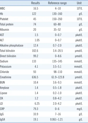
A 63-year-old man with renal failure was diagnosed as having IgG4-RD a year ago, confirmed by a kidney biopsy (tubulointer- stitial nephritis with increased interstitial lymphoplasmacytic in- filtration and >30 IgG4-positive plasma cells/HPF). He had vis- ited the outpatient clinic complaining of abdominal pain and a skin rash that had developed one month earlier. The computed tomography scan showed multiple lymphadenopathies, and the torso positron emission tomography images revealed hyperac- tive BM. Blood tests revealed leukocytosis, thrombocytopenia, marked eosinophilia (51% on white blood cell [WBC] differential count), and rare nucleated red blood cells (1/100 WBCs). Total serum IgG and IgG4 concentrations were increased (Table 1).
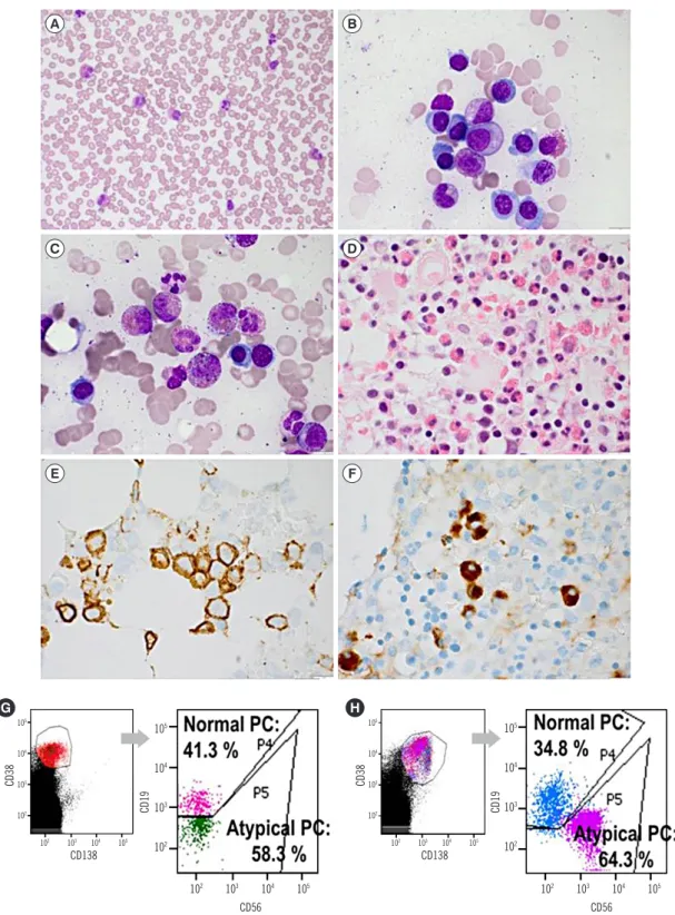
PB smear showed poikilocytosis and rouleaux formation (Fig.
1A). Serum protein electrophoresis indicated polyclonal gam-
Received: March 31, 2020 Revision received: May 19, 2020 Accepted: September 24, 2020
Corresponding author: Chan-Jeoung Park, M.D.
Department of Laboratory Medicine, University of Ulsan, College of Medicine and Asan Medical Center, 88 Olympic-ro 43-gil, Songpa-gu, Seoul 05505, Korea
Tel: +82-2-3010-4508, Fax: +82-2-478-0884 E-mail: [email protected]
© Korean Society for Laboratory Medicine
This is an Open Access article distributed under the terms of the Creative Commons Attribution Non-Commercial License (https://creativecommons.org/licenses/by-nc/4.0) which permits unrestricted non-commercial use, distribution, and reproduction in any medium, provided the original work is properly cited.
1 / 1 CROSSMARK_logo_3_Test
2017-03-16 https://crossmark-cdn.crossref.org/widget/v2.0/logos/CROSSMARK_Color_square.svg
Kim HJ, et al.
BM and flow cytometric findings in IgG4-RD
244 www.annlabmed.org https://doi.org/10.3343/alm.2021.41.2.243 mopathy.
The patient underwent a BM study because of unexplained thrombocytopenia with reversed albumin to globulin ratio. The BM aspirates showed increased number of plasma cells, which included immature forms (16.8% of plasma cells among the BM nucleated cells; Fig. 1B), as well as moderate eosinophilic hyperplasia (20.4% of eosinophils among the BM nucleated cells; Fig. 1C and D). The cellularity varied with an average of 50%. IHC staining of the BM biopsy showed increased num- bers of CD138-positive plasma cells with interstitial and clus- tered infiltration; however, clonal restriction for kappa or lambda light chains was not observed. IgG and IgG4 stains showed in- creased numbers of IgG- and IgG4-positive plasma cells; 31 IgG4-positive plasma cells/HPF and 35 IgG-positive plasma cells/HPF were observed (IgG4/IgG-positive plasma cell ra- tio=0.88; Fig. 1E and F). IgH gene rearrangement with BM as- pirate was not found.
Flow cytometric immunophenotyping was performed using a FACSCanto II flow cytometer (BD Biosciences, San Jose, CA, USA) and a CD38/CD138/CD19/CD45/CD56 panel. Plasma cells in the PB comprised 0.376% of the WBCs (62,040 cells/
mL); 58.3% of plasma cells were atypical plasma cells (low CD19 expression with CD38+/CD138dim/CD56−/CD45+). The im- munophenotype of the atypical plasma cells in the BM (64.3%
of the total BM plasma cells) was CD38+/CD138+/CD56+/CD- 19low/CD45low, slightly different from that of the PB cells (Fig. 1G and H). The difference in CD56 expression between PB and BM plasma cells is likely due to the nature of plasma cells in IgG4-RD. Among the heterogeneous plasma cells, only a few CD56-negative cells might migrate to the circulating system, as CD56 acts as an adhesion molecule. The patient was treated with high-dose steroids and discharged as his symptoms im- proved.
As IgG4-RD tends to form tumefactive lesions, patients are easily suspected of having malignancies. Additionally, BM find- ings can mimic other hematologic diseases such as lympho- mas, plasma cell neoplasms, and histiocytic disorders. However, IgG4-RD diagnosis is difficult based on BM studies because the core pathological features are rarely present in the BM. There- fore, if a patient suspected of having IgG4-RD has an abnormal complete blood count, a BM study with IHC staining for CD138, kappa and lambda light chains, IgG, and IgG4 and flow cyto- metric immunophenotyping of plasma cells in the PB and BM aspirates should be considered. Furthermore, when a patient undergoes a BM study for lymphoma workup and diagnosis of plasma cell neoplasms or histiocytic disorders, hematopatholo- gists should consider the possibility of IgG4-RD with BM involve- ment, if elevated serum IgG and IgG4 concentrations or other laboratory abnormalities are present.
ACKNOWLEDGEMENTS
None.
AUTHOR CONTRIBUTIONS
All authors have accepted their responsibility for the entire con- tent of this manuscript and approved submission.
CONFLICTS OF INTEREST
No potential conflicts of interest relevant to this paper were re- ported.
Table 1. Laboratory findings
Results Reference range Unit
WBC 16.5 4–10 109/L
Hb 122 130–160 g/L
Platelet 45 150–350 109/L
Total protein 74 60–80 g/L
Albumin 20 35–52 g/L
AST 1.5 0–0.7 μkat/L
ALT 1.05 0–0.7 μkat/L
Alkaline phosphatase 12.4 0.7–2.0 μkat/L
Total bilirubin 102.6 3.4–20.5 µmol/L
Direct bilirubin 99.2 0–8.6 µmol/L
Sodium 133 135–145 mmol/L
Potassium 4.1 3.5–5.1 mmol/L
Chloride 93 98–110 mmol/L
Creatinine 636.5 61.9–123.8 µmol/L
BUN 10.4 3.6–9.3 mmol/L
Amylase 1.4 0.5–1.8 μkat/L
Lipase 1.4 0.2–1.0 μkat/L
CK 1.2 0.8–4.2 μkat/L
LD 6.25 2.0–4.2 μkat/L
CRP 79.3 0–6 mg/L
IgG 33.9 7–16 g/L
IgG4 19.1 0.061–1.21 g/L
Abbreviations: WBC, white blood cell; Hb, hemoblobin; AST, aspartate ami- notransferase; ALT, alanine aminotransferase; BUN, blood urea nitrogen;
CK, creatine kinase; LD, lactate dehydrogenase; CPR, C-reactive protein;
IgG, Immunoglobulin G; IgG4, Immunoglobulin G4.
Kim HJ, et al.
BM and flow cytometric findings in IgG4-RD
https://doi.org/10.3343/alm.2021.41.2.243 www.annlabmed.org 245
Fig. 1. Morphologic and IHC findings of PB and BM samples, and flow cytometric immunophenotyping of PC. (A) Eosinophilia, poikilocytosis (schistocytes and spherocytes), and rouleaux formation (PB, Wright stain, ×400). (B) Increased numbers of PC including immature forms with slightly high NC ratio, nucleolus, and perinuclear halo (BM aspirate, Wright stain, ×1,000). (C) Increased number of eosinophils (BM aspirate, Wright stain, ×1,000). (D) Increased number of eosinophils and PC (BM biopsy, H & E stain, ×400). (E) Increased number of PC (BM clot section, IHC stain for CD138, ×1,000). (F) Increased numbers of IgG4+ PC (BM biopsy, IHC stain for IgG4, ×1,000). (G, H) Pres- ence of atypical PC with the immunophenotypes CD38+/CD138dim/CD56−/CD19low/CD45+ in PB and CD38+/CD138+/CD56+/CD19low/CD45low in BM aspirates.
Abbreviations: IHC, immunohistochemistry; PB, peripheral blood; BM, bone marrow; PC, plasma cells.
A
C
E
B
D
F
CD56 CD56
H G
CD138 CD138
105 104 103 102
105 104 103 102
105 104 103 102
105 104 103 102
CD38 CD38
CD19 CD19
102 103 104 105 102 103 104 105
102 103 104 105 102 103 104 105
Kim HJ, et al.
BM and flow cytometric findings in IgG4-RD
246 www.annlabmed.org https://doi.org/10.3343/alm.2021.41.2.243
RESEARCH FUNDING
None declared.
ORCID
Han Joo Kim https://orcid.org/0000-0002-0088-0488 Eunkyoung You https://orcid.org/0000-0002-6836-499X Seokchan Hong https://orcid.org/0000-0001-8722-3124 Chan-Jeoung Park https://orcid.org/0000-0003-4396-8348
REFERENCES
1. Kamisawa T, Zen Y, Pillai S, Stone JH. IgG4-related disease. Lancet 2015;385:1460-71.
2. Moutsopoulos HM, Fragoulis GE, Stone JH. Diagnosis and differential diagnosis of IgG4-related disease https://www.uptodate.com/contents/
diagnosis-and-differential-diagnosis-of-igg4-related-disease (Updated
on 20th May, 2019).
3. Stone JH, Zen Y, Deshpande V. IgG4-related disease. N Engl J Med 2012;366:539-51.
4. Umehara H, Okazaki K, Masaki Y, Kawano M, Yamamoto M, Saeki T, et al. Comprehensive diagnostic criteria for IgG4-related disease (IgG4- RD), 2011. Mod Rheumatol 2012;22:21-30.
5. Wallace ZS, Mattoo H, Carruthers M, Mahajan VS, Della Torre E, Lee H, et al. Plasmablasts as a biomarker for IgG4-related disease, indepen- dent of serum IgG4 concentrations. Ann Rheum Dis 2015;74:190-5.
6. Mattoo H, Mahajan VS, Della-Torre E, Sekigami Y, Carruthers M, Wal- lace ZS, et al. De novo oligoclonal expansions of circulating plasma- blasts in active and relapsing IgG4-related disease. J Allergy Clin Immu- nol 2014;134:679-87.
7. Ichiki A, Hashimoto N, Ueda T, Hiraiwa S, Tajiri T, Nakamura N, et al.
IgG4-related disease with bone marrow involvement. Intern Med 2016;
55:2295-9.
8. van den Elshout-den Uyl D, Spoto CPE, de Boer M, Leiner T, Leavis HL, Leguit RJ. First report of IgG4 related disease primary presenting as vertebral bone marrow lesions. Front Immunol 2019;10:1910.
9. Hwang SM, Paik JH, Lee JY. ImmunoglobulinG4-related disease mim- icking lymphoma. Ann Hematol 2019;98:2239-41.