355 http://dx.doi.org/10.4196/kjpp.2012.16.5.355
ABBREVIATIONS: IEC, ion exchange chromatography; RP-HPLC, reversed-phase high-performance liquid chromatography; PITC, phenylisothiocyanateor; OPA, o-phthalaldehyde; AQC, 6-aminoqui- nolyl-N-hydroxysuccinimidyl carbamate; AMQ, 6-aminoquinoline;
UV, ultraviolet; Glu, glutamate; Gly, glycine; Ala, alanine; BWI, body weight index; QC, quality control; RE, relative error; RSD, relative standard deviations.
Received July 20, 2012, Revised August 9, 2012, Accepted August 13, 2012
Corresponding to: Qing Zhong Li, Institute of Pharmacy and Department of Pharmacology, School of Pharmacy, Binzhou Medical University, Yantai 264003, China. (Tel) 86-535-6913216, (Fax) 86- 535-6913718, (E-mail) [email protected]
This is an Open Access article distributed under the terms of the Creative Commons Attribution Non-Commercial License (http://
creativecommons.org/licenses/by-nc/3.0) which permits unrestricted non-commercial use, distribution, and reproduction in any medium, provided the original work is properly cited.
Simultaneous Determination of Glutamate, Glycine, and Alanine in Human Plasma Using Precolumn Derivatization with 6-Aminoquinolyl- N-hydroxysuccinimidyl Carbamate and High-Performance Liquid Chromatography
Qing Zhong Li1, Qing Xian Huang2, Shu Cui Li1, Mei Zi Yang1, and Bin Rao3
1Institute of Pharmacy and Department of Pharmacology, School of Pharmacy, Binzhou Medical University, 2Department of General Surgery, Yantai Yuhuangding Hospital, Yantai 264003, 3Department of Quality Inspection, Hubei Provincial Bafeng Pharmaceuticals & Chemicals Share Co., Ltd, Hefeng 445800, China
A simple, sensitive and reproducible high-performance liquid chromatography (HPLC) method has been validated for determining concentrations of glutamate, glycine, and alanine in human plasma.
Proteins in plasma were precipitated with perchloric acid, followed by derivatization with 6-amino- quinolyl-N-hydroxysuccinimidyl carbamate (AQC). Simultaneous analysis of glutamate, glycine, and alanine is achieved using reversed-phase HPLC conditions and ultraviolet detection. Excellent linearity was observed for these three amino acids over their concentration ranges with correlation coefficients (r)> 0.999. The intra- and inter-day precision were below 10%. This method utilizes quality control samples and demonstrates excellent plasma recovery and accuracy. The developed method has been successfully applied to measure plasma glutamate, glycine, and alanine in twenty volunteers.
Key Words: Amino acids, AQC, HPLC, Plasma, Ultraviolet detection
INTRODUCTION
Several excitatory amino acids, such as endogenous glu- tamate, glycine, and alanine, which are involved in gluta- mate neurotransmission via NMDA receptors, are being in- vestigated as potential indicators of health status and dis- ease risk. Increased glutamate is closely related with neu- ronal damage by cerebral ischemia and plasma glutamate concentration is the most powerful predictor biomarker of lesion enlargement in the acute phase of ischemic stroke [1,2]. In some cases, the peripheral blood levels of gluta- mate, glycine, and alanine could be diagnostic, therapeutic or symptomatic biological markers [3-6]. Thus determi- nation of these amino acids plays an important role in diag- nosis and therapy of some psychiatric and neurological disorders. Moreover, the preparations of amino acids are increasingly used to treat a wide array of diseases in recent years. In china, glutamate, glycine, and alanine tablet or capsule is wildly used in the treatment of chronic prostati-
tis [7]. Simultaneous analysis of glutamate, glycine, and alanine may provide help for the study of the pharmacoki- netics of these preparations.
The method commonly employed for the analysis of free plasma amino acids is automated ion exchange chromatog- raphy (IEC) in combination with postcolumn ninhydrin detection. This method provides simplicity in sample prepa- ration and high reproducibility [8,9]. However, the long an- alytical run time is the main drawback [8,10]. Reversed- phase high-performance liquid chromatography (RP-HPLC) is used as an alternative due to its higher sensitivity and faster analysis. The most common RP-HPLC methods in- volve precolumn derivatization with phenylisothiocyanate (PITC) or o-phthalaldehyde (OPA) [11-14]. However, each reagent has its disadvantages or drawbacks: PITC needs a long derivatization time (20 min) and it is necessary to remove any excess reagent by drying, and OPA does not react with secondary amino acids, and glycine derivatives, for instance, are unable [15,16]. Precolumn derivatization utilizing 6-aminoquinolyl-N-hydroxysuccinimidyl carbamate (AQC) might constitute a feasible alternative, as to method characteristics that overcome these major drawbacks. In this procedure, primary and secondary amines react with the AQC reagent, yielding stable compounds. Excess re-
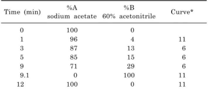
Table 1. Optimized gradient for the separation of amino acids at 37oC
Time (min) %A
sodium acetate
%B
60% acetonitrile Curve*
0 100 0
1 96 4 11
3 87 13 6
5 85 15 6
9 71 29 6
9.1 0 100 11
12 100 0 11
*Curve 6 is a linear segment; curve 11 is a step function.
agent is hydrolyzed to yield 6-aminoquinoline (AMQ), car- bon dioxide and N-hydroxysuccinimide [17]. The advantages of this method include a very simple derivatization proce- dure, stable derivatives, excellent separation, detection by either absorbance or fluorescence, and commercial avail- ability of reagents. Disadvantages include relatively long chromatography, high solvent consumption and no possi- bility of on-line derivatization, which seems limited com- pared to the defects of other methods.
In the present study, we describe a sensitive and rapid RP-HPLC method, using AQC derivatization and ultra- violet (UV) detection. The objective is to obtain a simple and rapid simultaneous measurement of glutamate, gly- cine, and alanine in human plasma.
METHODS Chemicals
AccQ.Fluor reagent kit (consisting of AQC reagent, aceto- nitrile and 0.2 mM sodium borate buffer, pH 8.8) as well as glass reaction tubes (6×50 mm) were provided by Waters (Milford, MA, USA). Amino acid standards (glutamate: Glu, glycine: Gly, alanine: Ala) were obtained from Sigma (St Louis, MO, USA). HPLC grade acetonitrile and triethyl- amine were from Merck (Darmstadt, Germany). Perchloric acid and sodium acetate were of analytical reagent grade and purchased from J.T. Baker (Phillipsburg, NJ, USA).
Ultrapure water was generated using a Milli-Q system pur- chased from Millipore (Bedford, MA, USA).
Chromatographic instruments and conditions
The HPLC system used was a Waters Alliance consisting of a 2,690 separation model, a thermostat-controlled col- umn oven, a system interface module, and a 2,487 Dual Wavelength UV Detector (all Waters components, Millipore, Milford, MA, USA). A Millennium Chromatography Mana- ger workstation running Millennium software controls sys- tem operation and results management. Separation was carried out using a 20×3.9 mm Sentry guard column (Nova- Pak C18 bonded silica) connected to a 4 μm AccQ-Tag C18
column (3.9×150 mm I.D.; both from Waters).
Working eluents
Eluent A was 0.14 M sodium acetate with 0.017 M trie- thanolamine (pH 4.95); eluent B was 60% (v/v) acetonitrile
aqueous solution. The gradient program is shown in Table 1.
The AccQ-Tag column was thermostated at 37oC and op- erated at a flow-rate of 1 ml/min. Detection was accom- plished by UV detector and the wavelength was set at 248 nm. For column regeneration, 3 min with 60% acetonitrile and 40% water was sufficient to wash out the column and before the next injection, 100% sodium acetate for 6 min was needed to equilibrate the system. Injections were made every 18 min (injection-to-injection), using an injection vol- ume of 10 μl.
Standard solutions
Stock solutions of Glu, Gly, and Ala needed in the method were prepared by dissolving appropriate amount of the compounds in 0.1 M hydrochloric acid and diluting to the appropriate volumes. These solutions were kept at 4oC for several days without noticeable change. The working sol- utions were prepared by appropriate dilutions with water as needed, and processed without delay.
Plasma preparation
Twenty healthy male volunteers were recruited for this study. They are 19∼25 years old with BWI (body weight index) between 19∼24 kg/m2. The study was performed in accordance with the Helsinki Declaration and the design and execution of the experiment were explained thoroughly to the participants, and informed consent was obtained.
Blood was collected, following an overnight fast, into evacuated lithium heparin gel tubes, immediately placed on ice, and centrifuged at 3,000 rpm for 15 min at 4oC.
Plasma was then used for the determination of Glu, Gly, and Ala without delay or stored at -70oC. Prior to analysis, a 100 μl portion of the plasma sample was added to 100 μl of 5% perchloric acid, and the solution was mixed with a vortex-mixer. After vortex-mixing, the precipitate was re- moved by centrifugation at 15,000 rpm for 10 min at 4oC and the supernatant was used for the analysis.
Quality control (QC) samples preparation
For validation of the method, QC samples were made by adding the appropriate amount of working standard sol- ution of Glu, Gly, and Ala to the plasma samples from an apparently healthy donor. The nominal concentrations of low, medium, and high QC samples were 40, 80, 160 μM for Glu; 160, 400, 640 μM for Gly and 200, 500, 800 μM for Ala, respectively.
Derivatization
In a typical analysis, 10 μl of standards or samples were mixed with 70 μl buffer solution (0.2 M borate buffer) and afterwards 20 μl derivatization reagent (2 mg/ml AQC) were added. After ten minutes response at 55oC, the deriv- atized solution was directly injected to separate the amino acids by HPLC.
Calibration and linearity
The retention time of each amino acid was studied using dilutions from stock solutions. Linearity was assured by calculating the regression coefficients for the calibration
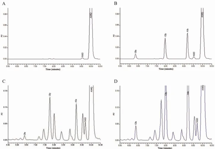
Fig. 1. Typical chromatograms of Glu, Gly, and Ala in plasma. (A) Chromatogram for derivatized blank; (B) chromatography for derivatized amino acid standard mixture; (C) representative chromatography of blank (unspiked) pooled human plasma sample; (D) chromatography of the high QC sample (spiked concentrations of Glu, Gly, and Ala were 160, 640, and 800 μM, respectively).
curves. Range of the analysis was determined by assuring that all the points on the calibration curve were charac- terised by acceptable precision and accuracy. For each curve, the peak area were calculated and plotted against concentration of the corresponding amino acid.
Precision and accuracy
Precision and accuracy were determined by the analysis of QC samples spiked at three concentrations listed above.
The intra-assay was determined by replicate analysis of a blank (unspiked) plasma sample (n=6) and a QC sample at each concentration (n=6) in a single run. The inter-assay was determined by replicate analysis of the same QC sam- ple stored at -70oC and analyzed 6 separate times over a period of 2 months. The calculated mean concentration relative to the nominal concentration (relative error: RE%) was used to express accuracy and relative standard devia- tions (RSD%) calculated from the blank sample and QC val- ues was used to estimate the inter- and intra-day pre- cision.
Recovery and stability
Recovery of Glu, Gly, and Ala from plasma was assessed
by analyzing pooled human plasma obtained from a healthy volunteer spiked at the three QC concentration levels listed above. Six replicate ‘control’ (unspiked) plasma samples and six spiked samples of each QC level were analyzed and recovery was determined by comparing the nominal amino acid concentration. The recovery of each amino acid was defined as 100%. Means, standard deviations (SD) and RSD% were calculated.
The stability of plasma samples stored at -70oC within 2 months was assessed. Also, QC samples were subjected to three freeze-thaw cycles (-70oC to room temperature) to evaluate sample stability. In addition, stability of proc- essed samples was assessed. Triplicate low and high QC samples were processed and analyzed, then re-injected and analyzed up to 24 h post-processing.
Application to determination of Glu, Gly, and Ala in human plasma
After the full validation, this method was successfully ap- plied to the determination of Glu, Gly, and Ala in plasma from twenty male volunteers.
Table 4. Plasma concentrations of Glu, Gly, and Ala from 20 healthy volunteers Amino
acids
Plasma concentration (μM) in 20 volunteers (No.1∼20) Mean±SD
1 2 3 4 5 6 7 8 9 10 11 12 13 14 15 16 17 18 19 20 (μM)
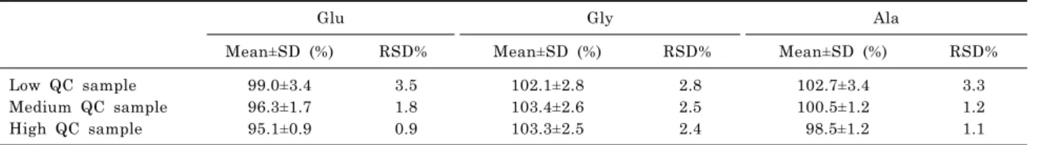
Glu 62.7 91.0 26.7 82.1 56.1 56.7 35.4 86.4 36.2 62.4 72.5 50.3 35.3 35.1 46.2 41.2 32.0 30.3 34.5 60.9 51.7±16.5 Gly 282.3 286.1 188.4 262.7 302.5 297.6 271.9 295.8 297.8 318.6 428.6 236.4 344.8 353.8 291.0 328.0 186.3 248.8 179.4 393.5 289.7±46.5 Ala 395.7 445.5 202.6 334.4 417.9 322.4 289.5 462.8 264.7 384.8 356.1 418.1 220.4 431.9 349.8 354.5 314.2 322.3 282.2 432.5 350.1±59.9 Table 3. Recovery rate (%) of Glu, Gly, and Ala from human plasma (n=6)
Glu Gly Ala
Mean±SD (%) RSD% Mean±SD (%) RSD% Mean±SD (%) RSD%
Low QC sample 99.0±3.4 3.5 102.1±2.8 2.8 102.7±3.4 3.3
Medium QC sample 96.3±1.7 1.8 103.4±2.6 2.5 100.5±1.2 1.2
High QC sample 95.1±0.9 0.9 103.3±2.5 2.4 98.5±1.2 1.1
Table 2. Intra- and inter-day precision and accuracy for Glu, Gly, and Ala (n=6)
Glu Gly Ala
Intra-day RSD%
Inter-day
RSD% RE% Intra-day
RSD%
Inter-day
RSD% RE% Intra-day
RSD%
Inter-day
RSD% RE%
Blank sample 6.2 9.5 2.7 4.0 5.7 4.7
Low QC sample 6.5 7.4 -3.3 3.3 8.0 -1.6 4.4 5.7 -5.0
Medium QC sample 6.9 6.7 -4.6 6.8 5.3 1.5 5.3 4.4 -4.3
High QC sample 4.9 4.1 -8.3 5.2 4.5 7.6 2.0 1.8 -7.1
RESULTS Chromatographic separation
Representative chromatograms of the derivatized blank, standard solution, blank (unspiked) plasma sample and QC plasma sample are depicted in Fig. 1. The method demon- strated good chromatographic separation of all the amino acids studied, and showed an excellent signal-to-noise ratio, allowing easy quantification. Retention times for Glu, Gly, and Ala were approximately 6.3, 7.9, and 9.3 min, respec- tively. The peaks of interest were well separated and there was no interference from other amino acids and endogenous compounds in plasma.
Linearity, precision and accuracy
Linear calibration curves were obtained for Glu, Gly, and Ala over the concentration ranges of 5∼320 μM, 18.75∼
1,200 μM, and 21.9∼1400 μM, respectively. The mean re- gression equations were y=1,414.2x+1791.3 for Glu, y=
1,590.7x+6,982.6 for Gly, and y=1,626.6x+7289.2 for Ala, with correlation coefficients (r)>0.999 for all three amino acids. The lower limit of quantitation (LOQ) for each amino acid demonstrated acceptable precision.
The intra- and inter-day RSD% were between 1.8% and 9.5% and most RSD% values calculated from the high QC samples of each amino acid were smaller than those from low and medium QC samples especially pronounced in Ala group. The accuracy, expressed as RE%, was within ±10%
(Table 2).
Stability and recovery
The plasma samples were stable for up to 2 months at
-70oC. Three freeze-thaw cycles had no effect on the stabil- ity of Glu, Gly, and Ala. Additionally, amino acid derivat- ives are stable at room temperature for 24 hours. The mean recoveries of all analytes from plasma at all concentrations ranged from 95% to 103%. At the same time, the RSD%
values were less than 5% which showed significant inverse relationship with the concentration of the analyte (Table 3).
Analysis of human plasma samples
The method described in this paper was applied for ami- no acid analysis in plasma samples from twenty male vol- unteers (Table 4). Free plasma amino acids exhibit wide variability between subjects. This probably reflects differ- ences in metabolic control since the amino acids belong to endogenous substances which can be influenced by physio- logic conditions of different volunteers as well as diet.
Plasma concentrations of Glu, Gly, and Ala were 51.7±16.5, 289.7±46.5, and 350.1±59.9 μM, respectively.
DISCUSSION
The increasing significance of plasma concentrations of Glu, Gly, and Ala in clinical fields, such as being used as diagnostic or therapeutic biomarkers, has prompted the
study of these amino acids analyses. In this study, we in- tended to develop a method of simultaneous determination of Glu, Gly, and Ala in plasma which would be highly accu- rate, but at the same time not so time-consuming. This method is based on the use of precolumn AQC derivatiza- tion as well as the optimized mobile phase and gradient for the separation. In addition, the sensitivity, linearity and detection limits of these amino acids by using UV detector were assessed strictly. The second step is method vali- dation, which verifies the performance of the entire ana- lytical process. Finally, plasma Glu, Gly, and Ala were de- termined using this method, and the concentrations of these amino acids were generally consistent with those re- ported in other studies [18,19].
As developed by the Waters Co., Ltd, the mobile phase used in the traditional AQC method [17] has been modified by increasing the proportion of eluent B (60% acetonitrile) while decrease the gradient time. The optimized gradient (Table 1) accelerated the separation of Glu, Gly, and Ala, which therefore shortened the time required for assay and the total run time is only about 18 min.
The other critical step in the whole procedure is sample preparation including the sample collection, storage and deproteinization. When deprotenization of the plasma sam- ples cannot be done immediately after centrifugation, it is recommended to store the samples at temperatures lower than -18oC to prevent further hydrolysis of proteins.
Previous studies demonstrated that the Glu increased mar- kedly when the storage temperature higher than -68oC, so the plasma samples were frozen immediately and stored at -70oC until analyzed in this study, and all the studied amino acids remained stable within 8 weeks. Another point regarding deproteinization should be emphasized as well.
The most wildly used methods for deproteinization of plas- ma samples include precipitation with acids or organic solvents. In our experiment, we tried acetonitrile, 5-sulfosa- licylic acid, and perchloric acid for the deproteinization of plasma samples and compared the efficacy of them. We found that the concentrations of Glu and Gly using acetoni- tril, were lower than those measured with 5-sulfosalicylic acid and perchloric acid. Also, we observed that perchloric acid protein precipitation was superior to 5-sulfosalicylic acid because 5-sulfosalicylic acid interfered with the chro- matography of Glu peaks, which was consistent with a pre- vious report [20]. For this reason, perchloric acid was used as the deproteinizing reagent in this analysis.
Our modified method has been successfully validated.
The mobile phase conditions produce excellent separation of Glu, Gly, and Ala in plasma (Fig. 1) with good sensitivity, i.e. lower LOQ for all analytes. After deproteinization with 5% perchloric acid, perfect recovery (95% to 103%; Table 3) of all analytes from plasma was achieved. Another im- portant characteristic of the method is high precision. The experimental results abstained from the intra- and in- ter-day precision and accuracy (Table 2) showed that RSD%
values of these three amino acids were below 10%.
Meanwhile, most RSD% values of high QC samples were smaller than those of low and medium QC samples, and similar results are also shown in Table 3. Precision relates to the random error of a measurement system and the ob- vious relationship between the precision of an analytical method and the concentration of the analyte was usually observed in many studies. The results of our study also demonstrated that smaller RSD%, which indicates higher precision, was achieved when the high QC samples were
analyzed.
In summary, the method described here, using a pre- column derivatization with AQC, is a rapid, sensitive, and selective technique for simultaneously analyzing Glu, Gly, and Ala in human plasma. The chromatographic conditions allow for the separation and quantification of amino acids using a simplified gradient. This assay shows good pre- cision and reproducibility, and it is a relatively simple and fast method. Finally, this method was used to determine Glu, Gly, and Ala in the plasma of twenty healthy human volunteers.
ACKNOWLEDGEMENTS
We would like to thank Dr. Ki-Wug Sung from the Catholic University of Korea for his kind and valuable suggestions.
REFERENCES
1. Lee GJ, Choi SK, Eo YH, Kang SW, Choi S, Park JH, Lim JE, Hong KW, Jin HS, Oh BS, Park HK. The effect of extracellular glutamate release on repetitive transient ischemic injury in global ischemia model. Korean J Physiol Pharmacol. 2009;13:23-26.
2. Castellanos M, Sobrino T, Pedraza S, Moldes O, Pumar JM, Silva Y, Serena J, García-Gil M, Castillo J, Dávalos A. High plasma glutamate concentrations are associated with infarct growth in acute ischemic stroke. Neurology. 2008;71:1862-1868.
3. Mitani H, Shirayama Y, Yamada T, Maeda K, Ashby CR Jr, Kawahara R. Correlation between plasma levels of glutamate, alanine and serine with severity of depression. Prog Neuro- psychopharmacol Biol Psychiatry. 2006;30:1155-1158.
4. Andreadou E, Kapaki E, Kokotis P, Paraskevas GP, Katsaros N, Libitaki G, Petropoulou O, Zis V, Sfagos C, Vassilopoulos D. Plasma glutamate and glycine levels in patients with amyotrophic lateral sclerosis. In Vivo. 2008;22:137-141.
5. Wesseldijk F, Fekkes D, Huygen FJ, van de Heide-Mulder M, Zijlstra FJ. Increased plasma glutamate, glycine, and arginine levels in complex regional pain syndrome type 1. Acta Anaesthesiol Scand. 2008;52:688-694.
6. Ohnuma T, Arai H. Significance of NMDA receptor-related glutamatergic amino acid levels in peripheral blood of patients with schizophrenia. Prog Neuropsychopharmacol Biol Psy- chiatry. 2011;35:29-39.
7. Zhao JL. Clinical therapeutic effects of sparfloxacin and glutamic acid, alanine acid and glycine acid capsules in the treatment of chronic prostatis. Chin Clin Prac Med. 2007;1:33- 8. Fekkes D. State-of-the-art of high-performance liquid chroma-35.
tographic analysis of amino acids in physiological samples. J Chromatogr B Biomed Appl. 1996;682:3-22.
9. Le Boucher J, Charret C, Coudray-Lucas C, Giboudeau J, Cynober L. Amino acid determination in biological fluids by automated ion-exchange chromatography: performance of Hitachi L-8500A. Clin Chem. 1997;43:1421-1428.
10. Fekkes D, Voskuilen-Kooyman A, Jankie R, Huijmans J.
Precise analysis of primary amino acids in urine by an automated high-performance liquid chromatography method:
comparison with ion-exchange chromatography. J Chromatogr B Biomed Sci Appl. 2000;744:183-188.
11. Turnell DC, Cooper JD. Rapid assay for amino acids in serum or urine by pre-column derivatization and reversed-phase liquid chromatography. Clin Chem. 1982;28:527-531.
12. Heinrikson RL, Meredith SC. Amino acid analysis by reverse- phase high-performance liquid chromatography: precolumn de- rivatization with phenylisothiocyanate. Anal Biochem. 1984;
136:65-74.
13. Anumula KR, Taylor PB. Quantitative determination of phenyl isothiocyanate-derivatized amino sugars and amino sugar alcohols by high-performance liquid chromatography. Anal Biochem. 1991;197:113-120.
14. Kutlán D, Presits P, Molnár-Perl I. Behavior and charac- teristics of amine derivatives obtained with o-phthaldialde- hyde/3-mercaptopropionic acid and with o-phthaldialdehyde/
N-acetyl-L-cysteine reagents. J Chromatogr A. 2002;949:235- 248.
15. Bosch L, Alegría A, Farré R. Application of the 6-amino- quinolyl-N-hydroxysccinimidyl carbamate (AQC) reagent to the RP-HPLC determination of amino acids in infant foods. J Chromatogr B Analyt Technol Biomed Life Sci. 2006;831:
176-183.
16. Cohen SA. Amino acid analysis using precolumn derivatization with 6-aminoquinolyl-N-hydroxysuccinimidyl carbamate. Methods
Mol Biol. 2000;159:39-47.
17. Cohen SA, Michaud DP. Synthesis of a fluorescent derivatizing reagent, 6-aminoquinolyl-N-hydroxysuccinimidyl carbamate, and its application for the analysis of hydrolysate amino acids via high-performance liquid chromatography. Anal Biochem.
1993;211:279-287.
18. Terrlink T, van Leeuwen PA, Houdijk A. Plasma amino acids determined by liquid chromatography within 17 minutes. Clin Chem. 1994;40:245-249.
19. Chih-Kuang C, Shuan-Pei L, Shyue-Jye L, Tuan-Jen W. Plasma free amino acids in Taiwan Chinese: the effect of age. Clin Chem Lab Med. 2002;40:378-382.
20. Frank MP, Powers RW. Simple and rapid quantitative high-performance liquid chromatographic analysis of plasma amino acids. J Chromatogr B Analyt Technol Biomed Life Sci.
2007;852:646-649.