약학회지 제 42권 제 5 호 473~479(1998i
Yakhak Hoeji
Vol. 42. No. 5
역상 고속액체크로마토그라프법을 이용한 혈장 및 뇨 중 로바스타틴의 정량
최 혜 진 * • 김 명 민 ** • 최 경 업 ****
삼성서울병원 약제부
*.
삼성생명과학연구소 임상약리학연구실**
{Received April 8. 1998)
Quantitative Analysis of Lovastatin in Hum an Plasma and Urine by Reversed-Phase High-Performance
Liquid Chromatography
Hye Jin Choi*, Myoung Min Kim** and Kyung Eob Choi* ***
*Diuision o f Pharm aceutical Services, S a m su n g M edical Center,
**Clinical Pharmacology Laboratory, S a m su n g B iom edical Research Institute, Seoul 135-230, Korea
Abstract— Lovastatin (LOVA). a fungal metabolite isolated from cultures of Aspergillus terreus . is a com
petitive HMG-CoA reductase inhibitor used for the treatment of primary hypercholesterolemia, and has also been shown to suppress growth in a variety of non-glioma tumor cell lines. A sensitive reversed- phase high-performance liquid chromatographic method with ultraviolet (UV) absorbance detection has been developed to quantitate LOVA in human plasma and urine samples using liquid-liquid ex
traction procedure. Baseline separation of LOVA and internal standard, simvastatin was achieved on a Novapak Ci« analytical coiumn with a mobile phase containing 0.025M NaH
2P
0 4 :ACN (35
:65. v/v%), ad
justed pH to 4.5. The flow rate was set at 1.5 m//min. and the column effluent was monitored by a UV detection at 238 nm. The limit of quantification was determined to be 0.5 [i-g/ml while extraction ef
ficiency of LOVA ranged from 73.4'
신2.9% at LOVA concentrations of 0.5 to 10 Good linearity with correlation coefficients greater than 0.999 was obtained in the range of LOVA concentrations from 0.5 to 10 M-g/m/. The accuracy and the precision were proven excellent with relative standard deviation (RSD, %) and relative error (RE. %) of less than 4.2 and 4.0. respectively. Intraday precision, evaluated at five LOVA concentrations (0.5, 1. 2. 5. 10 |ig/m/) and expressed as RSD ranged from Q-l.S2% while the interday precision at the same concentrations ranged from 0.7~ 10.5%. The analytical method des
cribed was then successfully employed for the determination of LOVA concentrations in plasma sam
ples obtained during a phase II clinical trial using high doses of LOVA (30—40 mg/kg/day). This method could be further utilized for the ongoing pharmacokinetic studies and therapeutic drug mon
itoring of the high-dose LOVA therapy in adenocarcinoma patients.
Keywords O Lovastatin. high-dose therapy. HPLC, human plasma, urine samples.
로바스타틴은
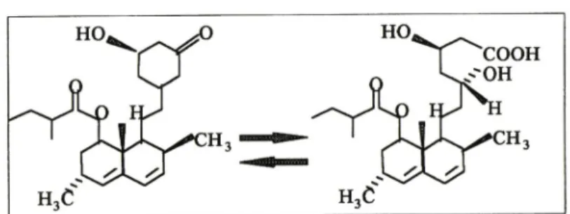
Aspergillus terreus균주의 배양액으로 부터 분리된 비활성형 탁론(Fig. 1)으로 화학식은[1S- [la(R*). 3a, ?P, 8p(2S=\ 4S*)-8ap]]-2 Methyl- butanoic acid 1.2,3.7.8,8a-hexahydro-3,7-dime-
" 본 논문에 관한 문의는 이 저자에게로 ( 전화) 02-3410-3360 ( 팩스) 02-3410-3399
thyl-8-(2-(tetrahydro~4-hydroxy-6-oxo-2H-py- ran-2-yl) ethyl] - 1-naphthalenyl ester이 다 .
로바스타틴은 prodmg으로서 경구투여 후 즉시 기■수
분해 되어 로바스타틴의 주 대사산물인 p-hydroxy
acid 형태의 로바스타틴 산 (Fig. 1)으로 번환된다. 로
바스타틴 산은 로바스타틴의 활성화된 물질로서 콜레
스테롤 생합성의 초기 율속단계 과정을 촉매하는 효소,
474 최 혜 진 • 김명민■최경업
Fig. 1 — Structural formula of lovastatin and its active metabolite, lovastatin acid, showing the inactive prodrug in the lactone form (left) and the active inhibitor in the hydroxyacid form (right).
HMG-CoA reductase를 저해하여 혈청 콜레스테롤치 를 저하시키는 작용을 한다.
로바스타틴의 약동학적 성상을 살펴보면. 경구투여 시 30% 정도만 흡수되 고, 혈장 단백 걸합율은 95% 이 상으로 나타난다. 동물실험에서 로바스타틴이 뇌관문 과 태반을 통과한다고 증명되었다. 대사는 주로 간에서 이루어지며, 로바스타틴 산과 최소 9개의 다른 대사물 질들이 발견되었다. 로바스타틴은 주로 담즙을 통해 배 설되며, 경구투여 72시간 후 분변으로 83%, 뇨중으로 10% 가 배설된다.**
로바스타틴은 Jakobisiak et al. (1991)®과 Keyo- marsi et al (1991)®에 의해 다양한 종양세포와 정상적 인 포유 세포주의 DNA 합성을 억제한다고 밝혀졌다.
마우스에 로바스타틴 5 mg/kg/hr을 7일동안 주입하였 을 경우 신경아세포종 세포주인 C1300-N2A의 성장을 억 제^*하였다. 이와같이 , 항암효과가 in vitro 및 in vivo 실험에서 밝혀졌으나, 그 작용기전에 대해서는 아직까 지 확실하게 알려지지 않았다. 일반적으로 로바스타틴 이 종양세포성장에 관여하는 다양한 중간물질(e.g., isoprenylated protein)과 최종산물의 생성을 억제함 으로써 항암효과를 나타낸다고 알려져 있다.
최근 Myers 등이 암환자를 대상으로 제 1상 임상시 험을 실시한 결과, 로바스타틴을 고용량인 35 mg/kg/
day로 투여하였을 때 항암효과를 관찰할 수 있었다 (not published). 로바스타틴외 주 활성대사체는 로 바스타틴 산이며, 다른 대사물질의 활성에 대해서는 아직까지 알려진 바가 없 다 상 용 량 (20 mg, qd)에서 의 로바스타틴의 약동학적 특성에 대한 연구가 많이 실시되지 않았으며 , 현재까지 로바스타틴의 혈장 농도 측정은 High-Performance Liquid .Chromatogra
phy (HPLC) 방법보다는 HMG-CoA reductase의 저 해활동을 측정하는 방법을 주로 사용하였다.* "'*" 효
소활성을 측정하여 농도를 구하는 방범은 비특이 적이 라는 단점이 있고, 기존 HPLC 분석법^은 샘폴 전처 리 과정이 복잡하다는 단점이 있다. 본 연구실에서는 비교적 신속하고 간단한 HPLC 분석법을 개발하여 고 용량 로바스타틴 치료를 받은 위암환자로부터 채취한 혈장 및 뇨중의 로바스타틴 정 량에 적용하였다.
실 험 방 법
시 약
- 로바스타틴 표준품은 중외제약에서, 내부 표 준물질로 사용된 심바스타틴 표준품은 한국 M SD에서 제공받았다. HPLC의 이동상 제조시 사용된 아세토니 트릴 (ACN) 은 HPLC grade이었으며, 물은 Milli-Q 이온 교환 여과 시스템(Waters, MA, USA)으로 정제 및 탈이온화된 것을 사용하였다. 샘폴 전처리 과정에서 주로 사용된 시약과 기타 시 약은 모두 reagent grade 로써 Sigma Co. (MO, USA)에서 구입하였다.
기 기
- 분석기기로는 Waters사 (MA, USA)외 HP
LC 시스템을 사용하였고, 이 시스템은 M717 Plus au- tosampler와 M510 펌프, 그리고 M486E Tunable UV 검출기로 구성되었다. 분석용 컬럼온 Novapak C,8 (150x3.9 mm, i.d.. Waters)을 사용하였으며, gu
ard 칼럼 (insert용 역상 C,8, Vydac. CA, USA)을 분석 용 칼럼 앞에 연결하여 사용하였다.
분 석 조 건
_ 이동상은 ACN과 0.025M NaHjPO*
(pH 4.5)를 65: 35 (v/v% )의 조성으로 분석할때마다 제조하였으며, 펄터 여과 및 초옴파기로 공기를 제거한 후 사용하였다. 유속은 1.5 m ; /m in로 하였으며, 샘폴 은 100 ^i/썩 주입하였다. 컬럼온도는 실온으로 하였고, UV 238 nm 에서 검출하였다.
로 바 스 타 틴 표 준 용 액 의 제 조
- 로바스타틴 스톡 용
액 (1.0 m g/m /)은 ACN/0.01M 초산 혼합액 (ACN 10
/.
Pharm. Soc. Korea역상 고속액체크로마토그라프법을 이용한 혈장 및 뇨 중 로바스타틴의 정량 475
(C)
(D)
Time Aiter Iqecticm (min)
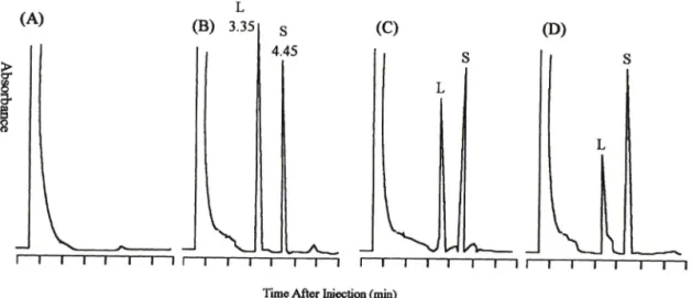
Fig- 2 — Chromatograms of lovastatin (L) from plasma standards and samples after the last dose. (A) blank hum an plasma
:(B) plasma standard spiked with 2 ^g/m/ of L and 10 M-g/m/ simvastatin (S
) :(C) plasma sample collected 2 hrs later after the last dose
:(D) plasma sample collected 6 hrs later after the last dose.
m / 에 6 나/의 빙초산을 가하여 제조)으로 제조하였다.
표준용액은 A C N : H 2 0 (8 0 : 20, v/v% ) 를 사용하여 10, 5, 2 및 1 ng/m /외 농도로 순차적으로 희석하여 제조하였다. 검 량선은 M ille n n iu m l® softw are p ro gra m (W aters, M A , U S A ) 의 Processing M e th o d 를 이용하여 구하였다.
심바스타틴 표준용액의 제조 - 로바스타틴과 같은 방법으로 제조한 내부 표준물질인 심바스타틴의 스톡 용액 (1.0 m g /m /) 을 A C N 으로 희석하여 10 lig/ml 농 도로 만든 다옴 사용하였다.
혈장 및 뇨중 로바스타틴 표준용액 spiking - 로바 스 타 틴 스톡 용액을 혈장 1 m ; 에 100 H/를 s p ik in g 한 후 이 것을 이용하여 혈장중 로바스타틴의 농도가 10, 5, 2, 1 및 0.5 n g /m / 가 되도록 혈장을 사용하여 단계적으 로 희석하여 사용하였다. 그리고. 또한 로 바 스 타 틴 스톡 용액을 어떠한 약물도 사용하지 않은 사람에서 채취한 b la n k urin e 1 m H l 100 를 sp ik in g 한 후 이 것을 이 용하여 뇨중 로 바 스 타 틴의 농도가 10, 5, 2, 1 및 0,5 ng/m /가 되도록 뇨를 사용하여 순차적으로 희석하여 사용하였다.
혈장 및 뇨 샘플 채취 - 고용량의 로바스타틴 (30~
40 m g /k g /d a y ) 을 처방에 따라 1 일 4 회로 7 일동안 투 여받온 위암환자 8명을 대상으로 혈액 및 뇨 샘폴을 채 취하였다. 혈액은 첫째날 첫 번째 용량을 투여하기 직전 및 투여 후 0.5, 1, 1.5, 2, 4 및 6 시간까지 채취하였고, 뇨는 0 ~ 2 시간 및 2 ~ 6 시간간격으로 모아 각 시간간격
의 총 뇨량을 정 확히 측정하고, 이 중 약 5 m /썩 분취하 여 분석할때까지 -80°C에서 보관하였다. 제 7일째 마지 막 용량을 투여한 다옴 혈액은 0.5, 1, 1.5, 2, 4, 6, 12, 24. 48 시간후에 채취하였고, 뇨 샘폴은 0~2, 2-6, 6-12, 12-24, 24~48시간간격으로 모아 각 시간간격 의 총 뇨량을 정 확히 측정 한 후 이중 약 5 m /씩 분취하 여 분석할때까지 -80°C에서 보관하였다. 채취 된 혈액 은 바로 헤파린 처리된 시험관에 넣어 원심분리를 한 후 혈장만 분리하여 1.5 m/ 에펜도르프 튜브에 250 H; 씩 소분한 후 분석할때까지 -80°C에서 보관하였다.
혈장 및 뇨 샘폴의 전처리 - 분석 직전 혈장과 뇨 샘 폴을 꺼내서 실온에서 서서히 녹인후. 다옴과 같은 액상 추출법으로 전처리를 하였다.
샘폴 250 |xZ씩 담긴 에펜도르프 튜브에 50 ^l/의 ACN/ 초산 0.01M 혼합액과 50 의 내부 표준용액(10 Hg/mO을 차례로 가한 후 진탕하였다. 여기에 100 [il 의 10% sodium dodecylsulfate(SDS)틀 가하여 재진 탕하고, 또 다시 500 의 ACN/ 초산 0.01M을 취하여 각각의 튜브에 가한 후 진탕하였다. 이것을 8,500 rpm 에서 15분동안 원심분리한 후 , 상등액을 1.2 m /썩 취 하여 시 험 관에 옮겼다. 각 시 험 관을 히트 볼록에 장착 하고 37°C에서 질소 가스를 가하면서 용매를 증발시킨 후 . 건조된 각각의 샘폴에 300 바의 ACN ■ ■ H20(20:
80)/0.01M 초산 혼합액 (2 m /의 ACN과 8 m /의 H
2O
를 가한 후 . 총 10 m ; 에 대하여 6 의 빙초산을 가하여
제조)을 가하여 재용해하였다.
476 최혜진■김 명민• 최경업
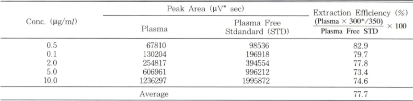
Assay validation - 액상 샘폴 처 리방법 의 추출 효율 을 구하기 위하여 혈장에서 처리한 로바스타틴의 피이 크 면적 에 추출비율을 곱한 후 혈장이 없는 표준용액에 서 의 로바스타틴 피 이크 면적으로 나누어 주었다.
검출력의 한계는 signal-tcrnoise 버를 3: 1로 하여 구하였고, 검량선은 회귀분석을 적용하여 로바스타틴 의 5가지 혈장 농도(0.5, 1, 2. 5. 10 ng/m ; ) 범위에서 의 상관계수 값 이 을 구하였다.
분석외 정밀도와 정확도룰 평가하기 위하여 산출된 검량선에서 각각에 대웅하는 농도의 상대적 표준편차 (RSD, % )와 상대적 오차(RE, % )를 계산하였다. In- ter-assay의 정밀도와 정확도는 3주동안외 기간에 5회 분석 한 결과로 나타냈으며 . intra-assay에서는 하루동 안 12시간 간격으로 실시하였다.
샘폴외 분리능(resolution, Rs)을 평가하고자 로바스 타틴과 심바스타틴의 capacity factor(k^ 와 분리 계수 (a )를 구하여 다음의 식에 대입하여 평가하였다.
Rs=1.18xAt/(W i + W
2)
At : difference in retention time between the two peaks
Wi, W
2: peak width at half height
결과 및 고찰
로바스타틴과 심바스타틴의 HPLC상 유지시간(re
tention time)은 각각 3.35 분 및 4.45분이었고, 분리
능 R„은 4.77로 나타났다. 혈장과 로바스타틴을 spike 한 혈장의 크로마토그램은 Fig. 2(A) 및 (B)에 나타내 었다. 과 a 값이 적합한 기준(2<k^<6. a> l)에 포함 되도록 이동상의 비율을 조절하여 최종적으로 ACN : 0.025M NaH2PC»4(v/v% )외 비율이 65: 35로 확립하 였고, 이에 따른 ka;, k /, 및 a 값은 각각 2.72, 3.94 및 1.45 이었다.
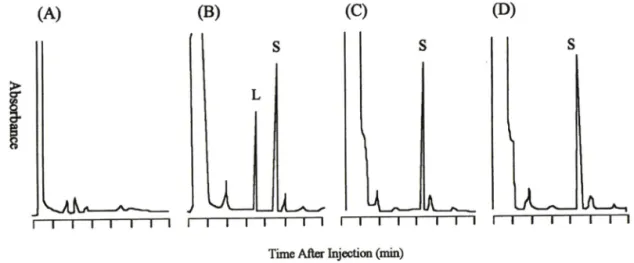
크로마토그램 작성 및 피이크 면적법을 이용한 농도 계산은 모두 Waters사의 Millennium® software pro- gram을 이용하여 실시하였다. Fig. 2(C) 및 (D)에 나타 낸 혈장 샘플 크로마토그램은 7일 투여기간 중 마지막 약물을 투여한 후 . 각각 2시 간과 6시 간 후에 나타난 것이 며, 6시간 후에 나타난 로바스타틴 피이크에서는 쇼울더 (shoulder)를 형성하고 있는 것을 볼 수 있었다. 이것은 로바스타틴의 대사물질로 사료된다. 샘폴 채취 시간을 48시간까지 실시하여 분석해 본 결과, 대부분 환자의 샘 폴에서 로바스타틴이 6시간 이후에는 noise보다 낮게 나 왔다. Fig. 3에 뇨 및 로바스타틴을 spike한 뇨 샘플의 피이크를 나타내고 있으나, 뇨 샘폴에서 로바스타틴은 거외 검출되지 않았다. 로바스타틴의 주요 대사물질인 로바스타틴 산은 prodrug인 로바스타틴보다 큰 극성을 지님으로 현분석조건에서 로바스타틴 앞부분에 나타날 것으로 추측된다.
추출효율은 로바스타틴 0.5~10 ng/m/ 농도 범위에 서 73.4~82.9%로 타났고, 평균 추출효율은 77.7%
였다(Table I). 검출'력외 한계는 signal-to-noise 비를 3 : 1로 하였을때 0.5 ng/m /로 나타났다. 검 량선은 로바
Time After Injection (min)
Fig. 3 — Chromatotograms from urine standards and samples after the last dose. (A) Blank urine
;(B) urine standard spiked with 2 |ig/m
;:(C) urine sample collected during hrs
:(D) urine sample collected during 2—6 hrs.
/.
Pharm. Soc. KoreaS
A—
(B)
I
Abs<x*bance
0.5 0.49±0.015 1.0 0.96±0.04 2.0 1.99+0.01 5.0 5.12±0.12 10.0 9.94±0.05
스타틴 0.5 - 1 0 [ig/ml 농도 범위에서 상관계수(r)가 0.9998 인 직선성을 나타내었다 (Y = 4 .6 6 X + 0 .1 3 , X : 샘폴 농도, Y : 괴이크 면적). 검량선의 정밀도와 정확 도는 각각 Table I I 에 R S D (% , R elative S ta n d a rd D e v iatio n ) 와 R Ei% , R elative E rro r) 로 나타내었다.
분석의 inter 및 in tr a d a y varia bility 시험결과를
Table I I— Precision and accuracy of the method with
human plasma standards Conc. Conc. Measured
(\ig/ml) (ng/m; )±SD
묘®°
네 묘®
네다옴과 같이 Table I I I 에 나타내었다. T able III~A 에 의하면 in te rd a y 의 정밀도는 0 .7 ~ 1 0 .5 % 의 R S D 로 0.5 U g /m / 에서만 10% 정도로 나타내었을 뿐 기타 농 도에서는 < 4 .0 % 차이를 보였다. 또한 , 0,5 U g /m ; 를 제외하고는 < 2 .0 % 의 R E 를 나타내어 분석의 정확성 을 확증해주었다. 한편, in tr a d a y 의 정밀도는 R S D 가 모두 2% 이하의 값을 보였다.
이상의 결과에 의하면, 로바스타틴을 상용량보다 80~ 100 배 정도의 고용량 (35 m g /k g /d a y ) 으로 투여하 였옴에도 불구하고 극히 미량(1 ng/m/. 미만) 만이 검 출된 것으로 보아, 대부분이 인체에 들어가자마자 활성 대사물질인 로바스타틴 산으로 대사된 것으로 추측된 다 . 이의 근거로는 6시간 이후의 혈장샘폴에서 로바스 타틴이 거의 검출되지 않은 것과, 혈장샘폴 분석중에 시
Table I II
~ Inter/intraday precision and accuracy of the method with human plasma standards of lovastatin A. 3-week period (Interday)
Conc. Measured (M-g/m/)
i^g/ral) S S ^ Mean SD RSD (%) RE (%)
Average 77.7
' Fraction in which 250 li/ of the sample was used for the extraction and later reconstituted with 300 \ xl of the sol
vent and of which 100 \il was finally injected for the analysis.
Table I
— — Peak areas of lovastatin at 0.5. 1, 2. 5 and 10 M-g/m/ and the corresponding extraction ratios
________________ Peak Area (nV* sec)_________________ Extraction Efficiency {%) Conc. (M-g/m/) Plasma Free (Plasma x 300*/350) ^
Plasma Stdandard (STD) Plasma Free STD
______________________ 역상 고속액체크로마토그라프법을 이용한 혈장 및 뇨 중 로바스타틴외 정량___________________
mB. 12-hr period (Intraday)
rinnr Conc. Measured (M-g/m/)
Oig/m/) 0 hr 12 hr later Mean SD RSD {%) RE (%) .5
.4 .6 3.1
4.2 0.5 2.3 0.5
o
5
4
2
3
4
4 <d
ci
^
^
■6 - 2 6 에 에
5
418
2 5
0
9
9
0
^ 1 1
l i 4
4
9
4
8 5
0
8
9
9 0 1 1 4 - 0^
0.56 1.04 1.93 5.02 .0.06
5
0
0
0
0
ci
h C 'j urS
^
0
0
0
6
4 4- 1- OA- L o
o
o
7
^ 4 - z 6
4
4
5
7 o
o
o1
o -o o- 7
1
6
2
4 5
0
9
9
0 l i
l i
4
» 4
2
9
0
7 6
0
8
9
0
^
±1 1± 4^
5
1
0
8
6 5
0
0
8
0 o-
L 2-
4.
o.
2
6
7
2
3 6
0
9
71 - n-
L
L4 - 8
6
9
2
5 4
9
9
1
9 o -
L 7
9
4
9
1 5
9
9
9 1-
4-
^ 5
0
0
0
0 o-
1
-9 -
o-
Q-
7
8
4
6 ox - 9-
7-
4^ 8
7
7
7
7 _/c
cc z- rN f 3
1 5
1 7 5
9
5
2
8 8
6
4
6
5
d비
&
&
^
9 1
3
9
9 0
4
7
1
7 l i
T—
I C.O0"i 8
2
0 0
9
2 7
0
4
6
6 6
3
5
0
3 1 2 6 2
0.1
2.0
5.0
10.0
478 최혜진■김명민■최경업
간이 지남에 따라 로바스타틴 피이크의 뒷부분에 쇼울 더가 크게 나타난 것을 예로 들 수 있다(Fig. 2(D) 참 조). 이번 연구에서는 이 대사물질로 추측되는 로바스 타틴 산 분리 정량을 못하였지만 앞으로 로바스타틴과 그 대사체인 로바스타틴 산외 분리 정량법을 확립할 예 정이다, 로바스타틴은 대부분 분번으로 배설되지만, 뇨 중으로도 10% 정도 배설되기 때문에, 뇨 중 로바스타 틴 분석이 가능하리라 추측하였으나. Fig. 3(C)에서와 같이 2시간때에도 검출되지 않았다. 이것으로 보아 뇨 로 배설되는 로바스타틴의 형태는 활성 대사물질인 것 으로 사료된다.
지금까지외 로바스타틴 분석이 비특이적인
효 소정 량법으로 이부어져 활성대사물질까지 동시에 분석되어 약동학적 특성을 나타내는데 있어 오류가 발생될수 있 었고, 기존의 HPLC 분석법®•에서 사용된 고형상 추출 법 (solid phase extraction)은 고가인 반면, 본 연구에 서 개발된 HPLC 분석법은 다량의 혈장 샘폴을 비교적 신속하고 정확하게 분석할 수 있으면서도 경제적으로 저렴한 편이다. 본 연구에서의 검출한계농도는 기존의 방법보다 우수하지 못했으나. 로바스타틴을 고용량으 로 투여한 후 채취한 샘폴에 적용하였기에 검출한계농 도가 그다지 중요하지 않았다고 사료된다. 본 분석법외 재현성은 기존의 것과 거의 동일하거나 우수하였다.
결 론
혈장 중 로바스타틴 농도 측정을 위해 HPLC 방법을 도입하여 정확하면서도 액상 추출법을 이용한 비교적 간편화된 분석법으로 확립하였다. 본 분석법의 특징은 다음과 같이 정리할 수 있다.
1. 분석용 칼럼은 Novapak Cis을 사용하였고, 이동 상은 ACN과 0.025M NaHjPOiCpH 4.5)를 65: 35(v/
v% )의 비율로 하여 유속 1.5 m//min 및 UV 238 nm 에서 검출하였다.
2. 혈장샘폴 전처리방법으로 액상추출법을 사용하여 다량의 샘플을 경제적으로 분석할 수 있었다.
3. k / = 2.72, k / = 3.94, a=1.45, R,=4.77로 산출 되었으며, 로바스타틴은 3.35분 , 심바스타틴은 4.45분 에 각각 검출되었다.
4. 평균 추출효율은 로바스타틴 0.5~10 ng/m; 농도 범위에서 77.7% 으로 나타났으며, 검출력의 한계는 sig- nal-to-noise비를 3: 1로 하였을 때 0.5 나g/m/이었다.
5. Interday 및 intraday외 정밀도는 로바스타틴 0.5-10 ng/m/ 농도 범위에서 각각 10.5% 및 1.82%
이하로 나타났다,
이와같은 신속하고 정확한 분석법은 임상연구에 적 합하게 적용될 수 있어 향후 암치료를 위한 로바스타틴 고용량 투여후의 약동학적 연구에 필수적인 요소가 되 리라고 본다. 또한, 본 HPLC 방법은 활성물질인 로바 스타틴 산(Lovastatin acid) 분석법 확립에 있어서 기 초를 제공하여 본 연구실에서 현재 개발단계에 있다.
갑사의 말씀
본 연구에 사용된 로바스타틴과 심바스타틴 표준물 질은 각각 중외제약(주)과 한국엠에스디(주)로부터 공 급받았고, 환자샘폴은 삼성의료원 혈액종양내과에 의 해 제공되었으며 이에 감사드럽니다.
문 헌
1) Henwood, J. M. and Heel. R. C. : Lovastatin. A preliminary review of its pharmacodynamic pro
perties and therapeutic use in hyperlipidaemia.
Drugs
36,429 (1988).
2) Jakobisiak, M., Bruno, S.. Skierski, J. S. and Darzynkiewicz, Z. '■ Cell cycle-specific effects of lovastatin. Proc. Natl. Acad. Sci. USA. 88, 3628 (1991).
3) Keyomarsi, K., Sandoval, L., Band, V., Pardee, A. B, : Synchronization of tumor and normal cells from G l to multiple cell cycles by lo
vastatin. Cancer Res.
51,3602 (1991).
4) Maltese, W. A., Defendini, R., Green, R. A., Sher
idan, K. M. and Donley, D. K. : Supression of murine neuroblastoma growth in vivo by mev- inolin. a competitive inhibitor of hydroxy-3- methylglutaiyl-coenzyme A reductase. /. Clin. In - vest.
76,1748 (1985).
5) Pan, H. Y., Devault, A. R., Wang-Iverson, D., Ivashkiv E., Swanson, B. N. and Sugerman, A.
A. : Comparative pharmacokinetics of pra
vastatin and lovastatin. I Clin. Pharmacol.
30,1128 (1990).
6) McKenney, J. M. : Lovastatin: A new cho- lesteroHowering agent. Clin Pharm.
7,21 (1988).
/.
Pharm. Soc. Korea역상 고속액체크로마토그라프법을 이용한 혈장 및 뇨 중 로바스타틴외 정량 479
7 ) K m k e m y e r , J . J . a n d T a l b e r t . R . L . : L o v a s t a t i n
: A n e w c h o l e s t e r o l - l o w e r i n g a g e n t .
Pharmaco
therapy 7,
1 9 8 ( 1 9 8 7 ) .8 ) M e r c k S h a r p a n d D o h m e : M e v a c o r ® p a c k a g e i n s e r t , i n P h y s i c i a n ' s D e s k R e f e r e n c e . O r a d e l l . N . J . .
Medical Economics
C o .,Inc.
1 4 1 2 ( 1 9 9 0 ) . 9 ) S t u b b s . R . J . . S c h w a r t z , M . a n d B a y n e . W . F . ;D e t e r m i n a t i o n o f m e v i n o l i n a n d m e v i n o l i n i c a c i d i n p l a s m a a n d b i l e b y r e v e r s e d - p h a s e h i g h - p e r f o r m a n c e l i q u i d c h r o m a t o g r a p h y .
I Chro
matogr. 383.
4 3 8 ( 1 9 8 6 ) .1 0 ) S e b t i . S . M . . T k a l c e v i c . G . T . a n d J a n i , J . P . ■ L o v a s t a t i n . a c h o l e s t e r o l b i o s y n t h e s i s i n h i b i t o r , i n h i b i t s t h e g r o w t h o f h u m a n H - r a s o n c o g e n e t r a n s f o r m e d c e l l s i n n u d e m i c e .
Cancer Commun.
3 . 1 4 1 . ( 1 9 9 1 ) .
1 1 ) V i n c e n t . T . S . . W u l f e r t , E . a n d M e r l e r . E . ■ I n h i b i t i o n o f g r o w t h f a c t o r s i g n a l i n g p a t h w a y s b y l o v a s t a t i n .
Biochem. Biophys. Res. Commun. 180.
1 2 8 4 ( 1 9 9 1 ) .
1 2 ) P e n t i k a i n e n . P . J . , S a r a h e i m o . M . . S c h w a r t z . J . I .. A m i n . R . D . . S c h w a r t z , M . S . . B r u n n e i니F e r b - e r . F . a n d R o g e r s . J . D . ■ C o m p a r a t i v e P h a r m a c o k i n e t i c s o f l o v a s t a t i n , s i m v a s t a t i n a n d p r a v a s t a t i n i n H u m a n s .
I Clin. Pharmacol. 32.
1 3 61 3 ) ( 1 9 9 2 ) .
C a r l u c c i , G . . M a z z e o , P . . B i o r . L . a n d B o l o g n a .
M .
'■
S i m u l t a n e o u s d e t e r m i n a t i o n o f s i m v a s t a t i n a n d i t s h y d r o x y a c i d f o r m i n h u m a n p l a s m a b y h i g h - p e r f o r m a n c e l i q u i d c h r o m a t o g r a p h y w i t h U V d e t e c t i o n . /.Pharm. Biomed. Anal. 10.
6 9 3 ( 1 9 9 2 ) .
1 4 ) V y a s , K . P . . K a i l P . H . , P i t z e n b e r g e r , S . M . , H a l p i n , R . A . . R a m j i t , H . G . , A r i s i o n . B . . M u r p h y , J . S . . H o f f m a n . W . F . . S c h w a r t z . M . S . . U l m . R . H . a n d D u g g a n , D .
K. '■
B i o t r a n f o i n i a - t i o n o f l o v a s t a t i n; I. S t r u c t u r e e l u c i d a t i o n o fin vivo
m e t a b o l i t e s i n t h e r a t a n d m o u s e .Drug Metab. Dispos. 18.
2 0 3 ( 1 9 9 0 ) .1 5 ) V y a s , K . P . . K a i l P . H ., P r a k a s h , S . R . a n d D u g g a n . D . E . : B i o t r a n s f o r m a t i o n o f l o v a s t a t i n: II.
In vitro
m e t a b o l i s m b y r a t a n d m o u s e l i v e r m i c r o s o m e s a n d i n v o l v e m e n t o f c y t o c h r o m e P - 4 5 0 i n d e h y d i 'o g e n a t i o n o f l o v a s t a t i n .Drug Metab. and Dispos. 18,
2 1 8 ( 1 9 9 0 ).1 6 ) W e j d e . J . . C a i 'lb e r g , M . . H j e i t m a n . M . a n d L a i ' s - s o n m . O . : I s o p r e n o i d r e g u l a t i o n o f c e ll g r o w t h:
I d e n t i f i c a t i o n o f m e v a l o n a t e - l a b e l l e d c o m p o u n d s i n d u c i n g D N A s y n t h e s i s i n h u m a n b r e a s t c a n c e r c e l l s d e p l e t e d o f s e m m a n d m e v a l o n a t e . /.
Cell Physiol. 155.
5 3 9 ( 1 9 9 3 ) .1 7 ) S n y d e r . L . R . , K i r k l a n d J . J . a n d G l a j c h , L . G . :