P Primary Calcified T-Cell Lymphoma of the Urinary Bladder: A Case Report
3
0
0
전체 글
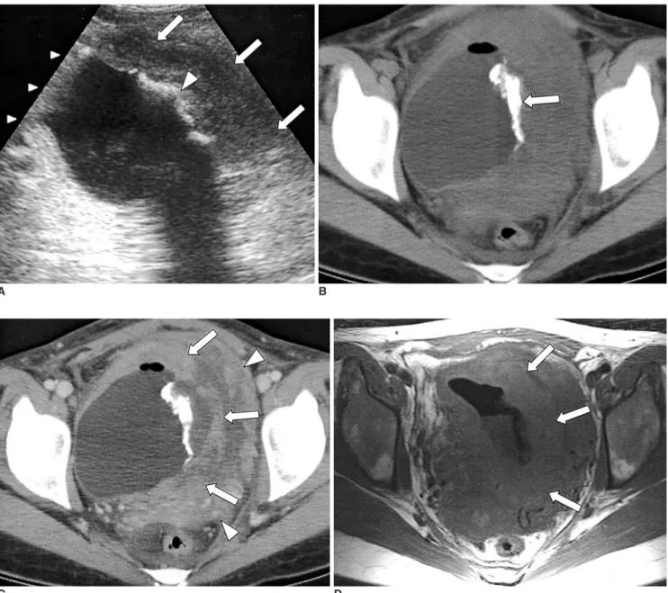
(2) Primary Calcified T-Cell Lymphoma of Urinary Bladder. ed that the neoplasm associated with chronic cystitis may originate in the lymphocytes present in the submucosa of the bladder as a result of an inflammatory or immune response (2). The radiologic findings of urinary bladder lymphoma vary, though from a review of the literature, certain patterns emerge. The tumor appears most frequently as a sessile solitary mass (66%); multiple sessile masses are less frequent (14%), and a polypoid mass is occasionally present (10%). Diffuse thickening of the bladder wall may. A. occur, but is infrequent (10%). Despite these patterns of occurrence, there are no characteristic findings which distinguish lymphoma of the urinary bladder from other tumors (1). In our case, ultrasonography and pelvic CT revealed diffuse bladder wall thickening, a finding which can frequently mimic transitional cell carcinoma (TCC) (1, 2). Pre-treatment calcification in lymphoma patients is extremely rare, and its pathogenesis is uncertain. Generally, tissue calcifications is metastatic or dystrophic (5 7); The. B. C D Fig. 1. A 30-year-old woman with primary lymphoma of the urinary bladder. A. Ultrasonogram shows diffuse left lateral wall thickening (arrows) and thick linear echogenic foci with posterior acoustic shadowing (arrowhead), suggesting calcification in the left lateral wall. B. Pre-contrast CT depicts irregular tubular-shaped calcification (arrow). C. Post-contrast CT demonstrates asymmetric wall thickening (arrows) with heterogeneous enhancement of the left lateral wall. Adjacent to this, displaced small bowel loops (arrowheads) are visible. D. T1-weighted spin-echo image (TR/TE = 500/8 msec) shows asymmetric thickening of the lateral wall, with low signal intensity (arrows). Korean J Radiol 4(4), December 2003. 253.
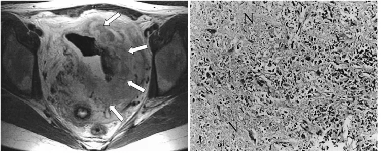
(3) Choi et al.. E F Fig. 1E. At T2-weighted fast spin-echo imaging (TR/TE = 3,500/78 msec), high signal intensity is observed (arrows). MR imaging, however, does not reveal the calcification depicted at CT. F. Photograph (original magnification, 200; hemotoxylin-eosin staining) of ultrasonography-guided biopsy specimen depicts infiltration of pleomorphic, hyperchromatic lymphoma cells, together with T-cell phenotype and focal necrosis (N).. former is caused by excessive or unstable calcium ion concentration in the blood. In our case, however, this level was normal, and this explanation was thus less likely. Dystrophic calcification, on the other hand, frequently occurs in degenerated or necrotic tissue (6), conditions which result from infarction and lead to calcification. Such infarction occurs typically in histologically aggressive lymphoma (7). In our case, high grade T-cell lymphoma accompanied by necrosis was diagnosed, and the pathogenesis for calcification was probably a dystrophic calcification. In TCC and urachal carcinoma, calcification occurs within the bladder wall. Infiltration appears to be more diffuse in lymphoma than in TCC, and in the former, hydronephrosis seems to be uncommon (8). Urachal carcinoma also commonly involves calcification, which typically occurs at the bladder dome. Schistosomiasis may produce mural calcification which shows with a typical thin arcuate pattern, and may also be associated with calcification in other parts of the urinary tract. Other entities that cause bladder wall injury can predispose to dystrophic calcification and include alkaline incrusted cystitis, cytotoxin cystitis, radiation therapy, and tuberculosis (9). Treatment of primary lymphoma of the urinary bladder is not uniform and the procedures employed have included radical cystectomy, radiation therapy, and chemotherapy. For prognosis, histologic grade is probably a useful indicator, and the overall prognosis appears to be good (2).. 254. In summary, we report a case of primary lymphoma of the urinary bladder associated with dense internal calcification. A case of this nature has not previously been reported in the literature in English.. References 1. Tasu JP, Geffroy D, Rocher L, et al. Primary malignant lymphoma of the urinary bladder: report of three cases and review of the literature. Eur Radiol 2000;10:1261-1264 2. Amin R. Case report: primary non-Hodgkin’s lymphoma of the bladder. Br J Radiol 1995;68:1257-1260 3. Mourad WA, Khalil S, Radwi A, Peracha A, Ezzat A. Primary T-cell lymphoma of the urinary bladder. Am J Surg Pathol 1998;22:373-377 4. Ohsawa M, Aozasa K, Horiuchi K, Kanamaru A. Malignant lymphoma of the bladder. Cancer 1993;72:1969-1972 5. Apter S, Avigdor A, Gayer G, Portnoy O, Zissin R, Hertz M. Calcification in lymphoma occurring before therapy. AJR Am J Roentgenol 2002;178:935-938 6. Ishikawa T, Kobayashi Y, Omoto A, et al. Calcification in untreated non-Hodgkin’s lymphoma of the jejunum. Acta Haematol 1999;102:185-189 7. Cleary KR, Osborne BM, Butler JJ. Lymph node infarction foreshadowing malignant lymphoma. Am J Surg Pathol 1982;6:435 -442 8. Kashi SH, Murphy JK, Britton JP, Whelan P. Primary lymphoma of the bladder: a clinicopathological study of 3 cases. Eur Urol 1990;17:186-188 9. Dyer RB, Chen MYM, Zagoria RJ. Abnormal calcification in the urinary tract. RadioGraphics 1998;18:1405-1424. Korean J Radiol 4(4), December 2003.
(4)
수치
관련 문서
We presently report the first case of hydronephrosis and hydroureter due to direct compression in the urinary bladder by silicon, which had been introduced by the patient himself 2
Extranodal natural killer (NK)/T-cell lymphoma, nasal type (ENKTCL) is a rare entity of non-Hodgkin lymphoma (NHL) that typically involves the nasal cavity and upper aerodigestive
In a computed tomography (CT) scan, fat deposition in the urinary bladder wall is seen as a linear hypoattenuating band surrounded by soft tissue density.. It is un- common, but
In the present case, dynamic contrastenhanced CT revealed a welldefined mass with moderate enhancement in the arterial phase, and washout of contrast media in the portal venous
In conclusion, we describe the first case of primary adrenal T-cell lymphoma with ocular and bone marrow metastasis in a young adult.. In the absence of clinical evidence to
Removal of foreign bodies embedded in the urinary bladder wall by a combination of laparoscopy and carbon dioxide cystoscopic assistance: Case report and literature review..
