© 2017 The Korean Academy of Medical Sciences.
This is an Open Access article distributed under the terms of the Creative Commons Attribution Non-Commercial License (http://creativecommons.org/licenses/by-nc/4.0) which permits unrestricted non-commercial use, distribution, and reproduction in any medium, provided the original work is properly cited.
pISSN 1011-8934 eISSN 1598-6357
Primary Pulmonary Extranodal Natural Killer/T-cell Lymphoma, Nasal Type Presenting as Diffuse Ground Glass Opacities: a Case Report
Extranodal natural killer (NK)/T-cell lymphoma, nasal type (ENKTCL) is a rare type of lymphoma that accounts for only 5%–18% of all cases of non-Hodgkin lymphoma (NHL).
In published series, 60%–90% of NK/T-cell lymphomas are localized to the nasal and upper airway. We describe a 55-year man who presented with cough, sputum, dyspnea on exertion, and a chest computed tomography scan shows diffuse ground glass opacities (GGOs), suggestive of an interstitial lung disease. He was treated with a corticosteroid and his symptoms improved. However, when the corticosteroid was tapered, his symptoms recurred. The patient underwent a surgical lung biopsy and ENKTCL was diagnosed. We present this case because ENKTCL involving only the lung is very rare but very informative.
To our knowledge, our patient is the first case that primary pulmonary ENKTCL is presented with GGOs.
Keywords: Lymphoma, Extranodal NK-T-Cell; Lung Involvement; Lung Diseases, Interstitial; Ground Glass Opacities
MyungJin Song,1 Ji-Ye Kim,2 Ji Soo Choi,1 Bora Yoon,1 MooHyun Kim,1
Soo-Jeong Kim,3 and Song Yee Kim1
1Division of Pulmonology, Department of Internal Medicine, Severance Hospital, Institute of Chest Diseases, Yonsei University College of Medicine, Seoul, Korea; 2Department of Pathology, Severance Hospital, Yonsei University College of Medicine, Seoul, Korea; 3Division of Hematology, Department of Internal Medicine, Severance Hospital, Yonsei University College of Medicine, Seoul, Korea Received: 10 February 2016
Accepted: 13 May 2016 Address for Correspondence:
Song Yee Kim, MD
Division of Pulmonology, Department of Internal Medicine, Severance Hospital, Institute of Chest Diseases, Yonsei University College of Medicine, 50-1 Yonsei-ro, Seodaemun-gu, Seoul 03722, Korea
E-mail: [email protected]
https://doi.org/10.3346/jkms.2017.32.10.1727 • J Korean Med Sci 2017; 32: 1727-1730
INTRODUCTION
Extranodal natural killer (NK)/T-cell lymphoma, nasal type (ENKTCL) is a rare entity of non-Hodgkin lymphoma (NHL) that typically involves the nasal cavity and upper aerodigestive tract. However, it can also occur at extranasal sites (the skin, liv- er, spleen, gastrointestinal tract, bone marrow, lung, and so on).
A primary presentation of ENKTCL in the lung and ENKTCL involving only the lung is rare (1,2). Furthermore, there is no re- port which primary pulmonary ENKTCL is presented with ground glass opacities (GGOs). Here, we describe a case of primary pul- monary ENKTCL presenting with pulmonary symptoms and GGOs.
CASE DESCRIPTION
A 55-year-old man without any past medical history presented with cough, sputum, and dyspnea on exertion for three months in March, 2015. He had a smoking history of 30 pack-years, and had quit smoking since his dyspnea developed. On physical ex- amination, he appeared acutely ill, and lung sounds were de- creased in bilateral lung fields. He had no peripheral lymphade- nopathy, organomegaly, or skin lesion. Laboratory findings were
within normal ranges, except for mild elevations of aspartate transaminase (AST) and alanine transaminase (ALT) (AST, 50 IU/L; ALT, 54 IU/L). An arterial blood gas analysis showed mild hypoxia (partial pressure of arterial oxygen, 60.3 mmHg; partial pressure of arterial carbon dioxide, 31.0 mmHg; and pH, 7.455).
The result of a pulmonary function test shows decreased diffus- ing capacity of the lungs for carbon monoxide (DLCO; 5.02 mmol/
min/kPa [58.66% of the predicted value]), forced vital capacity (FVC; 3.15 L [85.74% of the predicted value]), forced expiratory volume in 1 second (FEV1; 2.31 L [77.71% of the predicted val- ue]), and a ratio of FEV1/FVC (73.15).
His initial computed tomography (CT) scan shows diffuse GGOs in both lungs with underlying emphysema (Fig. 1). Bron- choalveolar lavage (BAL) and transbronchial lung biopsy (TBLB) via a flexible fiberoptic bronchoscope were performed. The BAL fluid contained 55% lymphocytes with a CD8/CD4 ratio of 1:1, 7% eosinophils, and pathology obtained from the TBLB showed multifocal interstitial and perivascular inflammatory infiltrates, which were predominantly lymphocytes. There was no evidence of infection in the results of TBLB and BAL. Although the pa- thology results were not diagnostic, and definite exposure to a known offending antigen did not exist, interstitial lung disease (ILD) such as hypersensitivity pneumonitis (HP) or desquama- CASE REPORT
Respiratory Diseases
1 / 1 CROSSMARK_logo_3_Test
2017-03-16 https://crossmark-cdn.crossref.org/widget/v2.0/logos/CROSSMARK_Color_square.svg
Song MJ, et al. • Primary Pulmonary NK/T Cell Lymphoma
1728 http://jkms.org https://doi.org/10.3346/jkms.2017.32.10.1727 tive interstitial pneumonia (DIP) was highly suggested based on the results of the CT scan. With the impression of ILD, he was treated with a corticosteroid (prednisolone 60-mg daily; Cima Labs Inc., Eden Prairie, MN, USA). His symptoms and imaging were improved at the beginning of corticosteroid treatment, but when the corticosteroid was tapered, his symptoms recurred and his imaging worsened. After three months of corticosteroid treatment, he took a follow-up CT scan and it shows diffuse GGOs aggravated compared to initial CT scan (Fig. 1). Therefore, the patient underwent wedge resections of the lung under video- assisted thoracoscopic surgery (VATS) for a definite diagnosis.
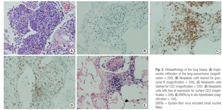
Wedge resections were done at lateral segment of right middle lobe and posterior basal segment of right lower lobe. The speci- men revealed diffuse lymphomatous infiltration, which was composed of medium-sized cells. Immunohistochemically, these cells showed positivity for CD56, granzyme B, CD2, and loss of surface CD3 expression. In situ hybridization for Epstein-Barr virus (EBV) encoded small nuclear RNAs (EBERs) was positive (Fig. 2). The pathology result was consistent with ENKTCL. Since most ENKTCL originates from the nose and paranasal area, in- cluding the upper aerodigestive tract, the patient underwent a further imaging work-up (paranasal sinus CT, neck CT, and pos- itron emission tomography-CT [PET-CT]) after pathologic con- firmation, even though he did not have any symptom or sign of nose or paranasal area involvement (3,4). PET-CT scan showed no significant fluorodeoxyglucose (FDG) uptake to suggest ma- lignancy (Fig. 3). The only organ that ENKTCL involved was the lung. The patient was treated with cyclophosphamide, doxoru- bicin, vincristine, and prednisolone (CHOP) chemotherapy.
DISCUSSION
This report concerns a patient with ENKTCL without other in- volvement, except for the lung, who was suspected as having ILD at the initial visit. ENKTCL commonly presents with mid- line facial destructive disease and shows a strong association with EBV (5). It occurs typically within the nasal cavity and up- per aerodigestive tract (3,4). ENKTCL arises in all geographic regions of the world, although it is much more common in Asian and Hispanic populations and rare in the USA and Europe (6).
Most cases are derived from the malignant transformation of NK cells that express CD56+ and a lack of surface CD3 expres- sion and T-cell receptor (TCR) gene rearrangements. More than 80% of ENKTCL affects the nose and paranasal area, so the ini- tial complaints of ENKTCL include local symptoms such as uni- lateral nasal cavity obstruction with purulent discharge and bleed- ing. Moreover, these symptoms can lead to early disease recog- nition (7).
However, ENKTCL can also occur predominantly at extrana- sal sites, which makes it hard to diagnose. The sites of predilec- tion of non-upper aerodigestive tract ENKTCL were the skin Fig. 1. Follow-up CT scan shows diffuse GGOs at both lungs with underlying emphy-
sema. Arrows indicate sites of wedge resections. Posterior basal segment of right lower lobe (A) and lateral segment of right middle lobe (B). (A, B) Axial view. (C) Coro- nal view.
CT = computed tomography, GGOs = ground glass opacities.
A
B
C
Song MJ, et al. • Primary Pulmonary NK/T Cell Lymphoma
http://jkms.org 1729
https://doi.org/10.3346/jkms.2017.32.10.1727
Fig. 2. Histopathology of the lung biopsy. (A) Angio- centric infiltration of the lung parenchyme (magnifi- cation × 200). (B) Neoplastic cells stained for gran- zyme B (magnification × 200). (C) Neoplastic cells stained for CD2 (magnification × 200). (D) Neoplastic cells with loss of expression for surface CD3 (magni- fication × 200). (E) EBERs by in situ hybridization (mag- nification × 100).
EBERs = Epstein-Barr virus encoded small nuclear RNAs.
A
D
B
E
C
Fig. 3. PET-CT scan shows no significant FDG uptake to suggest malignancy. The lung where ENKTCL is confirmed by biopsy also shows no FDG uptake.
PET-CT = positron emission tomography-computed tomography, FDG = fluorodeoxy- glucose, ENKTCL = extranodal natural killer/T-cell lymphoma, nasal type.
(37%), liver or spleen (31%), gastrointestinal tract (24%), bone marrow (22%), lung (14%), extremities (8%), and testis (5%) (3,4,8).
When ENKTCL is suspected, a histopathological examina- tion is necessary. The histopathology of ENKTCL comprises a mixed pattern ranging from small-to-medium sized atypical cells to large transformed cells. The tumor cells typically have the immunophenotype of CD56+, CD2+, cytoplasmic CD3+, and are negative to other T-cell antigens such as surface CD3, CD4, CD5, CD57, CD16, and B-cell antigens such as CD20. In addition, cytotoxic molecules such as granzyme B and T-cell intracellular antigen-1 (TIA-1) (76.2% of all ENKTCL), perforin, and the nm23-HI gene (42% of all ENKTCL) can be expressed in this type of lymphoma. ENKTCL seems to be caused by EBV, and EBER in situ hybridization is the most reliable way to dem- onstrate the presence of EBV, which can be achieved from par- affin-embedded tissues (4,7-9).
ENKTCL is rare, and has been recognized as an independent disease relatively recently; thus, the optimal therapeutic appro- ach and prognosis have not been fully defined yet. However, this entity is generally known to have an aggressive clinical course, and standard NHL treatment consisting of anthracycline-based chemotherapy is unsatisfactory (10).
The chest CT patterns of ENKTCL with lung involvement seems to be variable. In the previous reviews of ENKCL patients with lung involvement, patchy consolidations, nodules, and masses are the most frequent chest CT findings. GGOs are relatively rare and may represent an early stage of disease that develops into consolidations at the end stage (1,11-13).
We present this case because primary pulmonary ENKTCL is rare and chest CT finding of GGOs is especially rare. To our knowl-
Song MJ, et al. • Primary Pulmonary NK/T Cell Lymphoma
1730 http://jkms.org https://doi.org/10.3346/jkms.2017.32.10.1727 edge, our case is the first case that primary pulmonary ENKTCL
is presented with GGOs. Clinicians should know that ENKTCL can involve only the lung and can present as GGOs in CT scan.
Surgical lung biopsy should be considered when the clinical course is not according to initial impression.
DISCLOSURE
The authors have no potential conflicts of interest to disclose.
AUTHOR CONTRIBUTION
Investigation: Song M, Kim JY, Choi JS, Yoon B, Kim M, Kim SJ.
Visualization: Song M. Writing - original draft: Song M. Writing - review & editing: Kim SY.
ORCID
MyungJin Song https://orcid.org/0000-0003-2218-8959 Ji-Ye Kim https://orcid.org/0000-0003-4291-2967 Ji Soo Choi https://orcid.org/0000-0003-2716-6775 Bora Yoon https://orcid.org/0000-0002-8455-163X MooHyun Kim https://orcid.org/0000-0001-5793-3707 Soo-Jeong Kim https://orcid.org/0000-0001-8859-3573 Song Yee Kim https://orcid.org/0000-0001-8627-486X REFERENCES
1. Laohaburanakit P, Hardin KA. NK/T cell lymphoma of the lung: a case re- port and review of literature. Thorax 2006; 61: 267-70.
2. Morovic A, Aurer I, Dotlic S, Weisenburger DD, Nola M. NK cell lympho-
ma, nasal type, with massive lung involvement: a case report. J Hematop 2010; 3: 19-22.
3. Al-Hakeem DA, Fedele S, Carlos R, Porter S. Extranodal NK/T-cell lym- phoma, nasal type. Oral Oncol 2007; 43: 4-14.
4. Tse E, Liang RH. Natural killer cell neoplasms. Clin Lymphoma 2004; 5:
197-201.
5. Kanavaros P, Lescs MC, Brière J, Divine M, Galateau F, Joab I, Bosq J, Farcet JP, Reyes F, Gaulard P. Nasal T-cell lymphoma: a clinicopathologic entity associated with peculiar phenotype and with Epstein-Barr virus. Blood 1993; 81: 2688-95.
6. Chim CS, Ma SY, Au WY, Choy C, Lie AK, Liang R, Yau CC, Kwong YL. Pri- mary nasal natural killer cell lymphoma: long-term treatment outcome and relationship with the International Prognostic Index. Blood 2004; 103:
216-21.
7. Suzuki R, Takeuchi K, Ohshima K, Nakamura S. Extranodal NK/T-cell lymphoma: diagnosis and treatment cues. Hematol Oncol 2008; 26: 66- 72.
8. Aozasa K, Takakuwa T, Hongyo T, Yang WI. Nasal NK/T-cell lymphoma:
epidemiology and pathogenesis. Int J Hematol 2008; 87: 110-7.
9. Cheung MM, Chan JK, Wong KF. Natural killer cell neoplasms: a distinc- tive group of highly aggressive lymphomas/leukemias. Semin Hematol 2003; 40: 221-32.
10. Lee J, Kim WS, Park YH, Park SH, Park KW, Kang JH, Lee SS, Lee SI, Lee SH, Kim K, et al. Nasal-type NK/T cell lymphoma: clinical features and treatment outcome. Br J Cancer 2005; 92: 1226-30.
11. Lee BH, Kim SY, Kim MY, Hwang YJ, Han YH, Seo JW, Kim YH, Cha SJ, Hur G. CT of nasal-type T/NK cell lymphoma in the lung. J Thorac Imag- ing 2006; 21: 37-9.
12. Oshima K, Tanino Y, Sato S, Inokoshi Y, Saito J, Ishida T, Fukuda T, Wata- nabe K, Munakata M. Primary pulmonary extranodal natural killer/T-cell lymphoma: nasal type with multiple nodules. Eur Respir J 2012; 40: 795-8.
13. Fei W, Xiaohong W, Hong Z, Bei H. Pulmonary extranodal natural killer/
T-cell lymphoma (nasal type): a case report and radiological image review.
Medicine (Baltimore) 2015; 94: e1527.