bloodresearch.or.kr Blood Res 2017;52:218-33.
Letters to the Editor 227
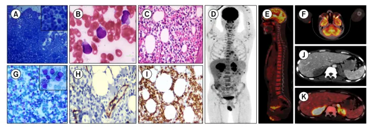
Fig. 1. Microscopic findings, immunohistochemical staining, and whole-body fluorine-18-fluorodeoxyglucose positron emission tomography /computed tomography (18F-FDG PET/CT) of primary adrenal T-cell lymphoma. (A) Aqueous humor aspiration showed infiltration of atypical lymphoid cells. (B) Bone marrow (BM) aspiration showed infiltration of atypical lymphoid cells with irregular nuclear membrane, coarse chromatin, basophilic cytoplasm and fine azurophilic granules. (C) BM biopsy showed infiltration of lymphoma cells with “fried egg” pattern. (D) FDG avid lesions in the adrenal glands. (E) Diffuse FDG uptake in the BM. (F) Absence of FDG avid intra-orbital and intra-ocular mass lesions. (G) Adrenal aspiration showed lymphoma infiltration. (H) CD34 immunohistochemistry (IHC) of the BM biopsy highlighting intra-sinusoidal pattern. (I) CD8 positivity in the BM lymphoid infiltrate. (J, K) Diffuse and intense FDG uptake in both the adrenal glands (A, B, & G: May-Grünwald Giemsa stain;
C: Hematoxylin and eosin stain).
Received on Oct. 9, 2016; Revised on Oct. 18, 2016; Accepted on Jan. 17, 2017 https://doi.org/10.5045/br.2017.52.3.224
AuthorsÊ Disclosures of Potential Conflicts of Interest No potential conflicts of interest relevant to this article were reported.
REFERENCES
1. Saygin C, Uzunaslan D, Ozguroglu M, Senocak M, Tuzuner N.
Dendritic cell sarcoma: a pooled analysis including 462 cases with presentation of our case series. Crit Rev Oncol Hematol 2013;88:253-71.
2. Vasef MA, Zaatari GS, Chan WC, Sun NC, Weiss LM, Brynes RK.
Dendritic cell tumors associated with low-grade B-cell malignancies. Report of three cases. Am J Clin Pathol 1995;104:696-701.
3. Cossu A, Deiana A, Lissia A, et al. Synchronous interdigitating dendritic cell sarcoma and B-cell small lymphocytic lymphoma in a lymph node. Arch Pathol Lab Med 2006;130:544-7.
4. Feldman AL, Arber DA, Pittaluga S, et al. Clonally related fol- licular lymphomas and histiocytic/dendritic cell sarcomas: evi- dence for transdifferentiation of the follicular lymphoma clone.
Blood 2008;111:5433-9.
5. Shao H, Xi L, Raffeld M, et al. Clonally related histiocytic/den- dritic cell sarcoma and chronic lymphocytic leukemia/small lymphocytic lymphoma: a study of seven cases. Mod Pathol 2011;24:1421-32.
6. Chen W, Lau SK, Fong D, et al. High frequency of clonal im- munoglobulin receptor gene rearrangements in sporadic histio- cytic/dendritic cell sarcomas. Am J Surg Pathol 2009;33:863-73.
7. Miyata-Takata T, Takata K, Yamanouchi S, et al. Detection of T-cell receptor γ gene rearrangement in paraffin-embedded T or natural killer/T-cell lymphoma samples using the BIOMED-2
protocol. Leuk Lymphoma 2014;55:2161-4.
8. O'Malley DP, Agrawal R, Grimm KE, et al. Evidence of BRAF V600E in indeterminate cell tumor and interdigitating dendritic cell sarcoma. Ann Diagn Pathol 2015;19:113-6.
9. Ma C, Zhang X, Zhao Y, Wang G, Zhang M. Detection and sig- nificance of BRAF gene in mature T/NK cell lymphoma.
Zhonghua Zhong Liu Za Zhi 2015;37:816-22.
10. Pokuri VK, Merzianu M, Gandhi S, Baqai J, Loree TR, Bhat S.
Interdigitating dendritic cell sarcoma. J Natl Compr Canc Netw 2015;13:128-32.
Primary adrenal T-cell lymphoma in a young adult presented with pseudo-hypopyon: a case report and literature review
TO THE EDITOR: A 26-year-old Indian man presented with weight loss for 6 months and blurred vision on his right eye for 2 weeks. He seemed to be pale, but had no lympha- denopathy or hepatosplenomegaly. Initial complete blood count showed a hemoglobin level of 10.7 g/dL, white blood cell count of 1.4×10⁹/L (neutrophils 44%, lymphocytes 43%, eosinophils 3%, and monocytes 10%), and platelet count of 40×109/L. Ophthalmic evaluation revealed a white me- niscus in the anterior chamber of the right eye. The corre- sponding aqueous humor aspirate showed infilatration of atypical lymphoid cells (Fig. 1). Whole body fluorine- 18-fluorodeoxyglucose (FDG) positron emission tomog- raphy/computed tomography (18F-FDG PET/CT) did not re-
Blood Res2017;52:218-33. bloodresearch.or.kr
228 Letters to the Editor
Table 1. Review of clinical and radiologic findings in patients with primary adrenal T-cell lymphoma.
This case Sampath et al. [4] Pimentel et al. [7] May et al. [8] Sfaxi et al. [9]
Age (yrs) 26 33 42 59 70
Gender Male Male Male Male Male
Diagnosis CD8 positive
extranodal T-cell lymphoma
Not sub-classified Large, cleaved T-cell
lymphoma Centroblastic T-cell
lymphoma Not sub-classified
Symptoms Weight loss,
blurred vision Weight loss, fever,
abdominal pain Weight loss, fever,
vomiting Asymptomatic Weight loss, fever
Adrenal insufficiency No No Yes No Yes
Side Bilateral Bilateral Bilateral Right adrenal Bilateral
Metastasis Anterior chamber
of the right eye No CNS (CSF positive) and rectus sheath of both eyes
No distant metastasis, but the mass was adherent to the IVC
No distal metastasis, but the left adrenal mass was adherent to the left renal pedicle
Treatment Conservative CHOP CHOP with intra-
thecal methotrexate Surgery and radiation Surgery and chemotherapy
Outcome Expired due to
intra-abdominal hemorrhage
After 3 cycles of therapy, patient was asymptomatic and gained 5 kg
Expired 4 months later due to disease progression
Remission for 8 years Expired due to multi-organ failure secondary to sepsis
Bone marrow infiltration Yes NA No No No
Abbreviations: CHOP, Cyclophosphamide 750 mg/m2 per day, vincristine 2 mg per cycle, doxorubicin 50 mg/m2 per cycle, prednisone 60 mg per day; CNS, central nervous system; CSF, cerebrospinal fluid; IVC, Inferior vena cava; NA, not available.
veal any FDG avid intra-ocular/intra-orbital lesions.
However, there were FDG avid soft tissue masses in both the adrenal glands (4.8×9.3×6.2 cm lesion with maximum standardized uptake value (SUVmax) of 22.7 in the right adrenal gland and 4.6×9.2×5.9 cm lesion with SUVmax of 17.6 in the left adrenal) suggesting a lymphomatous pathol- ogy (Fig. 1). CT guided fine needle aspiration from the adrenal glands revealed the infiltration of atypical lymphoid cells, which were CD3 positive and CD20 negative by immunocytochemistry. Bone marrow aspiration showed in- creased number (about 83%) of 15–20 μm sized atypical lymphocytes (Fig. 1). In flowcytometric analysis, these cells showed expression of CD45, CD2, CD3, and CD8, whereas no expression of CD1a, CD4, CD5, CD7, CD34, CD16, CD56, CD57, TCRαβ and TCRγδ. These lymphoid cells had clear cytoplasm, imparting a ‘fried-egg’ pattern of arrangement in bone marrow biopsy. On immunohistochemistry (IHC), these lymphoid cells were positive for CD8 and negative for CD3, CD5, CD4, and CD56. Negative staining for CD34 by IHC highlighted vascular proliferation and intra-sinus- oidal infiltration (Fig. 1). The patient was diagnosed with CD8-positive extranodal T-cell lymphoma, but the patient died on 2 days of admission, prior to initiating therapy.
Primary adrenal lymphoma (PAL) is a rare and aggressive malignancy and involves bilateral adrenals in 60-75% of cases [1, 2]. The most common PALs are diffuse large B-cell lymphomas (-80%), followed by peripheral T cell lymphoma (7%) [2-4]. Since the majority of intraocular lymphomas (IOL) are of B-cell origin, T-cell type IOLs are very rare and often secondary to cutaneous or adult T-cell lymphoma [5]. Except for a single case report showing choroidal involve-
ment, intraocular metastasis by PAL is extremely rare [6].
To the best to our knowledge, only 4 cases of T-cell type PAL have been reported in literature, but none of the cases had bone marrow involvement (Table 1) [4, 7-9].
The median age at diagnosis was 40 years (range, 31-70 yrs). The most common presentation is weight loss and most of the cases have poor outcome in spite of intensive chemotherapy.
In conclusion, we describe the first case of primary adrenal T-cell lymphoma with ocular and bone marrow metastasis in a young adult. In the absence of clinical evidence to suspect a primary adrenal pathology, our case reinforces the importance of thorough radiologic workup, especially
18F-FDG PET/CT, in the evaluation of occult lymphomas.
In addition, we also emphasize the role of morphological evaluation combined with immunophenotyping for definite diagnosis in lymphomas.
Karthik Bommannan1, Man Updesh Singh Sachdeva1, Aravind Sekar2, Rajender Kumar3, Pranab Dey2 Departments of 1Hematology, 2Cytopathology, 3Nuclear Medicine, Postgraduate Institute of Medical Education &
Research (PGIMER), Chandigarh, India Correspondence to: Man Updesh Singh Sachdeva Department of Hematology, Postgraduate Institute of Medical Education &
Research (PGIMER), Chandigarh 160012, India E-mail: [email protected],
bloodresearch.or.kr Blood Res 2017;52:218-33.
Letters to the Editor 229
Received on Dec. 8, 2016; Revised on Jan. 2, 2017; Accepted on Mar. 16, 2017 https://doi.org/10.5045/br.2017.52.3.227
AuthorsÊ Disclosures of Potential Conflicts of Interest No potential conflicts of interest relevant to this article were reported.
REFERENCES
1. Tsukahara T, Takasawa A, Murata M, et al. NK/T-cell lymphoma of bilateral adrenal glands in a patient with pyothorax. Diagn Pathol 2012;7:114.
2. Rashidi A, Fisher SI. Primary adrenal lymphoma: a systematic review. Ann Hematol 2013;92:1583-93.
3. Kasaliwal R, Goroshi M, Khadilkar K, et al. Primary adrenal lym- phoma: a single-center experience. Endocr Pract 2015;21:719- 24.
4. Santhosh S, Mittal BR, Shankar P, et al. (18)F-FDG PET/CT in bi- lateral primary adrenal T-cell lymphoma. Hell J Nucl Med 2011;14:166-7.
5. Levy-Clarke GA, Greenman D, Sieving PC, et al. Ophthalmic manifestations, cytology, immunohistochemistry, and molec- ular analysis of intraocular metastatic T-cell lymphoma: report of a case and review of the literature. Surv Ophthalmol 2008;53:285-95.
6. Schocket LS, Syed NA, Fine SL. Primary adrenal lymphoma with choroidal metastases. Am J Ophthalmol 2002;134:775-6.
7. Pimentel M, Johnston JB, Allan DR, Greenberg H, Bernstein CN.
Primary adrenal lymphoma associated with adrenal in- sufficiency: a distinct clinical entity. Leuk Lymphoma 1997;
24:363-7.
8. May F, Bachor R, Hack M, Gottfried HW, Hautmann RE. Primary adrenal nonHodgkin's lymphoma: long-term survival. J Urol 1998;160:487.
9. Sfaxi M, Bouzouita A, Bouasker I, et al. Primary bilateral adrenal T-cell lymphoma. A case report rarer than B-cell lymphoma. Ann Endocrinol (Paris) 2008;69:249-53.
Combined occurrence of
Bernard-Soulier syndrome and prekallikrein deficiency
TO THE EDITOR: Bernard-Soulier Syndrome (BSS) and pre- kallikrein (PK) deficiency are two rare genetic disorders with autosomal recessive transmission patterns, and the combined occurrence of these two disorders is rare [1, 2].
In BSS, abnormalities are caused by glycoprotein (GP) Ib/IX/V complex defects, which constitute the Von Willebrand factor (VWF) receptor on the platelet surface [1, 3, 4]. This condition is clinically characterized by throm- bocytopenia and prolonged bleeding time (BT) [5]. Common symptoms include easy bruising and gum and nose bleeding
episodes [6].
Coagulation factor XII (FXII), PK, and high-molecular weight kininogen (HMWK) are the three important plasma proteins of the kallikrein-kinin system. Deficiencies in any of these factors are rare and diagnosed when the results of routine coagulation tests show a prolonged activated parti- al thromboplastin time (aPTT) [2]. Most cases of PK defi- ciency are asymptomatic. However, there had been a few reports on the association between severe PK deficiency and thrombotic phenomena and recurrent pregnancy losses [7].
In this report, we describe the first combined occurrence of BSS and PK deficiency in a 3-year-old girl who presented with recurring epistaxis.
A 3-year-old girl, who was born out of consanguineous marriage through caesarean section with a birth weight of 2,650 g and gestational age of 38 weeks, was admitted to our hospital with a history of recurrent nose bleeding and body bruising within a one-year duration. She was hospitalized numerous times due to similar complaints. No significant history of trauma, jaundice, fever, or previous blood transfusions was recorded. The mother had a history of anemia and thrombocytopenia during pregnancy with a platelet count of 90,000/μL and did not have any known disease before pregnancy. In addition, she had no history of medication use. Her past medical history was negative for any thromboembolic phenomena. No family history of similar illness was obtained.
The results of the patient’s ENT (ear, nose, and throat) examination were not significant. She had active nose bleed- ing, and petechiae, purpura, and ecchymosis were observed in the limbs. Her systemic examination was essentially nor- mal with no organomegaly.
Blood test results revealed a hemoglobin level of 12.2 g/dL and white blood cell count of 5,600/μL, with normal differential counts. Her platelet count was 37×103/μL and platelet morphology showed numerous giant platelets.
Furthermore, the following laboratory tests were also ob- tained: BT (16 sec; control: 3–7 sec), prothrombin time (13.1 sec; control: 13.2 sec), aPTT (>180 sec; control: 34.1 sec), mixed PTT (35.3 sec), VWF (Ag=87%; VWF Ab=73%). The aggregating agent results were the following: ADP: 2 μmol/L;
(63; normal, 50–150); ristocetin: 1.5 mg/mL (0; normal, 50–
150); collagen: 2 μmol/L; (69; normal, 50–150); and arachi- donic acid: 0.5 mmol/L (53; normal, 50–150). The result of the platelet aggregation test confirmed the diagnosis of BSS, a condition wherein platelets do not aggregate in re- sponse to ristocetin and is characterized by thrombocytope- nia and giant platelets.
Factor VIII level was 62 mg/dL (normal, 60–150 mg/dL), factor IX level was 62 mg/dL (normal, 60–150 mg/dL), factor XI level was 72 mg/dL (normal, 50–110 mg/dL), and factor XII was 69 mg/dL (normal, 50–120 mg/dL). In addition, the absolute fibrinogen level was 2.4 mg/dL (normal, 1.5–4.5 mg/dL). The pre-incubation of plasma with the surface acti- vator, kaolin, caused a rapid shortening of the abnormal