Primary Pulmonary T-Cell Lymphoma:
a Case Report
Primary pulmonary T-cell lymphoma is an extremely rare malady, and we diag- nosed this in a 52-year-old male who was admitted to our hospital with cough for the previous two weeks. The chest CT demonstrated multiple variable sized mass-like consolidations with low density central necrosis in the peripheral por- tion of both the upper and lower lobes. Positron emission tomography (PET) showed multiple areas of hypermetabolic fluorodeoxyglucose (FDG) uptake in both lungs with central metabolic defects, which correlated with central necrosis seen on CT. The histological sample showed peripheral T-cell lymphoma of the not otherwise specified form. The follow-up CT scan showed an increased extent of the multifocal consolidative lesions despite that the patient had undergone chemotherapy.
rimary lymphoma of the lung is a rare disorder, and primary pulmonary lymphoma represents only 0.5-1% of all primary pulmonary malignan- cies, less than 1% of all the cases of non-Hodgkin’s lymphoma (NHL) and 3-4% of all the extranodal manifestations of NHL (1). Most of the cases of the
primary lymphoma of the lung originate from the B-cell lineage, and the disease is frequently located in the bronchus-associated lymphoid tissue. Very few cases of pulmonary T-cell lymphoma have been reported and the imaging features of this rare cancer have not been well characterized.
We report here on an extremely rare case of primary pulmonary peripheral T-cell lymphoma not otherwise specified (PTCLN), and we present the CT and positron emission tomography (PET) images.
CASE REPORT
A 52-year-old male was admitted to our hospital suffering with cough, fever and sweating for the previous two weeks. There was no history of treatment or medication for diabetes mellitus and hypertension. The patient was a smoker (45 pack-years) and he drank alcohol.
On the posteroanterior chest radiograph, variable sized nodules and masses were seen in both lower lobes and the right upper lobe (Fig. 1A). The contrast enhanced chest CT scan revealed variable sized masses in the right upper lobe and both lower lobes, and the masses displayed central necrosis (Fig. 1B-D). A small fluid collection was seen in the left hemithorax. No endobronchial lesion was visualized on bronchoscopy. A CT-guided percutaneous transthoracic needle biopsy (PTNB) was done for the mass in the left lower lobe.
The histologic examination of the percutaneous transthoracic needle biopsy Chung Hee Shin, MD1
Sang Hyun Paik, MD1 Jai Soung Park, MD1 Hee Kyung Kim, MD2 Sung Il Park, MD1 Jang Gyu Cha, MD1 Eun Suk Koh, MD2
Index terms : Lymphoma T-cell Peripheral
DOI:10.3348/kjr.2010.11.2.234
Korean J Radiol 2010;11:234-238 Received February 18, 2009; accepted after revision September 3, 2009.
Departments of 1Radiology and
2Pathology, Soonchunhyang University Bucheon Hospital, Gyeonggi-do 420-767, Korea
Address reprint requests to : Sang Hyun Paik, MD, Department of Radiology, Soonchunhyang University School of Medicine, 1174 Jung-dong, Wonmi-gu, Bucheon-si, Gyeonggi-do 420-767, Korea.
Tel. (8232) 621-5851 Fax. (8232) 621-5874 e-mail: [email protected]
P
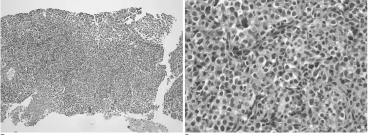
specimen showed diffuse infiltrates of large lymphoid cells (Fig. 1E). The tumor cells had pleomorphic, irregular nuclei and prominent nucleoli (Fig. 1F). Mitoses were easily observed, including some atypical forms.
Immunohistochemical staining demonstrated that the tumor cells were positive for CD3 (Fig. 1G) and they were negative for CD20, CD30 (Ki-1) and CD56. The Ki-67 labeling index was more than 70%. The pathologic diagno- sis was peripheral T-cell lymphoma not otherwise
specified.
We evaluated the patient for the presence of lymphoma involvement of other organs. Bone marrow aspiration and biopsy were both performed. The specimen showed nearly
normal, fully matured myeloids and an adequate number of megakaryocytes without lymphomatous involvement.
18F-fluoro-2-deoxyglucose positron emission tomogra- phy (FDG-PET) showed multiple hypermetabolic masses with photopenic defects (maxSUV 8.9) in both lungs (Fig.
1H). There was no evidence of mediastinal lymph node uptake or extrapulmonary uptake. Finally, we diagnosed the patient as suffering with primary pulmonary peripheral T-cell lymphoma not otherwise specified.
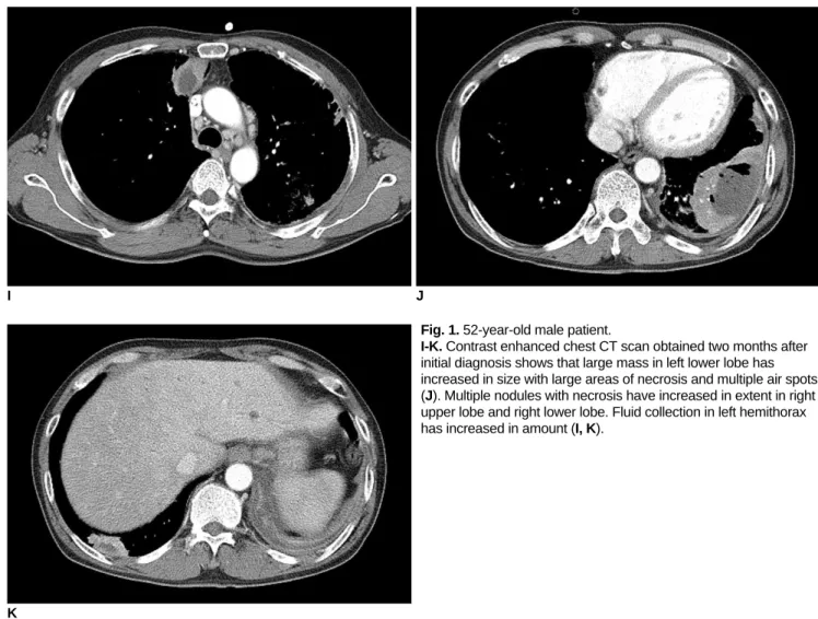
After chemotherapy, the B symptoms that included fever and sweating subsided. The follow-up contrast enhanced chest CT scan obtained two months after the initial diagno- sis showed aggravated T-cell lymphoma involvement in the lungs (Fig. 1I-K).
We observed multifocal irregular enhancing lesions in both parietal cortical areas on the brain CT and MRI obtained four months after the initial diagnosis and this all
A B
E F
Fig. 1. 52-year-old male patient.
E. Percutaneous transthoracic needle biopsy specimen shows diffuse infiltrates of large atypical lymphoid cells (Hematoxylin &
Eosin staining, ×100).
F. Tumor cells are large with pleomorphic, irregular nuclei and prominent nucleoli (Hematoxylin & Eosin staining, ×400).
G. Immunohistochemical staining of tumor cells revealed diffuse and strong positivity for cytoplasmic CD3 (Hematoxylin & Eosin staining, ×200).
H. Staging whole torso PET scan revealed intensely hypermeta- bolic lung mass with central metabolic defects. There was no evidence of mediastinal lymph node uptake or extrapulmonary uptake.
G
H
suggested multiple parenchymal and meningeal metastases.
Sadly, the patient eventually expired from pneumonia during ICU care after multiple episodes of neutropenic fever.
DISCUSSION
Primary pulmonary lymphoma is diagnosed according to
Peripheral T-cell lymphoma (PTCL) comprises a group of rare, aggressive cancers that develop from T-cells that are at different stages of maturity. The World Health
Organization (WHO) has divided the various types of PTCL into two main categories: 1) precursor T-cell neoplasms, which include precursor T-lymphoblastic lymphoma/leukemia; and 2) peripheral T-cell neoplasms, which are subcategorized as predominantly leukemic
I J
Fig. 1. 52-year-old male patient.
I-K. Contrast enhanced chest CT scan obtained two months after initial diagnosis shows that large mass in left lower lobe has increased in size with large areas of necrosis and multiple air spots (J). Multiple nodules with necrosis have increased in extent in right upper lobe and right lower lobe. Fluid collection in left hemithorax has increased in amount (I, K).
K
disease; the lymph nodes, liver and spleen may be involved. Most of the cases present with cough and dyspnea. The most common radiologic finding of PTCLN is generalized lymphadenopathy (3, 5). In patients with disseminated conditions, the imaging features are not distinguishable from those of the other subtypes of lymphoma in the disseminated state.
Our case shows a very unique feature of primary pulmonary PTCLN as the disease presented as multiple lung masses with central necrosis and there was no disease involvement at any other site on the detailed work up. The radiologic appearance of central necrosis in the consolida- tion or mass can also present in such benign conditions as abscess, lung infarction, c-ANCA (anti-neutrophil cytoplas- mic antibody)-associated granulomatous vasculitis and carcinomas. The thick wall and irregular inner margin are more frequently seen in malignant lesions, as was noted in our case. The cavitation in lymphoma is probably due to central ischemic necrosis. This may be due to the rapid tumor growth and it tends to occur in large nodules and masses. A cavity with air-fluid levels may be apparent when there is communication between an adjacent bronchus and a necrotic tumor mass.
There has been a report about extranodal PTCL showing a pattern of photopenic defect on PET scanning, like was seen in our case (6). This photopenic defect correlates well with central necrosis, yet necrosis is an uncommon PET scan finding for lymphoma. Tumor necrosis generally correlates with hypoxia and it is a predictor of a poor prognosis for patients with malignant tumor. Tumor hypoxia plays a major role in tumor progression and resistance to treatment, like what happened in our case (6).
For most of the subtypes of PTCL, the treatment regimen is typically CHOP-based chemotherapy (cyclophosphamide, doxorubicin, vincristine and
prednisone), or EPOCH (etoposide added to CHOP) in the frontline setting (7). Adult T-cell leukemia or lymphoma
has a poor prognosis due to the life-threatening complica- tions that include infections and hypercalcemia (8). The clinical course is usually aggressive, and relapses are more common for T-cell leukemia or lymphoma than for the B- cell lymphomas of a similar histologic grade (9, 10). We report herein on a rare case of PTCL and we present the CT and PET images of this often fatal neoplasm.
References
1. Cadranel J, Wislez M, Antoine M. Primary pulmonary lymphoma. Eur Respir J 2002;20:750-762
2. Cordier JF, Chailleux E, Lauque D, Reynaud-Gaubert M, Dietemann-Molard A, Dalphin JC, et al. Primary pulmonary lymphomas. A clinical study of 70 cases in nonimmunocompro- mised patients. Chest 1993;103:201-208
3. Lee HJ, Im JG, Goo JM, Kim KW, Choi BI, Chang KH, et al.
Peripheral T-cell lymphoma: spectrum of imaging findings with clinical and pathologic features. Radiographics 2003;23:7-26 4. Choi JW, Kim SS, Kim EY, Heran M. Peripheral T-cell
lymphoma in the neck: CT findings of lymph node involvement.
AJNR Am J Neuroradiol 2006;27:1079-1082
5. Okada F, Ando Y, Kondo Y, Matsumoto S, Maeda T, Mori H.
Thoracic CT findings of adult T-cell leukemia or lymphoma. AJR Am J Roentgenol 2004;182:761-767
6. Mavi A, Dhuriraj T, Cermik TF, Urhan M, Wasik M, Basu S, et al. Central photopenic lesions on FDG-PET scan in a patient with peripheral T cell lymphoma. Ann Nucl Med 2008;22:629- 633
7. Fisher RI, Gaynor ER, Dahlberg S, Oken MM, Grogan TM, Mize EM, et al. Comparison of a standard regimen (CHOP) with three intensive chemotherapy regimens for advanced non- Hodgkin’s lymphoma. N Engl J Med 1993;328:1002-1006 8. Senba M, Nakamura T, Kawai K, Senba MI. HTLV-I and acute
pancreatitis. Lancet 1991;337:1489
9. Kim JH, Lee SH, Park J, Kim HY, Lee SI, Park JO, et al.
Primary pulmonary non-Hodgkin’s lymphoma. Jpn J Clin Oncol 2004;34:510-514
10. Melnyk A, Rodriguez A, Pugh WC, Cabannillas F. Evaluation of the Revised European-American Lymphoma classification confirms the clinical relevance of immunophenotype in 560 cases of aggressive non-Hodgkin’s lymphoma. Blood 1997;89:4514-4520