363 https://e-kcj.org
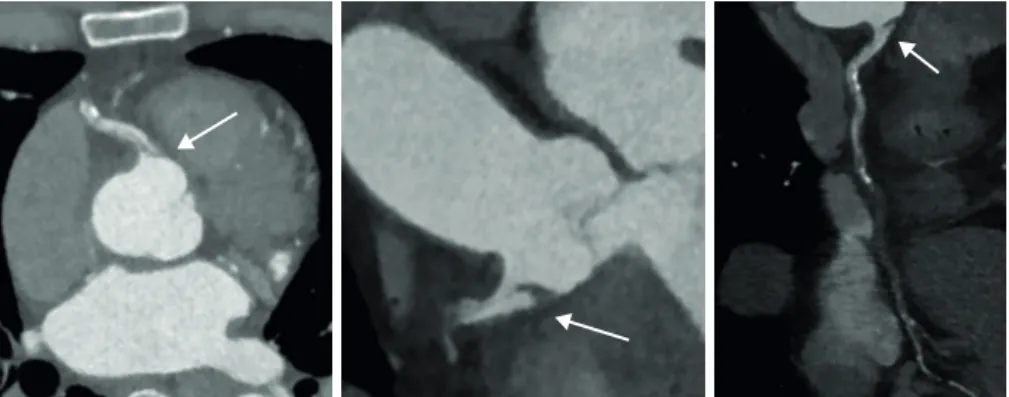
Unfortunately, right coronary artery (RCA) of a 55-year-old man with unstable angina was spirally dissected from ostium into mid-RCA during manipulation of a guiding catheter and extended to coronary sinus of aortic root (Figure 1). A guidewire could not be introduced into the true lumen. Intravascular ultrasonography-guided wiring could not be attempted due to emergent situation. After initiating medical therapy, chest pain and ST-segment elevation were resolved and hemodynamic status was stabilized. Computerized tomography (CT) performed on the third day showed extended dissection of aortic root was localized at sinus of Valsalva without propagation into ascending aorta (Figure 2).
The 7-day follow-up angiography showed collateral flow (Rentrop grade 2) to distal-RCA (Figure 3). Conservative management was maintained for aortic root dissection and delayed percutaneous coronary intervention (PCI) was planned for RCA dissected and totally occluded without surgery.
After 3 months, angiography showed no significant change of RCA dissection with slit-like true lumen (Figure 4A). PCI was strugglingly performed from distal-RCA to ostium with 4 stents (Resolute Onyx™) (Figure 4B and C). Then, effort angina was completely improved.
Korean Circ J. 2019 Apr;49(4):363-365 https://doi.org/10.4070/kcj.2018.0284 pISSN 1738-5520·eISSN 1738-5555
Received: Aug 16, 2018 Revised: Nov 26, 2018 Accepted: Feb 20, 2019 Correspondence to Pyung Chun Oh, MD
Cardiology Division, Department of Internal Medicine, Gil Medical Center, Gachon Cardiovascular Research Institute, Gachon University College of Medicine, 21, Namdong- daero 774 beon-gil, Namdong-gu, Incheon 21565, Korea.
E-mail: [email protected] Copyright © 2019. The Korean Society of Cardiology
This is an Open Access article distributed under the terms of the Creative Commons Attribution Non-Commercial License (https://
creativecommons.org/licenses/by-nc/4.0) which permits unrestricted noncommercial use, distribution, and reproduction in any medium, provided the original work is properly cited.
ORCID iDs Seok In Lee
https://orcid.org/0000-0002-8538-4511 Woong Chol Kang
https://orcid.org/0000-0003-4590-7178 Pyung Chun Oh
https://orcid.org/0000-0002-9955-223X Conflict of Interest
The authors have no financial conflicts of interest.
Seok In Lee , MD
1, Chul-Hyun Park, MD, PhD
1, Woong Chol Kang , MD, PhD
2, and Pyung Chun Oh , MD
21
Department of Thoracic and Cardiovascular Surgery, Gil Medical Center, Gachon Cardiovascular Research Institute, Gachon University College of Medicine, Incheon, Korea
2
Cardiology Division, Department of Internal Medicine, Gil Medical Center, Gachon Cardiovascular Research Institute, Gachon University College of Medicine, Incheon, Korea
Computerized Tomography is an Effective Modality to Evaluate
Iatrogenic Aortocoronary Dissection with Acute Myocardial Infarction
Images in
Cardiovascular Medicine
A B C
Figure 1. (A, B) Baseline coronary angiogram showed significant stenosis at the proximal left anterior descending artery and the mid RCA. (C) Coronary angiogram showed spiral dissection (arrow) of the RCA from ostium to mid portion with total occlusion. The dissection was extended to the right coronary sinus of the aortic root.
RCA = right coronary artery.
Author Contributions
Conceptualization: Lee SI, Kang WC, Oh PC; Data curation: Lee SI, Kang WC; Formal analysis: Lee SI, Park CH, Oh PC; Investigation:
Lee SI, Oh PC; Methodology: Lee SI, Park CH, Kang WC, Oh PC; Resources: Kang WC;
Supervision: Lee SI, Park CH, Oh PC; Writing - original draft: Lee SI; Writing - review & editing:
Park CH, Oh PC.
Iatrogenic dissection of coronary artery is an uncommon but life-threatening complication.
1)When dissection extends to the aortic root and ascending aorta, it should be meticulously evaluated and managed as quickly as possible.
2)3)CT is a helpful and effective tool to evaluate
364 https://e-kcj.org https://doi.org/10.4070/kcj.2018.0284
Cardiac CT for Iatrogenic Aortocoronary Dissection
Figure 2. Cardiac computerized tomogram. A flap formed by the right coronary artery dissection was identified at the ostium (arrow). Extended dissection flap of aortic root was localized at sinus of Valsalva without propagation into ascending aorta.
A B
Figure 3. (A) Follow-up coronary angiogram on the seventh hospital day showed no interval change of the spiral dissection of RCA. However, localized dissection of sinus of Valsalva was decreased. (B) Percutaneous coronary intervention was performed at the proximal left anterior descending artery with a drug-eluting stent (arrow) and left coronary angiogram showed collateral flow (Rentrop grade 2) from the left coronary artery to the distal RCA (dotted arrow).
RCA = right coronary artery.
C
A B
Figure 4. (A) After 3 months, follow-up angiography showed no significant change of the spiral dissection of RCA with slit-like true lumen (arrow). (B) Percutaneous coronary intervention was strugglingly performed from the distal portion (dotted arrow) to the ostium (arrow) of RCA with 4 drug-eluting stents. (C) Completion angiogram showed no visible dissecting flap and no residual stenosis at RCA with thrombolysis in myocardial infarction flow grade 3.
RCA = right coronary artery.
a range of extended dissection flap. If dissection flap is localized and hemodynamic status is stable, conservative management and delayed PCI would be a treatment option for aortocoronary dissection despite acute myocardial infarction.
4)5)REFERENCES
1. Dahdouh Z, Roule V, Lognoné T, et al. Iatrogenic bidirectional dissection of the right coronary artery and the ascending aorta: the worst nightmare for an interventional cardiologist. Korean Circ J 2012;42:504-6.
PUBMED | CROSSREF
2. Dunning DW, Kahn JK, Hawkins ET, O'Neill WW. Iatrogenic coronary artery dissections extending into and involving the aortic root. Catheter Cardiovasc Interv 2000;51:387-93.
PUBMED | CROSSREF
3. Li L, Cao Y. Extensive dissection to the coronary sinus of Valsalva during percutaneous intervention in right coronary artery—a case report and literature review. Clin Med Insights Cardiol 2011;5:41-4.
PUBMED | CROSSREF
4. Celik M, Yuksel UC, Yalcinkaya E, Gokoglan Y, Iyisoy A. Conservative treatment of iatrogenic left main coronary artery dissection: report of two cases. Cardiovasc Diagn Ther 2013;3:244-6.
PUBMED | CROSSREF
5. Shorrock D, Michael TT, Patel V, et al. Frequency and outcomes of aortocoronary dissection during percutaneous coronary intervention of chronic total occlusions: a case series and systematic review of the literature. Catheter Cardiovasc Interv 2014;84:670-5.
PUBMED | CROSSREF