Introduction
Acute myocardial infarction (AMI) is characterized by myo- cardial necrosis secondary to prolonged ischemia. Myocardial damage may lead to systolic and diastolic dysfunction, with the subsequent risk of left ventricular (LV) remodeling, neurohor- monal activation, and vascular dysfunction.1) Ischemic injury after AMI affects not only systolic but also diastolic LV function.
The phenomenon of myocardial stunning has also been shown to have both a systolic and a diastolic component.2) During the ischemic cascade, regional wall motion abnormalities appear early after the reduction in blood flow.3)4) Ischemia-induced dia- stolic dysfunction, or a delay in the onset of regional relaxation,
ORIGINAL ARTICLE J Cardiovasc Ultrasound 2015;23(3):150-157
has been demonstrated in the region perfused by the involved coronary artery in both animal and clinical models.5)6) LV dia- stolic dysfunction is an earlier, more sensitive sign of myocardi- al ischemia and persists longer than the systolic disturbance.7)8) The mechanism of diastolic dysfunction in patients with AMI may involve impaired diastolic relaxation, LV filling, or disten- sibility of the left ventricle, regardless of whether the LV ejection fraction (LVEF) is normal or abnormal.
For several decades, the recovery of LV systolic dysfunction after AMI and its prognostic implications have been the major focus of research. However, few studies have evaluated the im- pact of diastolic dysfunction on future clinical outcomes in pa-
• Received: May 22, 2015 • Revised: September 2, 2015 • Accepted: September 2, 2015
• Address for Correspondence: Kye Hun Kim, Department of Cardiovascular Medicine, Chonnam National University Hospital, 42 Jebong-ro, Dong-gu, Gwangju 61469, Korea Tel: +82-62-220-6266, Fax: +82-62-220-6264, E-mail: [email protected]
• This is an Open Access article distributed under the terms of the Creative Commons Attribution Non-Commercial License (http://creativecommons.org/licenses/by-nc/3.0) which permits unrestricted non-commercial use, distribution, and reproduction in any medium, provided the original work is properly cited.
Impaired Diastolic Recovery after Acute Myocardial Infarction as a Predictor of Adverse Events
Hyun Ju Yoon, MD, Kye Hun Kim, MD, Jong Yoon Kim, MD, Jae Young Cho, MD, Nam Sik Yoon, MD, Hyung Wook Park, MD, Young Joon Hong, MD, Ju Han Kim, MD, Youngkeun Ahn, MD, Myung Ho Jeong, MD, Jeong Gwan Cho, MD, and
Jong Chun Park, MD
Department of Cardiovascular Medicine, Chonnam National University Hospital, Gwangju, Korea
Background: To investigate the impact of left ventricular (LV) diastolic functional recovery on major adverse cardiac events (MACE) 6 months after acute myocardial infarction (AMI) in patients with preserved LV systolic function.
Methods: A total 463 patients with preserved LV systolic function at 6 months after an AMI were divided into two groups based on the extent of diastolic recovery assessed by echocardiography: group I (n = 241) showed improving diastolic function and group II (n = 222) did not. MACE included death, recurrent myocardial infarction, and rehospitalization due to heart failure, and these events were compared with the patients’ characteristics at baseline.
Results: Compared with group I, the patients in group II were older and had a higher prevalence of hypertension and diabetes.
Blood levels of hemoglobin and triglyceride were lower in group II, whereas the levels of N-terminal pro-B-type natriuretic peptide (NT-proBNP) and of high-sensitivity C-reactive protein were higher in this group than in group I. MACE were significantly more frequent in group II than in group I. Age, elevated NT-proBNP, and impaired diastolic recovery were significant independent pre- dictors of MACE.
Conclusion: Despite improvement in LV systolic function, LV diastolic function had not improved in 222 patients (47.9%) by the 6-month follow-up after the index AMI, and impaired diastolic functional recovery was found to be an independent predic- tor of MACE. Evaluation of diastolic function would be a useful way to stratify risk in patients discharged after an index AMI.
KEY WORDS: Myocardial infarction · Diastolic function · Prognosis.
tients with AMI in whom LV systolic function was preserved based on follow-up echocardiography. Therefore, the purpose of our study was to assess the role of diastolic functional recovery in predicting outcomes in such patients.
Methods
Study population
From August 2007 through July 2011, we identified a total of 2800 patients with AMI who underwent successful percu- taneous coronary intervention at our institution. Among these, a total of 463 patients [mean age (± standard deviation) 63.2
± 12.4 yr] who had preserved LV systolic function (defined as an LVEF greater than 50% on follow-up echocardiography at 6 months) were included in the final cohort. The patients were divided into two groups based on the presence or absence of diastolic functional recovery on echocardiography at 6 months after the index AMI: group I showed improvement (n = 241) and group II showed no improvement (n = 222). Improve- ment was defined as an increase of ≥ 1 grade in diastolic func- tion on echocardiography at 6 months, as compared with the initial echocardiographic result. Improvement in the E/e’ ratio (early filling/early diastolic mitral annular velocity ratio, as as- sessed by tissue Doppler imaging) from ≥ 10 on the baseline echocardiogram to < 10 at 6-month follow-up was also con- sidered to represent improvement, regardless of diastolic func- tional grading (see Materials and Methods section). Non-im- provement was defined as no change or worsening of diastolic function of ≥ 1 grade on 6-month follow-up echocardiography, as compared with the initial echocardiogram. The mean ages of groups I and II were 59.4 ± 12.2 and 67.3 ± 11.4 years, re- spectively, and there were 191 men in group I and 122 in group II. Major adverse cardiac events (MACE) included death, recurrent myocardial infarction (MI), and rehospitalization due to heart failure and were determined after the 6-month echocardiographic follow-up was completed. The study pro- tocol was approved by the Institutional Review Board at our institution (2010-05-092).
Definitions of hypertension, diabetes, dyslipidemia, MI, and heart failure
Subjects were considered to have hypertension if their blood pressure was ≥ 140/≥ 90 mm Hg9) or if they were being treated for hypertension. The American Diabetes Association crite- ria10) were used to define diabetes mellitus: fasting plasma glucose levels ≥ 126 mg/dL on two consecutive assessments or if the patient was being treated for diabetes. Dyslipidemia was diagnosed according to the 2004 update of the NCEP guide- lines.11) According to these guidelines, a high level of low-den- sity lipoprotein cholesterol (≥ 160 mg/dL), a low level of high- density lipoprotein cholesterol (≤ 40 mg/dL), and a high level of triglycerides (≥ 150 mg/dL) were included in our assessment.12)
An MI with ST-segment elevation was diagnosed if the pa-
tient had continuous chest pain lasting more than 30 minutes, new ST-segment elevation ≥ 2 mm on at least two contiguous electrocardiographic leads, and a creatine kinase MB fraction (CK-MB) or troponin I (Tn-I) level greater than three times normal values.13) The presence of non-ST-segment elevation MI was diagnosed if the patient had chest pain without new ST-segment elevation and was positive for one of these cardiac biomarkers.14) Infarct-related arteries were identified using a combination of electrocardiographic findings, LV wall motion abnormalities on two-dimensional echocardiography, and cor- onary angiography.
A positive family history meant early cardiovascular disease in immediate relatives. Clinical demographic features were ob- tained from a review of hospital records.
Laboratory tests
Routine laboratory studies were obtained at the time of hos- pital admission. Cardiac enzymes, including CK-MB and Tn- I, were checked serially, and the maximal values were used in our data analysis. Blood samples to assess the serum lipid pro- file and glucose levels were obtained on the morning following admission. High-sensitivity C-reactive protein (hs-CRP) was measured by means of the immunoturbidimetric CRP-Latex (II) high-sensitive assay using an Olympus 5431 autoanalyzer (Olympus America Inc., Melville, NY, USA). Serum N-termi- nal pro-B-type natriuretic peptide (NT-proBNP) was measured using an electrochemiluminescence sandwich immunoassay method with an Elecsys 2010 analyzer (Roche Diagnostics, Mannheim, Germany), having an analytic range that extended from 5 to 35000 pg/mL.15)
Echocardiographic examinations
Two-dimensional, M-mode, and Doppler echocardiographic examinations were performed (Vivid 7, GE, Milwaukee, WI, USA), with the image point at the time of initial admission (day 1 or 2) and at 6 months after the MI. LV volume and EF were measured using Simpson’s formula.16) The LV and left atrial (LA) volume indices were obtained by dividing volume by the body surface area. We used the mean values for three mea- surements taken by two independent observers of the techni- cally-speaking “best” cardiac cycles from each examination. In- tra-observer and inter-observer variabilities with Simpson’s method were 4 ± 5% and 5 ± 4%, respectively (absolute dif- ference divided by mean value of measurement).
The wall motion score index (WMSI) was derived for each patient. The left ventricle was divided according to a 17-segment model.17) For each segment, wall motion was scored from 1 (nor- mal) to 4 (dyskinetic). Recordings were stored digitally and analyzed offline with EchoPAC PC software (GE Vingmed Ultrasound, Horten, Norway). All segments were assessed by means of strain rate imaging and wall motion score (WMS) on two-dimensional echocardiography, according to the Ameri- can Society of Echocardiography guidelines. Because the num-
ber of infarcted segments per patient varied, WMSI was calculat- ed as the average score for the segments studied in each patient.
Doppler echocardiograms were recorded on a strip chart re- corder with a sweep speed of 100 mm/s. Early transmitral veloc- ity (E wave) was measured by pulsed-wave Doppler from the apical four-chamber view, with the sample volume located at the tip of the mitral leaflets. Early diastolic (e’), late diastolic (a’), and systolic (s’) velocities at the septal mitral annulus were ob- tained in this view by tissue Doppler imaging. LA volumes were measured using the multiple-discs method.18) The maxi- mal LA volume was obtained before mitral valve opening and the minimal volume before valve closure. Volume measure- ments were averaged over three to five cycles. Diastolic dysfunc- tion was considered normal, grade 1, grade 2, or grade 3 accord- ing to a multiparametric approach, including the E/A ratio, the E/e’ ratio, and LA volume based on current guidelines.19) An elevated E/e’ ratio and increased LA volume are associated with increased LV filling pressures.
Statistical analysis
The Statistical Package for Social Sciences for Windows, ver- sion 15.0 (SPSS Inc., Chicago, IL, USA) was used for all analy- ses. For each parameter, the mean, median, and standard devi- ation were calculated. Statistical significance between means for different groups was calculated by analysis of variance; statis- tical significance between frequencies was calculated by the chi-square test with Yates’ correction or, if the expected value was less than 5, by Fisher’s exact test. Relative risks (RR) and confidence intervals (CI) were also calculated. A p value of less than 0.05 was required to reject the null hypothesis. Variables found to be significant on the univariate analyses were entered into the multivariate models.
Results
Baseline clinical characteristics
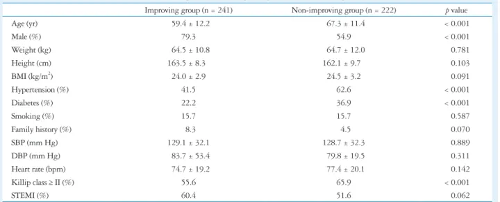
Table 1 summarizes the baseline clinical characteristics for
Table 1. Baseline clinical characteristics of patients in group I (improving) and group II (non-improving)
Improving group (n = 241) Non-improving group (n = 222) p value
Age (yr) 059.4 ± 12.2 067.3 ± 11.4 < 0.001
Male (%) 79.3 54.9 < 0.001
Weight (kg) 064.5 ± 10.8 064.7 ± 12.0 0.781
Height (cm) 163.5 ± 8.30 162.1 ± 9.70 0.103
BMI (kg/m2) 24.0 ± 2.9 24.5 ± 3.2 0.091
Hypertension (%) 41.5 62.6 < 0.001
Diabetes (%) 22.2 36.9 < 0.001
Smoking (%) 15.7 15.7 0.587
Family history (%) 08.3 04.5 0.070
SBP (mm Hg) 129.1 ± 32.1 128.7 ± 32.3 0.889
DBP (mm Hg) 083.7 ± 53.4 079.8 ± 19.5 0.311
Heart rate (bpm) 074.7 ± 19.2 077.4 ± 20.1 0.142
Killip class ≥ II (%) 55.6 65.9 < 0.001
STEMI (%) 60.4 51.6 0.062
BMI: body mass index, SBP: systolic blood pressure, DBP: diastolic blood pressure, STEMI: ST-segment elevation myocardial infarction
Table 2. Prescribed medications in groups I and II
Improving group (n = 241) Non-improving group (n = 222) p value
Aspirin (%) 99.6 100 0.285
Clopidogrel (%) 98.7 098.6 0.618
Cilostazol (%) 58.8 051.6 0.136
Beta blocker (%) 88.1 086.0 0.675
ACEI (%) 55.8 056.4 0.549
ARB (%) 38.5 037.2 0.775
CCB (%) 6.19 010.1 0.091
Diuretics (%) 20.6 033.3 0.007
Nitrate (%) 68.4 073.4 0.322
Inotropic agents (%) 28.9 028.5 0.921
Statin (%) 90.2 089.1 0.701
ACEI: angiotensin-converting enzyme inhibiter, ARB: angiotensin II-receptor blocker, CCB: calcium-channel blocker
groups I and II. Patients in group II were older and included more women than the patients in group I, and hypertension and diabetes were more prevalent in group II. Prescribed medi- cations did not differ between the groups except for the more frequent use of diuretics in group II (Table 2).
Laboratory, echocardiographic, and coronary angiographic findings
Table 3 summarizes the laboratory findings. Initial hemoglo- bin and triglyceride levels were lower, whereas the levels of NT- proBNP and hs-CRP were higher in group II than in group I.
Echocardiographic findings are summarized in Table 4. The parameters used to assess diastolic function, such as LA size, LA volume, e’, E/e’, and E/e’ at 6 months, were significantly
more impaired in group II than in group I. Coronary angio- graphic findings, summarized in Table 5, showed no significant differences between the groups.
Clinical outcomes
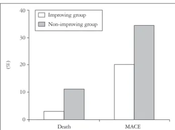
During the follow-up period (mean 910 ± 750 days), MACE occurred in 124 patients (31 deaths, 12 recurrent MIs, and 81 rehospitalizations due to heart failure) and were significantly more frequent in group II than in group I (76 vs. 48, respective- ly) (p = 0.001) (Fig. 1). Significantly more deaths occurred in group II than in group I (24 vs. 7, respectively) (p = 0.001), but the number of patients with recurrent MI (4 in group I vs.
8 in group II) as well as the number requiring rehospitalization (38 in group I vs. 43 in group II) did not differ significantly be-
Table 3. Laboratory findings in groups I (improving) and II (non-improving)
Improving group (n = 241) Non-improving group (n = 222) p value
WBC (mg/dL) 7277 ± 2287 7664 ± 2310 0.067
Hemoglobin (g/dL) 13.3 ± 2.10 12.8 ± 2.40 0.018
Glucose (g/dL) 178.0 ± 940.0 178.1 ± 9000. 0.990
Creatinine (mg/dL) 1.01 ± 0.60 1.03 ± 0.60 0.812
TC (mg/dL) 183.4 ± 38.50 181.9 ± 40.70 0.673
TG (mg/dL) 130.9 ± 99.90 115.4 ± 61.00 0.049
LDL-C (mg/dL) 119.5 ± 39.80 118.9 ± 37.40 0.883
HDL-C (mg/dL) 45.5 ± 12.2 43.9 ± 12.3 0.191
CK-MB (ng/mL) 69.9 ± 16.8 069.9 ± 123.6 0.668
Tn-I (ng/mL) 38.7 ± 41.7 47.1 ± 60.0 0.252
NT-proBNP (pg/mL) 1787 ± 4122 5258 ± 8818 < 0.001
hs-CRP (mg/dL) 1.55 ± 2.30 2.20 ± 3.80 0.038
HbA1c (%) 6.54 ± 1.50 6.64 ± 1.40 0.502
WBC: white blood cell count, TC: total cholesterol, TG: triglyceride, LDL-C: low-density lipoprotein cholesterol, HDL-C: high-density lipoprotein cholesterol, CK-MB: MB fraction of creatinine kinase, Tn-I: troponin I, NT-proBNP: N-terminal pro-B-type natriuretic peptide, hs-CRP: high-sensitivity C-reactive protein, HbA1c: glycosylated hemoglobin
Table 4. Echocardiographic findings in groups I and II
Improving group (n = 241) Non-improving group (n = 222) p value
LVEDV (mL) 160.8 ± 54.60 165.5 ± 57.60 0.160
LVESV (mL) 72.4 ± 35.1 76.8 ± 31.5 0.375
Initial EF (%) 56.3 ± 11.9 54.5 ± 12.1 0.100
Follow-up EF (%) 61.4 ± 6.80 60.1 ± 7.00 0.051
LAD (mm) 36.5 ± 5.30 39.7 ± 5.80 < 0.001
LAVI (mL/m2) 52.8 ± 17.1 63.8 ± 21.2 < 0.001
Initial WMS 21.2 ± 4.90 21.7 ± 5.10 0.253
Follow-up WMS 18.8 ± 3.80 18.8 ± 3.90 0.890
E (cm/sec) 0.64 ± 0.21 0.74 ± 0.26 < 0.001
A (cm/sec) 0.76 ± 0.18 0.83 ± 0.25 0.010
e’ (cm/sec) 0.065 ± 0.020 0.058 ± 0.020 0.013
Initial E/e’ ratio 10.97 ± 4.950 14.37 ± 7.060 < 0.001
Follow-up E/e’ ratio 11.5 ± 1.80 15.4 ± 6.44 < 0.001
LVEDV: left ventricular end-diastolic volume, LVESV: left ventricular end-systolic volume, EF: ejection fraction, LAD: left atrial dimension, LAVI: left atrial volume index, WMS: wall motion score, E: early diastolic mitral inflow velocity, A: late diastolic mitral inflow velocity, DT: deceleration time, e’: early diastolic velocity of mitral septal annulus
tween groups I and IIII, 38. Cumulative survival and MACE- free survival rates were significantly lower in group II than in group I on Kaplan-Meier analysis (Fig. 2).
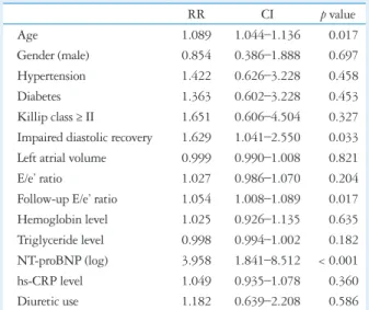
To identify independent predictors of mortality, we con- ducted a multivariate regression analysis using the variables of mortality found to be significant on univariate analysis. As shown in Table 6, age (RR = 1.089, CI = 1.044–1.136, p = 0.017), elevated NT-proBNP (RR = 3.958, CI = 1.841–8.512, p < 0.001), and impaired diastolic functional recovery (RR = 1.629, CI = 1.041–2.550, p = 0.033) were significant indepen- dent predictors of long-term mortality.
Discussion
The two main findings of this study are as follows:
1) Despite the recovery of LV systolic function after the index AMI, LV diastolic function had not improved by the 6-month follow-up in a significant proportion of patients.
2) Impaired diastolic functional recovery was an independent predictor of MACE after AMI.
AMI causes acute derangement of myocardial contraction and relaxation mechanics. In the case of myocardial ischemia,
diastolic dysfunction often precedes systolic dysfunction. To overcome these abnormal changes, the myocardial repair pro- cess is activated immediately after an index AMI, and these dynamic processes are regulated by a number of cytokines and neurohormones to prevent progressive dilation of the left ven- tricle.20)21) Various factors such as interstitial edema, fibrocellu- lar infiltration, and scar formation will also affect LV chamber stiffness,22)23) so many patients with AMI have a period of ad- vanced diastolic dysfunction, as well as systolic dysfunction, af- ter the acute episode.
Although acute diastolic and systolic dysfunction after AMI may improve with the successful restoration of coronary blood flow by means of a percutaneous coronary intervention, some patients show persistence or aggravation of systolic or diastolic functional abnormalities accompanied by chamber dilatation, so-called ventricular remodeling.24) Previous studies have shown that the persistent restrictive filling pattern at discharge may induce severe alterations in LV geometry or function, with a high risk for remodeling at 6 months,25) but the impact on clin- ical outcomes of impaired diastolic recovery during follow-up has been insufficiently evaluated.
In our study, diastolic function either did not improve or de- teriorated in about a half the patients with AMI even though systolic function was found to be improved on echocardiograph- ic follow-up at 6 months, and this persistent diastolic dysfunc- tion was a significant predictor of long-term MACE. There- fore, our results suggest that serial monitoring of diastolic function would be useful in the risk stratification of patients with AMI, regardless of systolic functional status.
Diastolic function as evaluated on Doppler ultrasound pro- vides important prognostic information that supplements in- formation about systolic function.26)27) A previous meta-analy- sis of results from several prospective postinfarction clinical trials revealed that 20% of a large cohort of patients with AMI had restricted LV filling.28) Diastolic function was reversible, es- pecially in patients with grade 3 and grade 4 diastolic dysfunc- tion. However, Doppler variables can change rapidly and are affected by factors such as the patient’s age, heart rate, and LV
Table 5. Coronary angiographic findings in groups I and II
Improving group (n = 241) Non-improving group (n = 222) p value
Infarct-related artery 0.154
LAD (%) 50.4 40.1
RCA (%) 13.3 20.3
LCX (%) 34.2 34.4
Left main (%) 0.42 1.51
Multivessel disease (%) 45.5 55.4 0.461
TIMI flow grade < 3
Pre-PCI (%) 68.8 70.1 0.788
Post-PCI (%) 00.0 00.0 1.182
LAD: left anterior descending coronary artery, RCA: right coronary artery, LCX: left circumflex coronary artery, TIMI: Thrombolysis in Myocardial Infarction, PCI: percutaneous coronary intervention
Fig. 1. Comparison of deaths and major adverse cardiac events (MACE) between group I (improving) and group II (non-improving).
40
30
20
10
0
Death MACE
(%)
Improving group Non-improving group
loading conditions, which explains why evaluating diastolic function with Doppler parameters alone is not sufficient. Be- cause LA volume is less influenced by acute changes and reflects subacute or chronic diastolic function, it may be more reason- able to include LA volume status when assessing diastolic func- tion in patients with AMI.29) In this study, we defined “im- provement” in terms of grade of diastolic dysfunction and a decrease in the E/e’ ratio to less than 10 at 6-month follow-up echocardiography, because patients with AMI reach a relative- ly steady state during this period of time.
The timing of echocardiography after infarction is critical in that LV chamber and myocardial stiffness may fluctuate before and after the repair process gets under way. The LV end-systol-
ic volume index, LVEF, infarct size, and progressive ventricular remodeling are known to be predictors of clinical outcome af- ter AMI. Although, as previously mentioned, risk stratification after AMI is often focused on LV systolic function, it appears that a more complete study of ventricular function that includes an assessment of LV diastolic function would be useful. In ad- dition, significant LV diastolic dysfunction might persist despite an improvement in LV systolic function. Sustained LV diastolic dysfunction may have a significant impact on exercise capacity and long-term outcomes in patients treated with cardiac resyn- chronization therapy.30) The results of our study indicate that patients in whom persistent diastolic dysfunction persists after an AMI have a worse prognosis than patients whose diastolic function has recovered. Moreover, the prognostic information provided by a diastolic assessment is independent of that derived from an evaluation of systolic function alone.
There are several limitations in the present study. First, it is a retrospective observational study. Second, we did not consider the possible effects of risk factor modification that might influ- ence clinical outcomes. Studies with longer follow-up periods are needed to determine such effects. Third, several factors that were not taken into account in the current analysis, such as coex- isting illnesses, could also have affected LV diastolic function.
In conclusion, we found that the failure to recover LV diastolic function is not uncommon after AMI despite LV systolic func- tional recovery, and impaired diastolic function 6 months after an index AMI is a significant predictor of long-term clinical out- comes such as MACE. Therefore, serial monitoring of diastolic function over a period of at least 6 months would be useful in predicting future clinical adverse events in patients with AMI.
• Acknowledgements
The current study was supported by a research grant from the Korean So- ciety of Echocardiography.
Table 6. Predictors of mortality on multivariate analysis
RR CI p value
Age 1.089 1.044–1.136 0.017
Gender (male) 0.854 0.386–1.888 0.697
Hypertension 1.422 0.626–3.228 0.458
Diabetes 1.363 0.602–3.228 0.453
Killip class ≥ II 1.651 0.606–4.504 0.327
Impaired diastolic recovery 1.629 1.041–2.550 0.033 Left atrial volume 0.999 0.990–1.008 0.821
E/e’ ratio 1.027 0.986–1.070 0.204
Follow-up E/e’ ratio 1.054 1.008–1.089 0.017
Hemoglobin level 1.025 0.926–1.135 0.635
Triglyceride level 0.998 0.994–1.002 0.182 NT-proBNP (log) 3.958 1.841–8.512 < 0.001
hs-CRP level 1.049 0.935–1.078 0.360
Diuretic use 1.182 0.639–2.208 0.586
RR: relative risk, CI: confidence interval, E: early diastolic mitral inflow velocity, e’: early diastolic velocity of mitral septal annulus, NT-proBNP: N- terminal pro-B-type natriuretic peptide, hs-CRP: high-sensitivity C-reactive protein
1.0
0.8
0.6
0.4
0.2
0.0
0 500 1000 1500 2000 Days
Log rank p = 0.001 MACE
Cumulative survival
Improving group
Non-improving group
B
1.0
0.9
0.8
0.7
0.6
0.5
0 500 1000 1500 2000 Days
Log rank p = 0.001 Mortality
Cumulative survival
Improving group
Non-improving group
A
Fig. 2. Kaplan-Meier curve analyses of cumulative survival (A) and of major adverse cardiac events (MACE) (B) in group I (improving) and group II (non-improving).
References
1. Møller JE, Pellikka PA, Hillis GS, Oh JK. Prognostic importance of diastolic function and filling pressure in patients with acute myocardial in- farction. Circulation 2006;114:438-44.
2. Nesto RW, Kowalchuk GJ. The ischemic cascade: temporal sequence of hemodynamic, electrocardiographic and symptomatic expressions of ischemia.
Am J Cardiol 1987;59:23C-30C.
3. St John Sutton M, Pfeffer MA, Plappert T, Rouleau JL, Moyé LA, Dagenais GR, Lamas GA, Klein M, Sussex B, Goldman S, Mena- pace FJ, Parker JO, Lewis S, Sestier F, Gordon DF, McEwan P, Ber- nstein V, Braunwald E. Quantitative two-dimensional echocardiographic measurements are major predictors of adverse cardiovascular events after acute myocardial infarction. The protective effects of captopril. Circulation 1994;
89:68-75.
4. Grigioni F, Enriquez-Sarano M, Zehr KJ, Bailey KR, Tajik AJ. Isch- emic mitral regurgitation: long-term outcome and prognostic implications with quantitative Doppler assessment. Circulation 2001;103:1759-64.
5. St John Sutton M, Lee D, Rouleau JL, Goldman S, Plappert T, Braunwald E, Pfeffer MA. Left ventricular remodeling and ventricular arrhythmias after myocardial infarction. Circulation 2003;107:2577-82.
6. Zhang Z, Friedman D, Dione DP, Lin BA, Duncan JS, Sinusas AJ, Sampath S. Assessment of left ventricular 2D flow pathlines during early diastole using spatial modulation of magnetization with polarity alternating velocity encoding: a study in normal volunteers and canine animals with myo- cardial infarction. Magn Reson Med 2013;70:766-75.
7. Cavalcante JL, Marwick TH, Hachamovitch R, Popovic ZB, Ald- weib N, Starling RC, Desai MY, Flamm SD, Kwon DH. Is there a role for diastolic function assessment in era of delayed enhancement cardiac magnetic resonance imaging?: a multimodality imaging study in patients with advanced ischemic cardiomyopathy. Am Heart J 2014;168:220-8.e1.
8. Hillis GS, Møller JE, Pellikka PA, Gersh BJ, Wright RS, Ommen SR, Reeder GS, Oh JK. Noninvasive estimation of left ventricular filling pressure by E/e’ is a powerful predictor of survival after acute myocardial in- farction. J Am Coll Cardiol 2004;43:360-7.
9. Chobanian AV, Bakris GL, Black HR, Cushman WC, Green LA, Izzo JL Jr, Jones DW, Materson BJ, Oparil S, Wright JT Jr, Roccel- la EJ; National Heart, Lung, and Blood Institute Joint National Committee on Prevention, Detection, Evaluation, and Treatment of High Blood Pressure; National High Blood Pressure Education Pro- gram Coordinating Committee. The Seventh Report of the Joint Nation- al Committee on Prevention, Detection, Evaluation, and Treatment of High Blood Pressure: the JNC 7 report. JAMA 2003;289:2560-72.
10. Report of the Expert Committee on the Diagnosis and Classification of Dia- betes Mellitus. Diabetes Care 1997;20:1183-97.
11. Grundy SM, Cleeman JI, Merz CN, Brewer HB Jr, Clark LT, Hun- ninghake DB, Pasternak RC, Smith SC Jr, Stone NJ; National Heart, Lung, and Blood Institute; American College of Cardiology Foundation; American Heart Association. Implications of recent clinical trials for the National Cholesterol Education Program Adult Treatment Panel III guidelines. Circulation 2004;110:227-39.
12. Hatzitolios AI, Athyros VG, Karagiannis A, Savopoulos C, Chara- lambous C, Kyriakidis G, Milidis T, Papathanakis C, Bitli A, Vogiat- sis I, Ntaios G, Katsiki N, Symeonidis A, Tziomalos K, Mikhailidis DP; IMPROVE Collaborative Group. Implementation of strategy for the management of overt dyslipidemia: the IMPROVE-dyslipidemia study. Int J Cardiol 2009;134:322-9.
13. O’Gara PT, Kushner FG, Ascheim DD, Casey DE Jr, Chung MK, de Lemos JA, Ettinger SM, Fang JC, Fesmire FM, Franklin BA, Grang- er CB, Krumholz HM, Linderbaum JA, Morrow DA, Newby LK, Ornato JP, Ou N, Radford MJ, Tamis-Holland JE, Tommaso JE, Tracy CM, Woo YJ, Zhao DX; CF/AHA Task Force. 2013 ACCF/
AHA guideline for the management of ST-elevation myocardial infarction:
executive summary: a report of the American College of Cardiology Founda- tion/American Heart Association Task Force on Practice Guidelines. Circu- lation 2013;127:529-55.
14. Wright RS, Anderson JL, Adams CD, Bridges CR, Casey DE Jr, Et- tinger SM, Fesmire FM, Ganiats TG, Jneid H, Lincoff AM, Peterson ED, Philippides GJ, Theroux P, Wenger NK, Zidar JP, Anderson JL, Adams CD, Antman EM, Bridges CR, Califf RM, Casey DE Jr, Chavey WE 2nd, Fesmire FM, Hochman JS, Levin TN, Lincoff AM, Peterson ED, Theroux P, Wenger NK, Zidar JP; American College of Cardiology Foundation/American Heart Association Task Force on Practice Guidelines. 2011 ACCF/AHA focused update incorporated into the ACC/AHA 2007 Guidelines for the Management of Patients with Unstable Angina/Non-ST-Elevation Myocardial Infarction: a report of the American College of Cardiology Foundation/American Heart Association Task Force on Practice Guidelines developed in collaboration with the Ameri- can Academy of Family Physicians, Society for Cardiovascular Angiography and Interventions, and the Society of Thoracic Surgeons. J Am Coll Cardiol 2011;57:e215-367.
15. Luchner A, Behrens G, Stritzke J, Markus M, Stark K, Peters A, Meisinger C, Leitzmann M, Hense HW, Schunkert H, Heid IM. Long- term pattern of brain natriuretic peptide and N-terminal pro brain natri- uretic peptide and its determinants in the general population: contribution of age, gender, and cardiac and extra-cardiac factors. Eur J Heart Fail 2013;
15:859-67.
16. Simonson JS, Schiller NB. Descent of the base of the left ventricle: an echo- cardiographic index of left ventricular function. J Am Soc Echocardiogr 1989;
2:25-35.
17. Cerqueira MD, Weissman NJ, Dilsizian V, Jacobs AK, Kaul S, Las- key WK, Pennell DJ, Rumberger JA, Ryan T, Verani MS; American Heart Association Writing Group on Myocardial Segmentation and Registration for Cardiac Imaging. Standardized myocardial segmentation and nomenclature for tomographic imaging of the heart. A statement for healthcare professionals from the Cardiac Imaging Committee of the Council on Clinical Cardiology of the American Heart Association. Circulation 2002;105:539-42.
18. Appleton CP, Galloway JM, Gonzalez MS, Gaballa M, Basnight MA.
Estimation of left ventricular filling pressures using two-dimensional and Doppler echocardiography in adult patients with cardiac disease. Additional value of analyzing left atrial size, left atrial ejection fraction and the differ- ence in duration of pulmonary venous and mitral flow velocity at atrial con- traction. J Am Coll Cardiol 1993;22:1972-82.
19. Nagueh SF, Appleton CP, Gillebert TC, Marino PN, Oh JK, Smiseth OA, Waggoner AD, Flachskampf FA, Pellikka PA, Evangelista A.
Recommendations for the evaluation of left ventricular diastolic function by echocardiography. J Am Soc Echocardiogr 2009;22:107-33.
20. French BA, Kramer CM. Mechanisms of Post-Infarct Left Ventricular Remodeling. Drug Discov Today Dis Mech 2007;4:185-96.
21. Eghbali M, Weber KT. Collagen and the myocardium: fibrillar structure, biosynthesis and degradation in relation to hypertrophy and its regression. Mol Cell Biochem 1990;96:1-14.
22. Cleutjens JP, Blankesteijn WM, Daemen MJ, Smits JF. The infarcted myocardium: simply dead tissue, or a lively target for therapeutic interventions.
Cardiovasc Res 1999;44:232-41.
23. Wijns W, Serruys PW, Slager CJ, Grimm J, Krayenbuehl HP, Hu- genholtz PG, Hess OM. Effect of coronary occlusion during percutaneous transluminal angioplasty in humans on left ventricular chamber stiffness and regional diastolic pressure-radius relations. J Am Coll Cardiol 1986;7:
455-63.
24. Yoon HJ, Jeong MH, Bae JH, Kim KH, Ahn Y, Cho JG, Park JC, Kang JC. Dyslipidemia, low left ventricular ejection fraction and high wall motion score index are predictors of progressive left ventricular dilatation af- ter acute myocardial infarction. Korean Circ J 2011;41:124-9.
25. Blomstrand P, Engvall M, Festin K, Lindström T, Länne T, Maret E, Nyström FH, Maret-Ouda J, Östgren CJ, Engvall J. Left ventricular diastolic function, assessed by echocardiography and tissue Doppler imaging, is a strong predictor of cardiovascular events, superior to global left ventricular longitudinal strain, in patients with type 2 diabetes. Eur Heart J Cardiovasc Imaging 2015 Mar 6. pii: jev027. [Epub ahead of print]
26. Fontes-Carvalho R, Sampaio F, Teixeira M, Rocha-Gonçalves F, Gama V, Azevedo A, Leite-Moreira A. Left ventricular diastolic dysfunction and E/E’ ratio as the strongest echocardiographic predictors of reduced exercise capacity after acute myocardial infarction. Clin Cardiol 2015;38:222-9.
27. Møller JE, Søndergaard E, Seward JB, Appleton CP, Egstrup K. Ra- tio of left ventricular peak E-wave velocity to flow propagation velocity as- sessed by color M-mode Doppler echocardiography in first myocardial infarction:
prognostic and clinical implications. J Am Coll Cardiol 2000;35:363-70.
28. Whalley GA, Gamble GD, Doughty RN. Restrictive diastolic filling predicts death after acute myocardial infarction: systemic review and meta- analysis of prospective studies. Heart 2006;92:1588-94.
29. Yoon HJ, Jeong MH, Jeong Y, Kim KH, Song JE, Cho JY, Jang SY, Jeong HC, Lee KH, Park KH, Sim DS, Yoon NS, Hong YJ, Park HW, Kim JH, Ahn Y, Cho JG, Park JC, Kang JC. Progressive dilation of the left atrium and ventricle after acute myocardial infarction is associated with high mortality. Korean Circ J 2013;43:731-8.
30. Fantoni C, Regoli F, Ghanem A, Raffa S, Klersy C, Sorgente A, Fal- etra F, Baravelli M, Inglese L, Salerno-Uriarte JA, Klein HU, Moccet- ti T, Auricchio A. Long-term outcome in diabetic heart failure patients treat- ed with cardiac resynchronization therapy. Eur J Heart Fail 2008;10:298- 307.