Veterinary Science
http://dx.doi.org/10.4142/jvs.2013.14.1.85
Received: 13 Feb. 2012, Revised: 8 Jun. 2012, Accepted: 28 Jun. 2012
Original Article
*Corresponding author: Tel: +82-2-880-1265; Fax: +82-2-880-8662; E-mail: [email protected]
ⓒ 2013 The Korean Society of Veterinary Science.
This is an Open Access article distributed under the terms of the Creative Commons Attribution Non-Commercial License (http://creativecommons.org/licenses/by-nc/3.0) which permits
unrestricted non-commercial use, distribution, and reproduction in any medium, provided the original work is properly cited.
Pulsed tissue Doppler imaging of the left ventricular septal mitral annulus in healthy dogs
Jihye Choi
1, Hyunwook Kim
2, Junghee Yoon
3,*
1
College of Veterinary Medicine, Chonnam National University, Gwangju 500-757, Korea
2
Haemaru Referral Animal Hospital, Seongnam 463-050, Korea
3
College of Veterinary Medicine, Seoul National University, Seoul 151-742, Korea
This study evaluated pulsed TDI variables including the isovolumic time interval and duration of the major wave in a population of large healthy dogs. Longitudinal myocardial motion at the septal mitral annulus was evaluated with pulsed TDI in 45 healthy adult dogs. Maximal myocardial velocities, isovolumic time intervals, and duration of the myocardial waves were measured. The correlation between time intervals and velocity variables was also investigated.
The mean maximal systolic velocity was 6.92 ± 1.78 cm/sec, the mean early diastolic velocity (Em) was 6.58 ± 1.81 cm/sec, the mean late diastolic velocity (Am) was 5.10 ± 2.00 cm/sec, the mean isovolumic contraction time (IVCT) was 53.61 ± 95.13 msec, and the mean isovolumic relaxation time (IVRT) was 26.74 ± 57.24 msec. The early diastolic mitral inflow velocity (E)/Em ratio was 10.94 ± 3.27 while the Em/Am ratio was 1.40 ± 0.40. There was a negative correlation between Am duration and Am amplitude, and a positive correlation between the IVRT and Em/Am ratio (p
< 0.05). The normal LV parameter using pulsed TDI method could be used as the reference range for identifying myocardial dysfunction in dogs.
Keywords: dog, longitudinal, pulsed, septal mitral annulus, tissue Doppler imaging
Introduction
Diagnoses of myocardial disorders have been predominantly based on two-dimensional (2D), M-mode, and Doppler mode, also called conventional echocardiography in human as well as veterinary cardiology. However, the ability to estimate functional alterations of the myocardium during the early stages of cardiac disease with conventional echocardiography
is critically limited. Tissue Doppler imaging (TDI) has emerged as one of the most sensitive and specific methods for noninvasive assessment of systolic and diastolic function in the early stages of cardiac disease [3,12,20,23].
It has been reported in humans that the power of TDI to predict fatalities attributed to cardiac disease by measuring the velocity of the mitral annulus in early diastole is greater than the predictive power of clinical data and conventional echocardiographic measurements [23].
TDI detects low velocity and high amplitude signals from the myocardium as opposed to blood flow signals with high velocity and low amplitude [3]. TDI has three modes including color 2D, color M-mode, and pulsed wave mode.
Color 2D and color M-mode are capable of analyzing multiple segments simultaneously and determining the velocity gradient of the myocardium. However, specialized software is needed for post-processing analysis, so TDI variables cannot be measured in real time. Pulsed TDI can estimate myocardial motion without post-processing procedures, but simultaneous analysis of multiple segments cannot be performed. Nevertheless, pulsed TDI has been used in humans to evaluate systolic and diastolic left ventricle (LV) function in healthy subjects and patients with various cardiac diseases such as dilated cardiomyopathy (DCM), hypertrophic cardiomyopathy (HCM), restrictive cardiomyopathy, constrictive pericarditis, ischemic cardiomyopathy, congestive heart failure (CHF), mitral valve (MV) regurgitation, atrial hypertension, and cardiac amyloidosis [16,23].
Many TDI studies, particularly ones on DCM and HCM, have been performed in dogs and cats since the technique was introduced to veterinary medicine [5-7,18,21].
However, a reliable normal range of TDI variables in dogs,
particularly for the pulsed wave mode, has not yet been
Fig. 1. (A) The correlations between Dur
Sm and myocardium systolic velocity of Sm, (B) between Dur
Sm and fractional shortening (FS), (C) between IVCT and Sm, and (D) between IVCT and FS. No significant correlations were observed.
Table 1. Pulsed tissue Doppler imaging (TDI) of the mitral valve septal annulus at the longitudinal axis in healthy dogs
Unit Number Mean SD 95% confidence interval
Sm Em Am IVCT IVRT Dur
Sm
Dur
Em
Dur
Am
cm/sec cm/sec cm/sec msec msec msec msec msec
45 45 45 23 23 23 23 23
6.92 6.58 5.10 53.61 26.74 175.15 118.15 89.62
1.78 1.81 2.00 95.13 57.24 34.17 31.00 30.11
6.39∼7.45 6.04∼7.13 4.50∼5.70 12.47∼94.75
1.99∼51.49 159.16∼191.14 103.64∼132.66 75.91∼103.33
Sm: systolic velocity of the myocardium, Em: early diastolic velocity of the myocardium, Am: late diastolic velocity of the myocardium,
IVCT: isovolumic contraction time, IVRT: isovolumic relaxation time, Dur
sm: Sm duration, Dur
Em: Em duration, Dur
Am: Am duration.
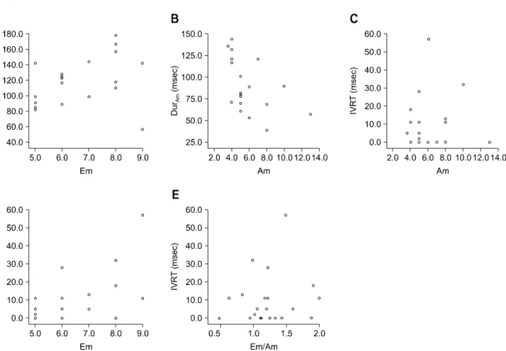
Dur
Em was not correlated with Em, but Dur
Am showed a
negative correlation with Am (p < 0.05). Discussion
TDI examination can be performed in the longitudinal or
radial axis of the myocardium. Depending on the axis, the