unctional mitral regurgitation (MR), which refers to MR in a dilated heart in the presence of anatomi-cally normal mitral leaflets, leads to additional mor-bidity and mortality beyond that expected from ischemic heart disease or left ventricular (LV) systolic dysfunction alone.1Surgical repair or replacement of the mitral valve at the time of surgical ventricular restoration (SVR) is possi-ble; however, the indication and efficacy of a concomitant mitral procedure needs to be clarified. The aim of this study was to report the surgical outcome in patients with or without MR in association with ischemic cardiomyopathy (I-CMP), and to compare the changes in LV function and dimensions after SVR and coronary artery bypass grafting (CABG) with or without concomitant mitral valve proce-dures.
Methods
Patient PopulationFrom July 2001 to June 2006, 49 consecutive patients with I-CMP underwent SVR for post-infarction dyskinetic
LV aneurysm or large non-aneurysmal akinetic LV (42 men, 7 women; mean age 59.8 years). All patients had previous myocardial infarction (MI) because of coronary artery disease, and were in New York Heart Association (NYHA) functional class III or IV; 5 patients required intravenous inotropic drugs preoperatively; 40 patients had multivessel; 1 patient was a surgical emergency and 5 patients were operated urgently because of hemodynamic instability.
To assess the efficacy of concomitant mitral procedures, the patients were divided into 2 groups: the non-MR group included 30 patients who had no or mild preoperative MR, and the MR group included 19 patients with MR grade 3 or more who required a concomitant mitral procedure during SVR. All these patients had functional MR diagnosed by transthoracic echocardiogram (TTE) performed preopera-tively. Functional MR was defined as the presence of sys-tolic restrictive leaflet motion, with or without annulus dilatation and absence of other mitral valve pathology.
Preoperative coronary angiography was performed in all patients, and the diagnosis and assessment of the LV was made by ventriculography. TTE was subsequently used to assess LV dimensions and function, as well as valvular function. Mean preoperative MR grade was 1.4 in the non-MR group, and 3.2 in the non-MR group (p<0.001). Mean pre-operative LV ejection fraction (LVEF) was 25.6% in the non-MR group and 23.6% in MR group (p=NS).
Surgical Technique
The operation was performed under standard cardiac anesthesia using conventional cardiopulmonary bypass
(Received March 26, 2007; revised manuscript received May 26, 2007; accepted June 14, 2007)
Departments of Thoracic and Cardiovascular Surgery, *Anesthesiolo-gy and Pain Medicine, Yonsei University College of Medicine, Seoul, Republic of Korea
Mailing address: Kyung-Jong Yoo, MD, Department of Thoracic and Cardiovascular Surgery, Yonsei University College of Medicine, 134 Shinchon-dong, Seodaemoon-gu, Seoul 120-752, Republic of Korea. E-mail: [email protected]
Changes in Left Ventricular Function and Dimension After
Surgical Ventricular Restoration With or Without
Concomitant Mitral Valve Procedure
Sak Lee, MD; Byung-Chul Chang, MD; Young-Nam Youn, MD;Young-Lan Kwak, MD*; Kyung-Jong Yoo, MD
Background An association of mitral regurgitation (MR) with ischemic cardiomyopathy (I-CMP) increases the risk of heart failure and its surgical management remains controversial.
Methods and Results Between July 2001 and June 2006, a total of 49 patients with I-CMP underwent surgical ventricular restoration (SVR) and coronary revascularization with or without concomitant mitral annuloplasty (MAP). The mean age was 59.8 years, and all patients had New York Heart Association (NYHA) class III or IV heart failure (mean left ventricular ejection fraction (LVEF) = 24.8%). Nineteen patients had MR >grade 3 (MR group). SVR and coronary artery bypass grafting were performed in all patients, and concomitant MAP was performed in the MR group. Echocardiography was performed preoperatively, postoperatively, and at mean of 19 months after operation. Preoperative left ventricular (LV) end-diastolic and end-systolic dimensions, left atrial volume index, and MR grade were statistically significantly increased in the MR group. On the early postopera-tive echocardiogram, mean LVEF was significantly improved, with reduction of LV dimensions, in both groups; however, at follow up, these parameters were more significantly improved in the MR group, but unchanged in non-MR group, reaching almost the same levels as the non-MR group.
Conclusion In patients with I-CMP, MR increases early and late mortality; however, after SVR and concomi-tant MAP, LV function seems to continuously improve with more significant reduction in the LV dimensions than in the non-MR group. (Circ J 2007; 71: 1516 – 1520)
Key Words: Cardiomyopathy; Coronary artery bypass grafting; Mitral valve
(CPB) and moderate systemic hypothermia. Transesophageal echocardiography was used to evaluate preoperative and postoperative LV and mitral valve functions. In all cases, except for 1 patient who had a severely calcified aorta, the aorta was cross-clamped, and myocardial protection was achieved with intermittent cold antegrade and retrograde blood cardioplegia. The aneurysm was incised parallel to the interventricular septum and left anterior descending coronary artery (LAD), and a circular patch plasty was per-formed. In cases of MR, exposure of the mitral valve was performed through a left atriotomy. Mitral annuloplasty (MAP) ring size was determined by standard measurement of the intertrigonal distance and anterior leaflet height, and then stringent downsizing by 2 ring sizes. In 13 of the 19 patients, ring annuloplasty was performed with size 28 mm or less (mean 28.5±1.5 mm, range: 26–32) of rigid or flexi-ble rings.
Follow-up and Statistical Analysis
All preoperative and hospital data of patients undergoing surgical procedures is recorded prospectively in a comput-erized database that has been in use since 1990. Follow-up data are collected through outpatient clinic reports assessed by the surgeon or the referring cardiologist. In the present patients serial TTE studies were performed (before surgery, within 1 month after surgery, and 1–2 years’ follow-up),
allowing for assessment of LV function and dimensions, as well as of the mitral valve. Early mortality was defined as death within 30 days of operation or death before discharge from the hospital.
SPSS software (version 12.0, SPSS Inc, Chicago, IL, USA) was used for the statistical analysis. Data are ex-pressed as mean ± standard deviation or counts and percent-ages when appropriate. Continuous variables were analyzed with 2-tailed Student’s t-test. Statistical significance was defined as p<0.05.
Results
Operative DataPreoperative characteristics of both groups are described in Table 1. In the preoperative echocardiographic data, the LV dimensions and left atrial (LA) volume index were sig-nificantly increased in the MR group, although both groups had similar LVEF. All patients underwent SVR by means of the Dor procedure or surgical anterior ventricular endo-myocardial restoration (SAVER), and 45 patients had con-comitant CABG with a mean of 2.4±1.2 (1–4) grafts. In 38 patients, the left internal thoracic artery was used as a con-duit to the LAD or a diagonal branch.
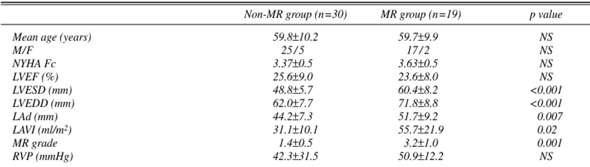
Non-MR group (n=30) MR group (n=19) p value
Mean age (years) 59.8±10.2 59.7±9.9 NS
M/F 25 / 5 17 / 2 NS NYHA Fc 3.37±0.5 3.63±0.5 NS LVEF (%) 25.6±9.0 23.6±8.0 NS LVESD (mm) 48.8±5.7 60.4±8.2 <0.001 LVEDD (mm) 62.0±7.7 71.8±8.8 <0.001 LAd (mm) 44.2±7.3 51.7±9.2 0.007 LAVI (ml/m2) 31.1±10.1 55.7±21.9 0.02 MR grade 1.4±0.5 3.2±1.0 0.001 RVP (mmHg) 42.3±31.5 50.9±12.2 NS
Table 1 Preoperative Data
MR, mitral regurgitation; NYHA Fc, New York Heart Association functional class; LVEF, left ventricular ejection fraction; LVESD, left ventricular end-systolic dimension; LVEDD, left ventricular end-diastolic dimension; LAd, left atrial dimension; LAVI, left atrial volume index; RVP, right ventricular pressure.
Non-MR group (n=30) MR group (n=19) p value Operation time (min) 330.3±62.2 367.0±93.5 NS CPB time (min) 159.9±47.2 187.5±50.0 0.037 ACC time (min) 105.5±37.3 118.6±47.7 NS Ventilator time (days) 2.0±3.5 3.8±6.8 NS
ICU stay (days) 5.3±7.3 6.4±8.4 NS
Hospital stay (days) 15.4±6.2 23.5±16.2 0.033 Inotropic therapy duration (days) 2.5±1.3 4.3±2.8 0.004 Postop. Cx IABP insertion 0 (0%) 1 (5%) NS Arrhythmia 2 (7%) 1 (5%) NS MI 0 (0%) 1 (5%) NS CVA 1 (3%) 0 (0%) NS ARF 2 (7%) 1 (5%) NS LCO 3 (10%) 1 (5%) NS Bleeding 0 (0%) 1 (5%) NS Infection 3 (10%) 1 (5%) NS Respiratory Cx 3 (10%) 1 (5%) NS
Table 2 Operative and Postoperative Data
CPB, cardiopulmonary bypass; ACC, aortic cross-clamping; ICU, intensive care unit; Postop., postoperative; Cx, complication; IABP, intra-aortic balloon pump; MI, myocardial infarction; CVA, cerebrovascular accident; ARF, acute renal failure; LCO, low cardiac output. Other abbreviation see in Table 1.
Early Postoperative Findings
Early mortality was 3.3% (1/30) in the non-MR group, and 10.5% (2/19) in the MR group. The patient in the MR group who had prior renal allograft transplantation and severe total aortic calcification, and who underwent opera-tion without aortic cross-clamping and CPB instituted through the femoral artery and vein, died during surgery from heart failure and intractable ventricular arrhythmia. Perioperative data and early postoperative complications of both groups are described in Table 2. CPB time, duration of hospital stay, and duration of inotropic therapy were sig-nificantly longer in the MR group; however, there were no
statistically significant differences between the 2 groups regarding overall operation time, aortic cross-clamping time, duration of ventilator support, and duration of stay in the intensive care unit.
Follow-up Data
The immediate postoperative and late follow-up echocar-diograms showed similar results for all variables. The mean LVEF increased more significantly in the non-MR group (from 25.6% to 39.2%, p<0.001) than in the MR group (from 23.6% to 31.1%, p=0.025), resulting in a significant difference between the 2 groups (p=0.002) in the early
post-Fig 1. Mean left ventricular ejection fraction (LVEF) significantly increased in both groups early after surgery (p<0.001 in the non-mitral regurgitation (MR) group vs p=0.03 in the MR group). Patients with MR had further improvement in LVEF during the follow-up (f/u) period, but those with MR had a nonsignificant decrease.
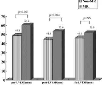
Fig 3. Mean left ventricular end-systolic dimensions (LVESD) de-creased early after surgery in both groups but without statistical sig-nificance, and remained constant afterwards. The difference between the 2 groups, which was significant before surgery, became insignifi-cant after surgery and during follow-up (f/u). MR, mitral regurgitation.
Fig 2. Mean left ventricular end-diastolic dimension (LVEDD) sig-nificantly decreased early after surgery in the mitral regurgitation (MR) group (p=0.03) and remained constant afterward in both groups. The difference between the 2 groups, which was significant before sur-gery, became insignificant after surgery and during follow-up (f/u).
Fig 4. Mean left atrial volume index (LAVI) was significantly in-creased in the mitral regurgitation (MR) group compared with the non-MR group (p=0.019). Whereas the LAVI remained constant in the non-MR group, it gradually decreased in the MR group, but without statistical significance (p=NS), and the difference between the 2 groups, which was significant before and early after surgery, became insignificant during follow-up (f/u).
operative period. However, during follow-up, the LVEF increased gradually in the MR group to 33.1% and slightly decreased to 36.2% in the non-MR group, resulting in simi-lar LVEF in both groups (p=NS) (Fig 1). The preoperative LV end-diastolic dimension (LVEDD) was significantly smaller in the non-MR group, and decreased in both groups early after surgery (from 62.1 mm to 58.7 mm in the non-MR group, p=NS vs from 71.8 mm to 64.4 mm in the non-MR group, p=0.03), with a still significantly smaller LVEDD in the non-MR group. However, during the follow-up period, a greater reduction in the LV dimensions occurred in the MR group, resulting in similar LVEDDs in both groups (p= 0.061) (Fig 2). The LV end-systolic dimension, LA volume index, and MR grades were similar (Figs 3–5). Mean right ventricular pressure (RVP) decreased in both groups in the early postoperative period, and although it gradually increased it remained lower than the preoperative level in both groups (Fig 6).
Mean follow-up duration of surgical survivors (n=46) was 28.6±23.4 (2–113) months (non-MR group: 28.1±18.9 vs MR group: 29.4±30.1 months, p=0.43). There was 1 late death in the non-MR group because of congestive heart failure, and 2 late deaths in the MR group because of ven-tricular arrhythmia in 1 patient and congestive heart failure in the other. No patients in the MR group required reopera-tion for residual MR during follow-up. Follow-up NYHA functional class improved from 3.37±0.50 to 1.52±0.63 in the non-MR group, and from 3.63±0.49 to 1.88±0.96 in the MR group.
Discussion
MR in I-CMP develops in less than 15% of patients after MI and is associated with increased cardiac morbidity and mortality.1 Although it is well accepted that severe MR should be repaired at the time of CABG in cases of ischemic MR, there is less consensus about the indications for surgery in patients with grade 2 or 3 MR, and it is uncertain if concomitant MAP during SVR is beneficial in patients with I-CMP and MR.2–4
Because ischemic MR results from remodeling of the ischemic LV, a standard therapeutic approach to relieve
ischemic MR is ring annuloplasty, which reduces the mitral annular area by bringing the dilated posterior annulus ante-riorly to reduce the anterior – posterior dimension and thus bring the leaflets into apposition.5–7Bolling et al8performed MAP using an undersized circumferential ring to reduce the annulus in severe MR associated with I-CMP, and based on their mid-term results they emphasized that MAP in end-stage cardiomyopathy was a new and effective strategy. However, many recent studies report recurrent or persistent MR early after MAP as a result of progressive remodeling of the dilated failing LV.5,9–11
In most of cases of I-CMP, anterolateral wall involvement is the main cause promoting LV remodeling, and because SVR excludes the anteroseptal wall, LV function improves and prevents further LV remodeling by improving the LV shape and volume restoration, thus giving further benefit to preventing recurrence of MR after MAP. Isomura et al12 reported that aggressive combination of mitral operation in addition to CABG and LV restoration resulted in improve-ment of clinical symptoms and quality of life after surgery, although early and midterm survival was higher in patients without MR. The present study showed similar results, and although hospital mortality was higher in the MR group, all the parameters reflecting LV function and dimensions con-stantly improved in patients who underwent concomitant MAP.
The RVP, which indirectly reflects pulmonary artery pres-sure, was measured in this study and showed somewhat dif-ferent results from other parameters. Although it decreased in the early postoperative period, it then constantly increased in both groups to the preoperative level. The reason for increased pulmonary artery pressure after SVR has been proposed in several reports.13,14Dor13stated that in 25% of cases, postoperative pulmonary artery pressure is increased at 1 year, and tcould be caused by a lack of diastolic compli-ance or a continuous process in LV remodeling, mainly in patients operated many years after MI. Tanoue et al14 reported that although LV contractility and efficiency im-proves after the Dor procedure in patients with a dyskinetic anterior LV aneurysm, the afterload does not change. There-fore, the use of appropriate afterload-reducing therapy may play an important role in the postoperative management of
Fig 5. Mean grade of mitral regurgitation (MR) significantly de-creased in both groups early after surgery (p=0.04 in the non-MR group vs p<0.001 in the MR group), and remained constant in the MR group without recurrence. MR, mitral regurgitation. f/u, follow-up.
Fig 6. Mean right ventricular pressure (RVP) was significantly de-creased in the mitral regurgitation (MR) group after surgery (p=0.01), but gradually increased to the preoperative level during follow-up (f/u) in both groups.
these patients, especially after concomitant MAP.
In the present study, I-CMP with more than moderate MR showed similar good short- and mid-term results, compared with I-CMP with less than mild MR, when MR was repaired during SVR. Furthermore, we think that I-CMP with MR should be repaired even if it is grade II. This study and others have shown that RVP and the LV dimen-sions increase with time because of several factors as mentioned above. It suggests that MR may increase during long-term follow-up even though patients’ clinical symp-toms improve, and that MR and the LA volume index decrease continuously in short- and mid-term follow-up. Therefore, aggressive MR repair and appropriate postop-erative medical therapy will favor good long-term results in patients with I-CMP and MR.
Study Limitations
Despite prospective collection of operative data, this study is retrospective and thus suffers from the potential limitations of any retrospective analysis and the smallness of the 2 unbalanced sample groups. Although the results showed higher hospital mortality in the MR group, because of small sample size and imbalance in the groups it is diffi-cult to say whether this is significant. In addition, the pres-ence of advanced degrees of MR can lead to an overestima-tion of ventricular funcoverestima-tion based on LVEF, which is still the most widely reliable and applicable clinical indicator of LV function. Finally, SVR by definition reduces ventricu-lar volume; therefore using LV volume and LVEF may be an unreliable means of assessing postoperative ventricular function. The method of SVR (Dor or SAVER) may affect the result of the study; however, we did not differentiate these methods because it was determined only by the surgeon’s preference.
Conclusion
In patients with I-CMP, MR increases early and late mor-tality, and preoperative LV function is poorer with larger LV dimensions; however, after SVR and concomitant MAP, LV function continuously improves with more significant re-duction of the LV than in a non-MR group. Because I-CMP is a disease of the ventricle, vessels, and valves, surgical cor-rection of all 3 lesions may improve surgical results as well as long-term survival. However, in patients with none or mild MR, only SVR may result in significant improvement of LV function and dimensions. Long-term follow-up and a
larger randomized prospective trial may be necessary to clarify the indication and feasibility of MAP in such patients.
References
1. Kongsaerepong V, Shiota M, Gillinov AM, Song JM, Fukuda S, McCarthy PM, et al. Echocardiographic predictors of successful versus unsuccessful mitral valve repair in ischemic mitral regurgita-tion. Am J Cardiol 2006; 98: 504 – 508.
2. Grossi EA, Crooke GA, DiGiorgi PL, Schwartz CF, Jorde U, Applebaum RM, et al. Impact of moderate functional mitral insuffi-ciency in patients undergoing surgical revascularization. Circulation 2006; 114(Suppl I): I-573 – I-576.
3. Wong DR, Agnihotri AK, Hung JW, Vlahakes GJ, Akins CW, Hilgenberg AD, et al. Long-term survival after surgical revasculari-zation for moderate ischemic mitral regurgitation. Ann Thorac Surg 2005; 80: 570 – 577.
4. Duarte IG, Shen Y, MacDonald MJ, Jones EL, Craver JM, Guyton RA. Treatment of moderate mitral regurgitation and coronary disease by coronary bypass alone: Late results. Ann Thorac Surg 1999; 68: 426 – 430.
5. Hung J, Papakostas L, Tahta SA, Hardy BG, Bollen BA, Duran CM, et al. Mechanism of recurrent ischemic mitral regurgitation after annuloplasty: Continued LV remodeling as a moving target.
Circula-tion 2004; 110(Suppl II): II-85 – II-90.
6. Smolens IA, Pagani FD, Bolling SF. Mitral valve repair in heart fail-ure. Eur J Heart Fail 2000; 4: 365 – 371.
7. Romano MA, Bolling SF. Update on mitral repair in dilated cardio-myopathy. J Card Surg 2004; 19: 396 – 400.
8. Bolling SF, Deeb GM, Brunsting LA, Bach DS. Early outcome of mitral valve reconstruction in patients with end-stage cardiomyopa-thy. J Thorac Cardiovasc Surg 1995; 109: 676 – 683.
9. Moainie SL, Guy TS, Gorman JH 3rd, Plappert T, Jackson BM, St John-Sutton MG, et al. Infarct restraint attenuates remodeling and reduces chronic ischemic mitral regurgitation after postero-lateral in-farction. Ann Thorac Surg 2002; 74: 444 – 449.
10. Hein S, Arnon E, Kostin S, Schonburg M, Elsasser A, Polyakova V, et al. Progression from compensated hypertrophy to failure in the pressure-overloaded human heart: Structural deterioration and com-pensatory mechanisms. Circulation 2003; 107: 984 – 991.
11. Kuwahara E, Otsuji Y, Iguro Y, Ueno T, Zhu F, Mizukami N, et al. Mechanism of recurrent/persistent ischemic/functional mitral regurgi-tation is the chronic phase after surgical annuloplasty: Importance of augmented posterior leaflet tethering. Circulation 2006; 114(Suppl I): I-529 – I-534.
12. Isomura T, Suma H, Yamaguchi A, Kobashi T, Yuda A. Left ventricu-lar restoration for ischemic cardiomyopathy-comparison of presence and absence of mitral valve procedure. Eur J Cardiothorac Surg 2001; 19: 684 – 689.
13. Dor V. The endoventricular circular patch plasty (“Dor procedure”) in ischemic akinetic dilated ventricles. Heart Fail Rev 2001; 6: 187 – 193.
14. Tanoue Y, Ando H, Fukumura F, Umesue M, Uchida T, Taniquchi K, et al. Ventricular energetics in endoventricular circular patch plasty for dyskinetic anterior left ventricular aneurysm. Ann Thorac Surg 2003; 75: 1205 – 1208.