158 https://e-jcvi.org
A 19-year-old male was referred to us for evaluation of heart murmur. The patient was asymptomatic and in good physical condition. On examination, his blood pressure was 126/80 mmHg, and pulse rate was 80 beats/min. Grade IV/VI systolic murmur was heard at the left sternal border.
His electrocardiogram (ECG) showed a normal sinus rhythm. A two-dimensional transthoracic echocardiogram was performed in the usual manner with a Vivid S5 General Electric
(Milwaukee, WI, USA) ultrasound system and a 3 MHz transducer. The result showed situs solitus and normal arrangement of atria and ventricles with no atrioventricular or ventricular- arterial discordance. Transthoracic echocardiography showed a large perimembranous ventricular septal defect (VSD) partially obstructed by a septal leaflet of the tricuspid valve (Figure 1, Movies 1, 2, 3). A mobile filamentous structure was also seen attached to the ventricular side of the base of the anterior mitral valve leaflet with chordal attachment from anterior papillary muscles. This structure was moving in systole into the left ventricular outflow tract (LVOT) (Figure 1, 2, Movies 1, 4, 5). No relevant subaortic obstruction was demonstrated with a 14.16 mmHg maximum gradient across the LVOT (Figure 3).
J Cardiovasc Imaging. 2020 Apr;28(2):158-160 https://doi.org/10.4250/jcvi.2019.0095 pISSN 2586-7210·eISSN 2586-7296
Images in
Cardiovascular Disease
Received: Oct 13, 2019 Revised: Nov 5, 2019 Accepted: Nov 10, 2019 Address for Correspondence:
Deepak Agrawal, MD
Department of Cardiology, Jaipur Heart Institute, Lal Kothi, Near S. M. S. Stadium, Tonk Road, Jaipur 302015, India.
E-mail: [email protected] Copyright © 2020 Korean Society of Echocardiography
This is an Open Access article distributed under the terms of the Creative Commons Attribution Non-Commercial License (https://
creativecommons.org/licenses/by-nc/4.0/) which permits unrestricted non-commercial use, distribution, and reproduction in any medium, provided the original work is properly cited.
ORCID iDs Ashok Garg
https://orcid.org/0000-0001-9991-0538 Deepak Agrawal
https://orcid.org/0000-0002-2448-2687 G L Sharma
https://orcid.org/0000-0002-3710-4511 Conflict of Interest
The authors have no financial conflicts of interest.
Ashok Garg , MD1, Deepak Agrawal , MD2, and G L Sharma , MD2
1Department of Preventive and Non-Invasive Cardiology, Jaipur Heart Institute, Jaipur, India
2Department of Cardiology, Jaipur Heart Institute, Jaipur, India.
Non-Obstructive Accessory Mitral Valve Tissue in the Left Ventricular Outflow Tract with
PerimembranousVentricular Septal Defect:A Rare Entity
C D
B A
Figure 1. Apical five-chamber view; arrows point to filamentous accessory mitral valve tissue (A), aortic valve (B), perimembranous ventricular septal defect (C), and septal leaflet of the tricuspid valve (D).
The patient underwent elective surgery for a diagnosis of perimembranous VSD and non- obstructive accessory mitral valve tissue. The operation showed successfully closure of the VSD and excision of accessory mitral tissue in the LVOT.
SUPPLEMENTARY MATERIALS
Movie 1
Apical five-chamber view showing filamentous AMVT moving in the LV outflow tract with a perimembranous VSD. AMVT: accessory mitral valve tissue, Ao: aorta, LA: left atrium, LV: left ventricle, RA: right atrium, RV: right ventricle, VSD: ventricular septal defect.
Click here to view Movie 2
Parasternal long-axis view showing colored flow through the VSD. Ao: aorta, LA: left atrium, LV: left ventricle, RV: right ventricle, VSD: ventricular septal defect.
Click here to view
159 https://e-jcvi.org https://doi.org/10.4250/jcvi.2019.0095
Rare Non-Obstructive AMVT in LVOT with VSD
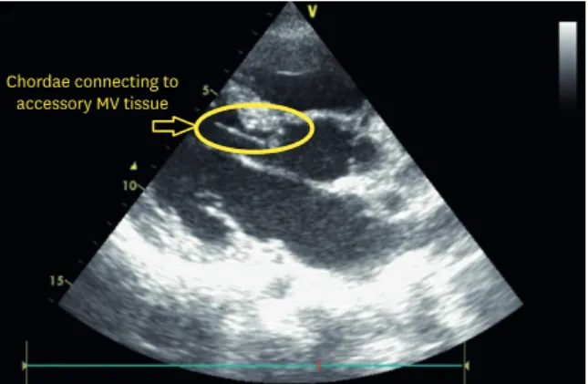
Chordae connecting to accessory MV tissue
Figure 2. Parasternal long-axis view showing accessory mitral valve tissue attached to the chordae.
Figure 3. Apical five-chamber view with continuous width Doppler showing a maximum pressure gradient of 14.16 mmHg through the left ventricular outflow tract.
Movie 3
Apical five-chamber view showing turbulent colored flow through the perimembranous VSD partially obstructed by a septal leaflet of the tricuspid valve. Ao: aorta, LA: left atrium, LV: left ventricle, RA: right atrium, RV: right ventricle, VSD: ventricular septal defect.
Click here to view Movie 4
Parasternal long-axis view showing filamentous AMVT moving in the LV outflow tract. AMVT:
accessory mitral valve tissue, Ao: aorta, LA: left atrium, LV: left ventricle, RV: right ventricle.
Click here to view Movie 5
Parasternal long-axis view showing filamentous accessory mitral valve tissue attached to the chordae.
Click here to view
160 https://e-jcvi.org https://doi.org/10.4250/jcvi.2019.0095
Rare Non-Obstructive AMVT in LVOT with VSD