A Case of Successful MitraClip for Severe Mitral Regurgitation with Left Ventricular Dysfunction in Korea
3
0
0
전체 글
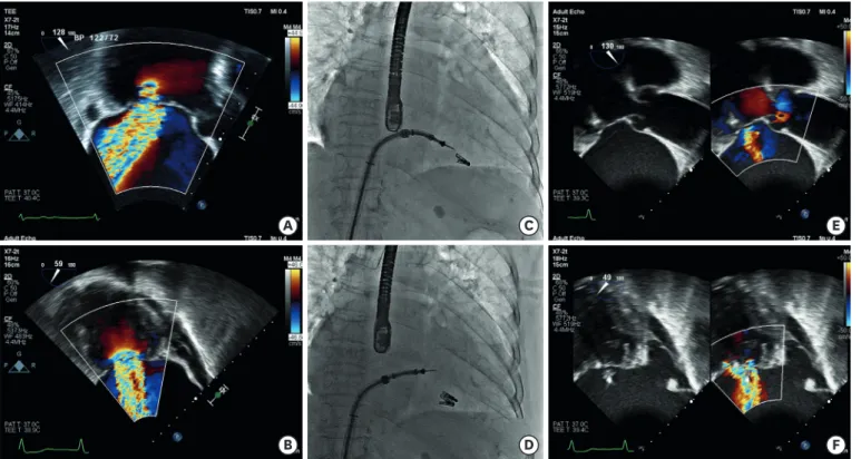
(2) Successful MitraClip in Korea. Copyright © 2020. The Korean Society of Cardiology This is an Open Access article distributed under the terms of the Creative Commons Attribution Non-Commercial License (https:// creativecommons.org/licenses/by-nc/4.0) which permits unrestricted noncommercial use, distribution, and reproduction in any medium, provided the original work is properly cited. ORCID iDs Jung-Joon Cha https://orcid.org/0000-0002-8299-1877 Sung-Jin Hong https://orcid.org/0000-0003-4893-039X Jung-Sun Kim https://orcid.org/0000-0003-2263-3274. MR ERO MR RV. 0.68 cm2 73 mL. degenerative mitral valve with a prolaptic motion of anterior mitral leaflet with tethering (Supplementary Videos 1 and 2). There was incomplete coaptation of A2-P2. This resulted in severe mitral regurgitation (MR) with an effective regurgitant orifice area (EROA) of 0.68 cm2, the regurgitant volume of 73 mL. She had enlarged LV dimensions and LV ejection fraction of 42% due to old myocardial infarction. The Society of Thoracic Surgeons score for mortality was 12.3%. After a Heart Team discussion, the patient was offered the transcatheter mitral-valve repair according to recent European Society of Cardiology guidelines (Class IIb).1)-3) The first grasp was performed centrally with careful attention to grasp both leaflets (A2-P2) adequately. With a single clip, the MR was reduced from IV to III. Thus, the second grasp was performed at the lateral side parallel to the first clip. After the second grasp (Supplementary Videos 3), MR was moderate in severity with EROA of 0.21 cm2 and regurgitant volume of 36 mL (Supplementary Videos 4 and 5). Additionally, TR was reduced from IV to III. She was discharged with improved functional status from New York Heart Association Classification IV to II.. A. B. F. G. C. D. H. I. E. MR ERO MR RV. 0.25 cm2 42 mL. J. Figure 2. (A, B) Transthoracic echocardiographic parasternal long axis and (C,D) apical 3-chamber views showing degenerative mitral valve with severe MR. (E) pre-procedural EROA and regurgitant volume is 0.68 cm2 and 73 mL, respectively. After the MitraClip, (F, G) transthoracic echocardiographic parasternal long axis and (H, I) apical 3-chamber views showing degenerative mitral valve with severe MR. (J) post-procedural EROA and regurgitant volume is 0.21 cm2 and 36 mL, respectively. MR = mitral regurgitation, EROA = effective regurgitant orifice area.. https://e-kcj.org. https://doi.org/10.4070/kcj.2020.0062. 837.
(3) Successful MitraClip in Korea. Jiwon Seo https://orcid.org/0000-0002-7641-3739 Seung Hyun Lee https://orcid.org/0000-0002-0311-6565 Sak Lee https://orcid.org/0000-0001-6130-2342 Chi Young Shim https://orcid.org/0000-0002-6136-0136 Geu-Ru Hong https://orcid.org/0000-0003-4981-3304 Conflict of Interest The authors have no financial conflicts of interest. Author Contributions Conceptualization: Cha JJ, Hong SJ, Kim JS; Data curation: Seo J, Shim CY, Hong GR; Investigation: Hong SJ, Kim JS; Supervision: Kim JS, Hong GR; Visualization: Seo J, Shim CY, Hong GR; Writing - original draft: Cha JJ, Kim JS, Hong GR;Writing - review & editing: Kim JS, Hong GR, Shim CY, Hong SJ, Lee SH, Lee S.. SUPPLEMENTARY MATERIALS Supplementary Video 1 Transthoracic echocardiography demonstrated severe mitral regurgitation with a prolaptic motion of anterior mitral leaflet. Click here to view. Supplementary Video 2 Transesophageal echocardiography showed severe mitral regurgitation due to incomplete coaptation of A2-P2. Click here to view. Supplementary Video 3 Successful MitraClip was performed with two clips. Click here to view. Supplementary Video 4 Transthoracic echocardiography demonstrated mitral regurgitation was reduced from IV to II. Click here to view. Supplementary Video 5 Transesophageal echocardiography showed the clips were well positioned with reduced mitral regurgitation. Click here to view. REFERENCES 1. Baumgartner H, Falk V, Bax JJ, et al. 2017 ESC/EACTS Guidelines for the management of valvular heart disease. Eur Heart J 2017;38:2739-91. PUBMED | CROSSREF. 2. Stone GW, Lindenfeld J, Abraham WT, et al. Transcatheter mitral-valve repair in patients with heart failure. N Engl J Med 2018;379:2307-18. PUBMED | CROSSREF. 3. Choi JY, Hong GR. Transcatheter mitral valve repair: growing evidence regarding it's efficacy and optimal indication. Korean Circ J 2019;49:542-4. PUBMED | CROSSREF. https://e-kcj.org. https://doi.org/10.4070/kcj.2020.0062. 838.
(4)
수치

관련 문서
The Recovery of Left Ventricular Function after Coronary Artery Bypass Grafting in Patients with Severe Ischemic Left Ventricular Dysfunction: Off-pump Versus On-pump.. Jae Hyun
Cardiac Resynchronization Therapy Using a Dual Chamber Pacemaker in Patients with Severe Left Ventricular Dysfunction.. and a Left Bundle
There was an error in the article, “Graft strategy for coronary artery bypass grafting in patients with severe left ventricular dysfunction [1].” After the
We present a case of successful surgical resection of a giant left ventricular (LV) pseudoaneurysm that developed 5 yr after mitral valve replacement (MVR).. A 59-yr-old female
A 75-yr-old man debilitated with New York Heart Association (NYHA) functional class III-IV due to severe left ventricular systolic dysfunction received LVAD implantation as
MR, mitral regurgitation; EF, ejection fraction; ACC/AHA, American College of Cardiology/American Heart Association; TIMI, Thrombolysis In Myocardial Infarction; CABG, coronary
