Phylogenetic Diversity and Community Structure of Microbiome Isolated from Sargassum Horneri off the Jeju Island Coast
Kyung-Mi Moon, So-Hyun Park and Moon-Soo Heo*
Marine applied Microbes and Aquatic Organism Disease Control Lab, Department of Aquatic Biomedical Sciences, School of Marine Biomedical Sciences & Marine and Environmental Research Institute, Jeju-National University, Jeju-si 63243, Korea
Received May 9, 2018 /Revised May 31, 2018 /Accepted June 4, 2018
Recently, Sargassum horneri, the marine weed inhabiting the shoreline, beach, and littoral sea area, has caused serious damage to intensive aquaculture farms particularly those around Jeju Island, South Korea. The purpose of this study was to investigate the diversity of microorganisms in Sargassum horneri and to provide basic data on ecological problems by identifying microbial functions. A total of 88 isolates were identified by 16S rRNA sequencing. Proteobacteria was the dominant phylum ac- counting for 88%, including class α-proteobacteria, six genera, and ten species. The dominating genus, Pseudobacter, accounted for 40% in Pseudorhodobacter, 20% in Paracoccus, and the remaining at 10%
each were Rhizobium, Albirhodobacter, Skermanella, and Novosphingobium. Class β-proteobacteria in- cluded five genera and ten species. Genus Hydrogenophaga accounted for 50%, while genus Azoarcus accounted for 20%, and the remaining Oxalicibacterium, Duganella, and Xenophilus were 10% each.
Class γ-proteobacteria with 13 genera and 57 species, accounted for 74% in phylum Proteobacteria, 23% in Shewanella, 19% in Cobetia, 12% in Pseudomonas, 4% each in Vibrio and Serratia, and 2%
each in Rheinheimera, Raoultella, Pantoea, Acinetobacter, Moraxella, and Psychrobacter genera. In ad- dition, Actinobacteria with two species of Nocardioides genera accounted for 50%, and Bacteroidetes accounted for 33%, with three genera and five species that included Lacihabitans and Mariniflexile.
The remaining Dyadobacter, Cellulophaga, and Ferruginibacter genera each accounted for 11%.
Key words : 16S rDNA, antibacterial activity, Sargassum horneri, systematics, strain diversity
*Corresponding author
*Tel : +82-64-754-3473, Fax : +82-64-756-3493
*E-mail : [email protected]
This is an Open-Access article distributed under the terms of the Creative Commons Attribution Non-Commercial License (http://creativecommons.org/licenses/by-nc/3.0) which permits unrestricted non-commercial use, distribution, and reproduction in any medium, provided the original work is properly cited.
Journal of Life Science 2018 Vol. 28. No. 10. 1179~1185 DOI : https://doi.org/10.5352/JLS.2018.28.10.1179
서 론
괭생이모자반(Sargassum horneri)은 갈조강(Phaeophyceae), 모자반목(Fucale), 모자반과(Sargassaceae)에 속하는 황갈색의 식물체로 얇고 주걱 모양의 잎을 지닌 조간대의 갈조류이다 [29]. 이와 같은 거대 해조류는 미생물의 도움을 받아 성장하 며, 바다 생물의 안전한 공간이 될 수도 있다[7]. 이들은 주로 동쪽 중국연안에서 볼 수 있고[15], 항산화[10], 뼈의 신진대사 [31], 오염방지 활성[3], 효소추출 활성[9], 항알레르기[35], 항 염증[14], 항 응고[1] 및 라디칼 소거제[23]와 같은 다양한 연구 분야에서 적용되고 있다. 그리고 2007년[16] 및 2010년[28]의 동중국해에서 괭생이모자반의 유조(Drifiting seaweed)가 보 고되었는데 이러한 현상은 기생충 제거 및 어류 초기 발달 기간과 생활사에 도움을 준다고 알려져 있다[2, 41]. 그러나 유조 현상이 대량으로 발생할 경우 해안을 뒤덮고 오랜 기간
방치하게 되면 썩은 냄새로 인근 지역 주민의 불편을 사며, 생태계학적으로도 문제를 일으킨다고 알려져 있다[26].
최근 제주 인근 지역의 해변가와 해안 및 연근해 양식장에 2015년을 기준으로 매년 대량의 괭생이모자반의 출현으로 막 대한 피해가 속출되고 있는 추세이다. 국립해양조사에 따르면 매년 유입되고 있는 괭생이모자반은 중국 저우산군도에 서식 하고 있는 종과 99.9% 이상의 상동성을 지녔으며, 이러한 현상 은 중국의 동중국해 바다와 생태계 복원 사업 진행에 따른 피해 현상으로 간주되고 있다. 또한 2009년 Pang 등에 따르면 동중국 연안 인구 밀도 수가 높은 해안 지역에서는 조류의 손실이 커서 복원을 위한 양식 개발에 집중하고 있음을 시사 했다[25].
본 연구에서는 제주 연안에 대량 출현된 괭생이모자반에 서식하고 있는 미생물을 분리하고 동정하였다. 그리고 어류질 병세균과 인체유해세균에 대한 항균 활성을 탐색하고자 하였 다. 이러한 연구는 제주 연안에 재발생 우려가 큰 괭생이모자 반의 유입에 따른 생태학적 기초자료로 활용되는데 도움될 것이라 사료된다.
재료 및 방법
시료 채취
본 실험에 사용 된 괭생이모자반은 2017년 5월 9일 제주도
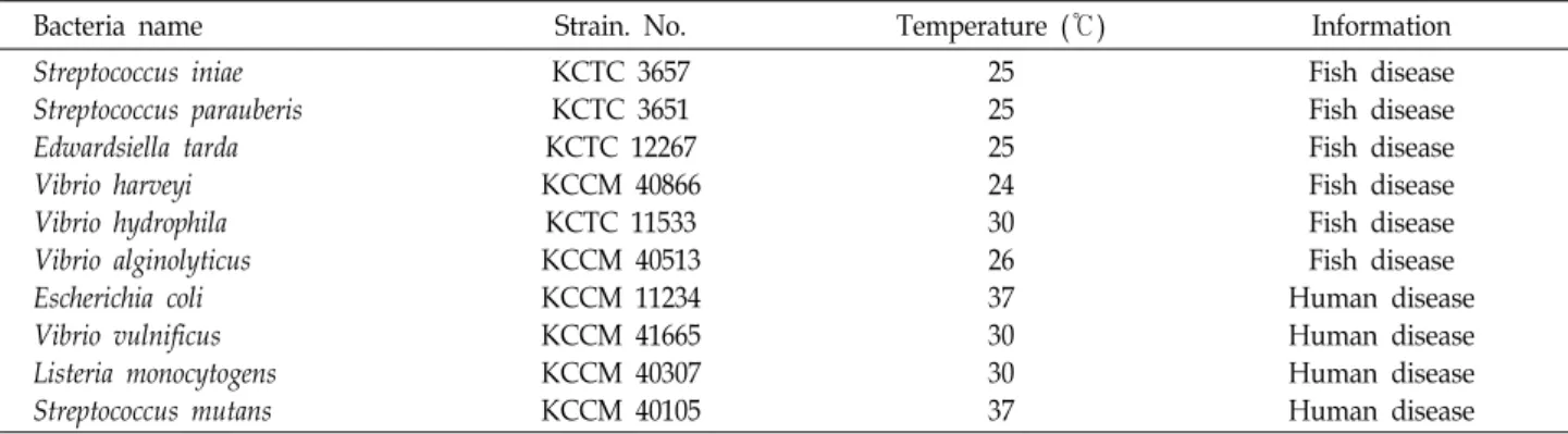
Table 1. List of isolated strains used for antibacterial assay
Bacteria name Strain. No. Temperature (℃) Information
Streptococcus iniae Streptococcus parauberis Edwardsiella tarda Vibrio harveyi Vibrio hydrophila Vibrio alginolyticus Escherichia coli Vibrio vulnificus Listeria monocytogens Streptococcus mutans
KCTC 3657 KCTC 3651 KCTC 12267 KCCM 40866
KCTC 11533 KCCM 40513 KCCM 11234 KCCM 41665 KCCM 40307 KCCM 40105
25 25 25 24 30 26 37 30 30 37
Fish disease Fish disease Fish disease Fish disease Fish disease Fish disease Human disease Human disease Human disease Human disease
제주시 인근 연안(GPS정보; 33.497293, 126.450918)에서 채집
하여 아이스박스에 담아 -4℃를 유지 시키며 운송 시킨 후 실 험에 사용하기 전까지 -80℃에서 보관하였다
균주 분리
괭생이모자반에 서식하는 균주 분리를 위해 멸균 된 해수로 한 번 세척하고 난 뒤 멸균 된 가위로 잘게 잘라내었다. 다음 0.85% 생리식염수 9 ml에 잘려진 괭생이모자반 1 g을 넣어 단계별(10
-1, 10
-2)로 희석시켜 R2A agar (R2A, Difco., USA), 1% R2A agar (1% R2A, Difco., USA), Marine agar (MA, Difco., USA)에 각각 100 μl씩 도말하였다. 그리고 25℃에서 일주일 동안 배양시킨 뒤, 괭생이모자반에서 서식하는 균체를 계수한 후, CFU/g 단위로 환산하였다. 그리고 다 자라난 균 중 단일 콜로니를 순수 분리시켜 위와 동일한 배지에 48시간 동안 재 배양하였다. 분리 된 균주는 25%(v/v) glycerol에 현 탁 시켜 -80℃에서 보관하였다.
분리 균주의 항균 활성 탐색
항균 활성 탐색을 위해 사용 된 어류 및 인체 유해 세균은 미생물자원센터(Korean Collection for Type Cultures, KCTC) 와 한국미생물보존센터(Korean Culture Center of Microor- ganisms, KCCM)에서 분양 받은 것을 사용하였고(Table 1), -80℃에서 보관 되었던 균주를 재활성화시켜 사용하였다. 각 각의 유해세균은 적절한 배지에서 배양되었고(Table 2), 항균 탐색을 위해 괭생이모자반에서 분리 된 균주는 R2A와 1%
R2A, MA에 접종 시켜 25℃에서 48시간 동안 배양하였다. 배 양 된 균주는 멸균된 0.85% 생리식염수에 현탁시켜 14,240x g로 원심 분리하여 상등액과 균체를 각각 나누어 항균 활성에 사용되었다. 그리고 분리 된 상등액 1 ml은 0.45 μm syringe filter (Whatman, UK)를 통해 여과시키고, 균체는 0.85% 생리 식염수 1 ml에 현탁시켜 사용하였다. 여과 된 상등액과 현탁 된 균체는 멸균 된 8 mm paper disc (ADVANTEC, Japan)에 50, 100 μl씩 각각 분주하여 25℃에서 24시간 동안 건조시켰다.
유해균들은 MacFarland turbidity 0.4로 조절한 뒤 Muller Hinton Agar (MHA, Difco, USA)에 도말 하여 배양 온도에
따라 각각 48시간 동안 형성된 억제환을 측정하였다.
16S rRNA 유전자 염기서열 분석
분리 된 균주는 Genomic DNA Extraction Kit (Bioneer, Korea)를 사용하여 DNA를 추출하였다. 추출 된 genomic DNA 1 ul은 27F/1492R universal primer 각각 0.5 μM primer 와 DNA polymerase, dNTPs, reaction buffer가 함유된 20 ul PreMix (Bioneer, Korea)를 사용하여 PCR을 수행하였다. PCR 을 위한 반응 조건은 Initial denaturation (95℃, 2분), Denatu- ration (95℃, 30초), Annealing (55℃, 30초), Extension (72℃, 30초)으로 총 30 cycle로 수행하였고, 마지막으로 다시 한번 Extension (72℃, 5분) 하였다. 증폭 된 PCR product는 Red safe (Intron, USA)를 첨가하여 1% agarose (promegaCo., USA) gel에서 전기 영동하여 확인하였다. 다음 AccuprepTM PCR purification Kit (Bioneer, Korea)를 사용하여 PCR prod- uct에 남은 primers, nucleotides, polymeraes, salts를 제거 및 정제하여 elution buffer (10 mM Tris-Cl, pH 8.5) 30 μl로 elu- tion 하였다. 그리고 PCR product의 염기서열을 분석하기 위 해 (주)솔젠트(Daejeon, Korea)에 의뢰하여 결과를 얻었다. 염 기서열은 EzTaxone (http://eztaxon-e.ezbiocloud.net/)과 NCBI 를 이용하여 표준균주와의 상동성을 조사하였다. ClustalX로 multiple alignment를 수행한 뒤, MEGA 6.0로 계통도 (phylo- genetic tree)를 작성하였다.
결과 및 고찰
분리균주의 항균활성 탐색
괭생이모자반의 균주 다양성 확인을 위해 평판배지에서 colony를 순수 분리하여 배양한 결과, R2A는 5.8×10
-3CFU/
g, 1% R2A에서는 4.1×10
-4, 3.0×10
-3CFU/g, MA는 5.6×10
-4CFU/g로 계수 됐다. 각각의 배지에 따른 세균 수는 큰 차이를 나타내지 않았으나, 1% R2A와 MA에 비해 R2A에서 여러 속 (Genus)을 관찰할 수 있었다.
Alpha-proteobacteria강에 속하는 Novosphingobium속은 나
노 입자의 생합성 기술을 통해 Escherichia coli, Vibrio para-
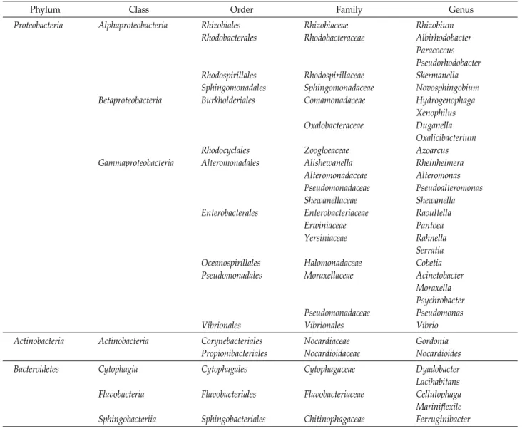
Table 2. Classification of microbial isolates originated from Sargassum horneri
Phylum Class Order Family Genus
Proteobacteria Alphaproteobacteria
Betaproteobacteria
Gammaproteobacteria
Rhizobiales Rhodobacterales
Rhodospirillales Sphingomonadales Burkholderiales
Rhodocyclales Alteromonadales
Enterobacterales
Oceanospirillales Pseudomonadales
Vibrionales
Rhizobiaceae Rhodobacteraceae
Rhodospirillaceae Sphingomonadaceae Comamonadaceae Oxalobacteraceae
Zoogloeaceae Alishewanella Alteromonadaceae Pseudomonadaceae Shewanellaceae Enterobacteriaceae Erwiniaceae Yersiniaceae
Halomonadaceae Moraxellaceae
Pseudomonadaceae Vibrionales
Rhizobium Albirhodobacter Paracoccus Pseudorhodobacter Skermanella Novosphingobium Hydrogenophaga Xenophilus Duganella Oxalicibacterium Azoarcus Rheinheimera Alteromonas Pseudoalteromonas Shewanella Raoultella Pantoea Rahnella Serratia Cobetia Acinetobacter Moraxella Psychrobacter Pseudomonas Vibrio Actinobacteria Actinobacteria Corynebacteriales
Propionibacteriales
Nocardiaceae Nocardioidaceae
Gordonia Nocardioides Bacteroidetes Cytophagia
Flavobacteria Sphingobacteriia
Cytophagales Flavobacteriales Sphingobacteriales
Cytophagaceae Flavobacteriaceae Chitinophagaceae
Dyadobacter Lacihabitans Cellulophaga Mariniflexile Ferruginibacter
haemolyticus 등 다양한 병원체에서 항균 활성을 보였다[36].
또한 Gamma-proteobacteria강 Rheinheimera속도 항균 작용을 갖고 있다고 알려져 있으며[37], Alteromonas속은 polyanionic 와 polysaccharide의 항생제 물질을 합성한다고 알려져 있다 [38]. 그리고 Pseudoalteromonas속은 그람 양성 세균에만 항균 작용을 나타낸다고 보고되었다[39]. 특히 Rahnella속은 근두암 종병이라 불리는 Agrobacterium vitis의 성장을 억제하며[11], 이 외에도 다양한 식물 병원성 세균에 항균 활성을 나타내었다 [45]. Acinetobacter속은 endolysin을 분리하여 약물 내성 세균 에 의한 감염을 효과적으로 방어할 수 있는 lysozyme으로 치 료와 소독제로서의 좋은 역할을 할 수 있다고 보고되었다[40].
그러나 본 연구에서 분리된 31속 88종의 모든 균주는 어류 질병세균 및 인체유해세균에 대한 억제환이 측정되지 않았다 (자료 미제시). 이러한 점을 보아 괭생이모자반에서 서식하고 있는 균주를 이용한 유해 세균의 질병 제어 또는 예방을 위한 유용미생물로서의 이용가능성은 없다 사료된다.
16S rRNA 염기서열의 계통학적 분석
16S ribosomal RNA PCR로 증폭 된 88종은 EZBiocloud의 Identify을 통해 3문(Phylum) 8강(Class) 16목(Order) 22과 (Family) 31속(Genus)으로 분석되었으며(Table 2), 계통수는 Fig. 1에서 보는 바와 같다.
Proteobacteria는 괭생이모자반에서 분리 된 균주 중 88%를 차지한 우점문으로 16S와 23S ribosomal RNA의 특이적 부위 에 따라 alpha-, gamma-, beta-subclasses로 나뉘는데[22], 본 연구에서 alpha-proteobacteria강과 beta-proteobacteria강은 각각 10%, gamma-proteobacteria강은 65%로 우세하게 나타 났다. 다음 Bacteroidetes문은 10%를 차지했고, 그 중 Cyto- phagia강, Flavobacteria강이 각각 4%, Actinobacteria강 2%, Sphingobacteriia강이 1%로 나타났다. 마지막 Actinobacteria 문은 2%를 차지했고 Actinobacteria강이 2%로 나타났다(Fig.
2).
Proteobacteria문 alpha-proteobacteria강은 97-99%의 상동
Fig. 2. Pie-diagram showing various genus of bacterial isolated from Sargassum horneri.
Fig. 1. Neighbour-joining phylogenetic tree determined from the 16S rDNA sequences of bacteria from the Sargassum horneri. GenBank accession numbers given in paren- theses. Boostrap values (>50%) based on 1,000 repli- cations are shown.
성을 나타냈고 6속 10종으로 Pseudorhodobacter속이 40%를 차 지했다. 이 속은 그람 음성균으로 세균 엽록소(Bacteriochlor- ophyll)가 존재하지 않고 non-photosynthetic으로 알려져 있 다[30]. 나머지 Paracoccus속은 20%, Rhizobium, Albirhodobacter,
Skermanella 및 Novosphingobium 속은 각각 10%를 차지했다.Paracoccus속은 그람 음성균으로 다양한 신진대사를 나타내며
[20], 유독물질을 분해한다고 알려져 있고[33], 괭생이모자반 에서 분리된 이 종은 97%의 상동성을 나타내어 추후 신종 실 험이 진행되어야 한다. Rhizobium속은 식물 뿌리에 부착 되는 세균으로 식물 성장 및 molecular mechanism에 관여하며 농 업에선 중요 세균으로 인식되고 있다[6]. Albirhodobacter속은 해안가에서 주로 분리 되며[24], 전세계적으로 문제되고 있는 해조류 증식을 억제되는 것을 방지할 수 있다고 알려져 있어 [18] 추가 연구를 통해 현재 대량으로 발생되고 있는 유조 현상 을 막는데 도움이 될 것이라 사료된다.
beta-proteobacteria강은 97-98%의 상동성을 나타냈고 5속 10종으로 Hydrogenophaga속이 50%, Azoarcus속 20%, 나머지
Oxalicibacterium, Duganella, Xenophilus속은 각각 10%를 차지했다. Hydrogenophaga속은 폐수[4, 34]나 섬유 하수 처리장[8], 쓰레기[42] 등과 같은 오염된 곳에서 쉽게 분리되고 질산염을 질소로 탈질화 시켜준다고 알려져 있다[19]. 그리고 식물 근권 세균으로 알려진 Azoarcus속은 생태계의 식물 성장을 억제할 수 있는 질소를 감소시키고 알코올 탈수소 효소를 조절한다고 알려져 있으며[11, 17], 괭생이모자반에서 분리된 Azoarcus속 은 Azoarcus communis (AF011343) 종과 97%의 상동성을 지녀 추가 신종 실험이 필요한다 여겨진다.
gamma-proteobacteria강의 상동성은 98-100%였고 13속 57
종으로 Proteobacteria문에서 74%로 우점강에 속한다. Shewa-
nella는 23%를 차지한 속으로 저온에서 보관 된 해산물에서
쉽게 찾아볼 수 있고 H
2S를 생성하는 균주로 알려져 있다[32].
또한 Cobetia, Pseudomonas, Pseudoalteromonas속 등은 해양에서 우점속으로 분리된다는 기존 보고 내용과 일치하였다[5].
Actinobacteria문은 1997년 Stackebrandt 등에 의해 처음 분류 되었고[27], Gordonia와 Nocardioides속은 방선균으로 다양한 생리 활성 화합물을 지니고 있어 일부 약물로 개발되기도 했 고 천연 약제로 이용되고 있다[19].
Bacteroidetes문 Cytophagia강은 99-99% 상동성을 나타냈 고 1속 2종으로 우점속인 Lacihabitans속은 담수나 강에서 분리 되어 보고된 바 있으나 추가 연구가 미비한 실정이다[12].
Flavobacteria강은 상동성 98-99%, 1강 2속으로 Cellulophaga,
Mariniflexile로 동정되었고 Sphingobacteriia강은 Ferrugini- bacter속 Ferruginibacter paludis (KC690141)종과 95%의 상동성을 나타내었고, 추후 표준균주와 함께 신종 실험이 진행되어 야 할 것이며, 이들은 주로 갈조류나 해수 및 담수와 같은 곳에 서 발견되었다[13, 21, 43].
이처럼 괭생이모자반에 서식하고 있는 미생물은 주로 식물 이나 해수 또는 담수 심지어 오염된 지역에서도 분리되는 것 을 확인할 수 있었다. 또한, 이 중 분리된 몇몇 균주는 환경적 으로 문젯거리가 되고 있는 부분에 관한 연구가 진행되고 있 었다.
본 연구에서 분리된 균주는 질병 제어나 예방을 위한 이용 가치가 없었지만 추후 추가 연구를 통한 유용미생물로서의 이용가치는 무궁무진하다 사료된다.
감사의 글
본 논문은 정부의 재원으로 한국 연구재단의 지원을 받아 수행된 지역 신 산업 선도인력양성사업과 중견연구사업의 성 과임(2016H1D5A1911152 & 2107R1A2B4005688).
References
1. Athukorala, Y., Lee, K. W., Kim, S. K. and Jeon, Y. J. 2007.
Anticoagulant activity of marine green and brown algae col- lected from Jeju Island in Korea. Bioresour. Technol. 98, 1711-1716.
2. Cho, J. Y., Kwon, E. H., Choi, J. S., Hong, S. Y., Shin, H.
W. and Hong, Y. K. 2001. Antifouling activity of seaweed extracts on the green alga Enteromorpha prolifera and the mussel Mytilus edulis. J. Appl. Phycol. 13, 117-125.
3. Cho, S. H., Myoung, J. G., Kim, J. M. and Lee, J. H. 2001.
Fish fauna associated with drifting seaweed in the coastal area of tongyeong, Korea. T. Am. Fish. Soc. 130, 1190-1202.
4. Contzen, M., Moore, E. R. B., Blumel, S., Stolz, A. and Kampfer, P. 2000. Hydrogenophaga intermedia sp. nov., a 4-aminobenzene-sulfonate degrading organism. Syst. Appl.
Microbiol. 23, 487-493.
5. Dang, H., Zhu, H., Wang, J. and Li, T. 2009. Extracellular hydrolytic enzyme screening of culturable heterotrophic bacteria from deep-sea sediments of the southern okinawa trough. world. J. Microbiol. Biotechnol. 25, 71-79.
6. Ditta, G., Stanfield, S., Corbin, D. and Donald, R. 1980.
Broad host range DNA cloning system for gram-negative bacteria: construction of a gene bank of Rhizobium meliloti.
Proc. Natl. Acad. Sci. USA. 77, 7347-7351.
7. Du, J., Singh, H. and Yi, Y. H. 2017. Biosynthesis of silver nanoparticles by Novosphingobium sp. THG-C3 and their an- timicrobial potential. Artif. Cells. Nanomed. Biotechnol. 45, 211-217.
8. Egan, S., Harder, T., Burke, C., Steinberg, P., Kjelleberg, S.
and Thomas, T. 2012. The seaweed holobiont: understand- ing seaweed-bacteria interactions. Fems. Microbiol. Rev. 37, 462-476.
9. Gan, H. M., Shahir, S., Ibrahim, Z. and Yahya, A. 2011.
Biodegradation of 4-aminobenzenesulfonate by Ralstonia sp.
PBA and Hydrogenophaga sp. PBC isolated from textile wastewater treatment plant. Chemosphere 82, 507-513.
10. Gauthier, M. J. and Flatau, G. N. 1976. Antibacterial activity of marine violet-pigmented Alteromonas with special refer- ence to the production of brominated compounds. Can. J.
Microbial. 22, 1612-1619.
11. Guo, Y. B., Li, J., Chen, F., Wu, W., Wang, J. and Wang, H. 2009. Mutations that disrupt either the pqq or the gdh gene of Rahnella aquatilis abolish the production of an anti- bacterial substance and result in reduced biological control of grapevine crown gall. Applied and environmental. Appl.
Environ. Microbiol. 75, 6792-6803.
12. Gupta, H. K., Gupta, R. D., Singh, A., Chauhan, N. S. and Sharma, R. 2011. Genome sequence of Rheinheimera sp.
strain A13L, isolated from pangong lake, india. J. Bacteriol.
193, 5873-5874.
13. Heo, S. J., Lee, K. W., Song, C. B. and Jeon, Y. J. 2003.
Antioxidant activity of enzymatic extracts from brown seaweeds. Algae 18, 71-81.
14. Heo, S. J., Park, E. J., Lee, K. W. and Jeon, Y. J. 2005. Antiox- idant activities of enzymatic extracts from brown seaweeds.
Bioresource. Technol. 96, 1613-1623.
15. Hurek, T., Reinhold-Hurek, B., Montagu, M. V. and Kellen- berger, E. 1994. Root colonization and systemic spreading of Azoarcus sp. strain BH72 in grasses. J. Bacteriol. 176, 1913- 1923.
16. Joung, Y., Seo, M. A., Kang, H., Kim, H., Ahn, T. S., Cho, J. C. and Joh, K. 2015. Emticicia aquatica sp. nov., a species of the family Cytophagaceae isolated from fresh water. Int.
J. Syst. Evol. Microbiol. 65, 4358-4362.
17. Jung, Y. T. and Yoon, J. H. 2013. Mariniflexile jejuense sp.
nov., isolated from the junction between seawater and a freshwater spring, and emended description of the genus Mariniflexile. Int. J. Syst. Evol. Micr. 63, 1329-1334.
18. Kim, M. E., Jung, Y. C., Jung, I., Lee, H. W., Youn, H. Y.
and Lee, J. S. 2015. Anti-inflammatory effects of ethanolic extract from Sargassum horneri (turner) C. Agardh on lip- opolysaccharide-stimulated macrophage activation via NF-
κB pathway regulation. Immunol. Invest. 44, 137-146.
19. Komatsu, T., Matsunage, D., Mikami, A., Sagawa, T., Boisnier, E., Tatsukawa, K., Ishida, K., Takashige, H. T. and Sugimoto, T. 2008. Abundance of drifting seaweeds in east- ern east china sea. J. Appl. Phycol. 20, 801-809.
20. Komatsu, T., Tatsukawa, K., Filippi, J. B., Sagawa, T., Matsu- naga, D., Mikami, A., Ishida, K., Ajisaka, T., Tanaka, K., Aoki, M., Wang, W. D., Liu, H. F., Zhang, S. D., Zhou, M.
D. and Sugimoto, T. 2007. Distribution of drifting seaweeds in eastern east china sea. J. Mar. Syst. 67, 245-252.
21. Krause, A., Julich, H., Mankar, M. and Reinhold-Hurek, B.
2017. The regulatory network controlling ethanol-induced expression of alcohol dehydrogenase in the endophyte Azoarcus sp. strain BH72. Mol. Plant Microbe Interact. 30, 778-785.
22. Kristyanto, S. and Kim, J. 2016. Isolation of marine algicidal bacteria from surface seawater and sediment samples asso- ciated with harmful algal blooms in Korea. Kor. J. Microbiol.
52, 40-48.
23. Lai, M. J., Lin, N. T., Hu, A., Soo, P. C., Chen, L. K. and Chang, K. C. 2011. Antibacterial activity of Acinetobacter bau- mannii phage ΦAB2 endolysin (LysAB2) against both gram- positive and gram-negative bacteria. Appl. Microbiol.
Biotechnol. 90, 529-539.
24. Lambo, A. J. and Patel, T. R. 2006. Cometabolic Degradation of polychlorinated biphenyls at low temperature by psy- chrotolerant bacterium Hydrogenophaga sp. IA3-A. Curr.
Microbiol. 53, 48-52.
25. Lee, J. H., Kim, Y. S., Choi, T. J., Lee, W. J. and Kim, Y.
T. 2004. Paracoccus haeundaensis sp. nov., a gram-negative, halophilic, astaxanthin-producing bacterium. Int. J. Syst.
Evol. Microbiol. 54, 1699-1702.
26. Lim, J. H., Baek, S. H. and Lee, S. T. 2009. Ferruginibacter alkalilentus gen. nov., sp. nov. and Ferruginibacter lapsinanis sp. nov., novel members of the family ‘Chitinophagaceae’ in the phylum Bacteroidetes, isolated from freshwater sediment.
Int. J. Syst. Evol. Microbiol. 59, 2394-2399.
27. Manz, W., Amann, R., Ludwig, W., Wagner, M. and Schlei- fer, K. H. 1992. Phylogenetic oligodeoxynucleotide probes for the major subclasses of proteobacteria: problems and solutions. Syst. Appl. Microbiol. 4, 593-600.
28. Nakai, M., Kageyama, N., Nakahara, K. and Miki, W. 2006.
Phlorotannins as radical scavengers from the extract of Sargassum ringgoldianum. Mar. Biotechnol. 8, 409-414.
29. Nupur., Vaidya, B., Tanuku, N. R. S. and Pinnaka, A. K.
2013. Albirhodobacter marinus gen. nov., sp. nov., a member of the family Rhodobacteraceae isolated from sea shore water of visakhapatnam, india. Antonie. Van. Leeuwenhoek. 2, 347- 355.
30. Pang, S. J., Ti, F. L., Shan, F., Gao, S. G. and Zhang, Z. H.
2009. Cultivation of the brown alga Sargassum horneri: sexual reproduction and seedling production in tank culture under reduced solar irradiance in ambient temperature. J. Appl.
Phycol. 21, 413-422.
31. Shnit-orland, M., Sivan, A. and Kushmaro, A. 2012. Antibac
terial activity of Pseudoalteromonas in the coral holobiont.
Microbial. Ecol. 64, 851-859.
32. Smetacek, V. and Zingone, A. 2013. Green golden seaweed tides on the rise. Nature 504, 84-88.
33. Stackebrandt, E., Rainey, F. A. and Ward-rainey, N. L. 1997.
Proposal for a new hierarchic classification system, Actinobacteria classis nov. Int. J. Syst. Bacteriol. 47, 479-491.
34. Tifeng, L. S., Pang, S. S. and Li, J. 2017. Analyses of the genetic structure of Sargassum horneri in the yellow sea: im- plications of the temporal and spatial relations among float- ing and benthic populations. J. Appl. Phycol. 1, 1-8.
35. Uchida. T. 1993. The life cycle of Sargassum horneri (phaeo- phyta) in laboratory culture. Eur. J. Phycol. 29, 231-235.
36. Uchino, Y., Hamada, T. and Yokota, A. 2002. Proposal of Pseudorhodobacter ferrugineus gen. nov., comb. Nov., for a non-photosynthetic marine bacterium, Agrobacteruim ferrugi- neum, related to the genus Rhodobacter. J. Gen. Appl. Microbiol.
48, 309-319.
37. Uchiyama, S., Hashizume, M., Hokari, Y., Nakagawa, T., Igarashi, A. and Yamaguchi, M. 2004. Characterization of active component in marine alga Sargassum horneri extract in stimulating bone calcification in vitro. J. Health Sci. 50, 634-639.
38. Voge, B. F., Venkateswaran, K., Masataka, S. and Lone, G.
2005. Identification of Shewanella baltica as the most im- portant H2S-producing species during iced storage of danish marine fish. Appl. Environ. Microbiol. 71, 6689-6697.
39. Xu, G., Zheng, W., Li, Y., Wang, S., Zhang, J. and Yan, Y.
2008. Biodegradation of chlorpyrifos and 3,5,6-trichloro-2- pyridinol by a newly isolated Paracoccus sp. strain TRP. Int.
Biodeter. Biodegr. 62, 51-56.
40. Yoon, J. H., Kang, S. J., Ryu, S. H., Jeon, C. O. and Oh, T. K. 2008. Hydrogenophaga bisanensis sp. nov., isolated from wastewater of a textile dye works. Int. J. Syst. Evol. Micr.
58, 393-397.
41. Yoshioka, H., Ishida, M., Nishi, K., Oda, H., Toyohara, H.
and Sugahara, T. 2014. Studies on anti-allergic activity of Sargassum horneri extract. J. Funct. Foods 10, 154-160.
42. Kokita, T. and Omori, M. 1998. Early life history traits of the gold-eye rockfish, sebastes thompsoni, in relation to suc- cessful utilization of drifting seaweed. Mar. Biol. 132, 579- 589.
43. Hoven, R. N. V. and Santini, J. M. 2004. Arsenite oxidation by the heterotroph Hydrogenophaga sp. str. NT-14: the arsen- ite oxidase and its physiological electron acceptor. Biochim.
Biophys. Acta. 1656, 148-155.
44. Barbeyron, T., L’Haridon, S., Michel, G. and Czjzekm, M.
2008. Mariniflexile fucanivorans sp. nov., a marine member of the Flavobacteriaceae that degrades sulphated fucans from brown algae. Int. J. Syst. Evol. Microbiol. 58, 2107-2113.
45. Chen, F., Li, J. Y., Guo, Y. B., Wang, J. H. and Wang, H.
M. 2009. Biological control of grapevine crown gall: purifica- tion and partial characterization of an antibacterial sub- stance produced by Rahnella aqquatilis strain HX2. Eur. J.
Plant. Pathol. 124, 427-437.