Copyright ⓒ 2012, The Microbiological Society of Korea
제주도에서 채집한 해양 해면, Asteropus simplex의 공생세균에 관한 계통학적 분석
정인혜․박진숙*
한남대학교 생명시스템과학과
Phylogenetic Analysis of Bacterial Diversity in the Marine Sponge, Asteropus simplex, Collected from Jeju Island
In-Hye Jeong and Jin-Sook Park*
Department of Biological Science and Biotechnology, Hannam University, Daejeon 305-811, Republic of Korea (Received December 15, 2012 / Accepted December 26, 2012)
Culture-dependent RFLP and culture-independent DGGE were employed to investigate the bacterial community associated with the marine sponge Asteropus simplex collected from Jeju Island. A total of 120 bacterial strains associated with the sponge were cultivated using modified Zobell and MA media. PCR amplicons of the 16S rDNA from the bacterial strains were digested with the restriction enzymes HaeIII and MspI, and then assigned into different groups according to their restriction patterns. The 16S rDNA sequences derived from RFLP patterns showed more than 94% similarities compared with known bacterial species, and the isolates belonged to five phyla, Alphaproteobacteria, Gammaproteobacteria Actinobacteria, Bacteroidetes, and Firmicutes, of which Gammaproteobacteria was dominant. DGGE fingerprinting of 16S rDNAs amplified from the sponge-derived total gDNA showed 12 DGGE bands, and their sequences showed more than 90% similarities compared with available sequences. The sequences derived from DGGE bands revealed high similarity with the uncultured bacterial clones.
DGGE revealed that bacterial community consisted of seven phyla, including Alphaproteobacteria, Betaproteobacteria, Gammaproteobacteria, Deltaproteobacteria, Actinobacteira, Chloroflexi, and Nitrospira.
Alphaproteobacteria, Gammaproteobacteria, and Actinobacteria were commonly found in bacteria associated with A. simplex by both RFLP and DGGE methods, however, overall bacterial community in the sponge differed depending on the analysis methods. Sponge showed more various bacterial community structures in culture- independent method than in culture-dependent method.
Keywords: Asteropus simplex, bacterial diversity, DGGE, RFLP, sponge
*For correspondence. E-mail: [email protected]; Tel.: +82-42-629-8771;
Fax: +82-42-629-8769
해면은 열대에서 극지에 이르기까지 넓은 분포를 나타내는 해양환경의 주된 구성 생물로써, 높은 생물량과 다양성 그리고 저서계와 표영계 서식지를 연결하는 능력으로 인하여 생태학적 으로 매우 중요하다(Webster et al., 2011). 또한, 여과섭식 동물 로써 주변의 해수 혹은 담수의 여과를 통하여 먹이원을 공급받 으며, 이때 미생물과의 공생을 통하여 영양원을 얻거나 골편의 안정화, 노폐물의 가공을 수행하고 혹은 미생물 감염에 저항하 기 위하여 많은 종류의 이차대사산물을 생산하는 것으로 알려져 있다(Thomas et al., 2010). 해면으로부터 생산되는 많은 독특한 천연물들이 해면에 공생하는 미생물에 의해 생산되는 것으로 알 려지면서 해면 공생미생물은 미생물학자들 뿐만 아니라 천연물
연구자들에게도 각광을 받고 있다. 최근 수년에 걸쳐 해면 공생 미생물에 관한 연구는 급격히 증가되고 있다(Taylor et al., 2011). 최근의 연구에 따르면, 해면은 제약산업에서 신약을 위한 잠재적인 원천으로서 2차 대사물질 분리 및 생명공학적인 응용 의 관점에서도 많은 연구가 이루어지고 있다(Bergman et al., 2011).
Demospongiae 강(class)에 속하는 A. simplex는 우리나라 제 주도를 비롯하여 세계의 해양 환경에 널리 분포하는 해면으로, 이 해면에 의해 생산되는 생리활성물질로 asteropterin이 있으며 이물질은 암의 진행에 관련하는 리소솜 단백질 분해효소 (lysosomal protein-degrading enzyme)인 cathepsin B에 대하여 억제 작용을 갖는 것으로 보고되어 있다(Murayama et al., 2008). A. simplex의 광범위한 서식지 분포를 고려할 때 미생물 다양성에 관한 연구는 천연물 생산 등을 위한 기초 연구로 중요
성을 갖는다. 그러나 이 해면에 관한 미생물 군집에 관한 보고는 거의 이루어진 바 없으며 미생물 군집 구성에 관한 연구는 이제 시작 단계이다.
미생물 군집구조의 분석에는 배양(culture-dependent)과 비배 양(culture-independent)에 근거하는 분자적 방법들이 많이 이용 되고 있으며, 공생 세균 군집 구조를 이해하는 데 매우 중요하다.
배양에 근거한 PCR-RFLP (polymerase chain reaction- restriction fragment length polymorphism)는 증폭된 16S rDNA에 특정 유 전자 부위를 인식하는 제한효소로 처리하여 절단된 유전자의 패 턴을 분석하는 방법으로 신속하며 간편하여 배양 가능한 해면 공생세균의 다양성을 용이하게 파악할 수 있는 기술이다(Botstein et al., 1980). 비 배양에 근거한 PCR-DGGE (polymerase chain reaction-denaturing gradient gel electrophoresis)는 특정 유전자 부위를 PCR로 증폭하여 젤 상에서 전기영동에 의해 확인하는 방법으로 하나 이상의 염기서열이 달라도 서로 다른 밴드로 구 분되므로 자연환경의 미생물을 배양하지 않고 군집 구조를 쉽게 파악할 수 있는 기술이다(Fischer and Lerman, 1983). 세균의 군 집은 매우 다양하며 풍부한 것으로 알려져 있지만 배양 가능한 공생 세균은 오직 1%에 불과한 것으로 알려져 있어(Hentschel et al., 2003), 비배양 방법에 의한 해면 공생미생물의 다양성 및 군집구조의 연구는 중요하다. 비배양에 의한 미생물 군집 분석 을 통해 많은 해면 특이적인 새로운 속(genus)과 종(species)의 미생물 검출이 가능하였다(Jackson et al., 2012).
본 연구는 제주도 무릉아치에서 해양 해면 A. simplex를 채집 하여 배양에 의한 PCR-RFLP 방법과 비배양에 의한 16S rDNA-DGGE에 의해 해면 공생세균의 다양성을 조사하고 결과 를 비교하였다.
재료 및 방법
해면시료의 채집
해양 해면 A. simplex에 공생하는 세균의 다양성을 조사하기 위하여 제주도 무릉아치에서 2011년 8월 22일에 스쿠버 다이빙 을 이용하여 약 15 m 깊이의 바다에서 해면을 채집하였다. 채집 한 해면은 멸균된 인공해수로 3회 세척 후 4℃에서 운반 후 24시 간 이내에 실험에 사용하였다.
해면 공생세균의 배양 및 분리
해면 조각을 멸균된 인공해수로 3회 세척 후, 해면의 안쪽을 1 cm3 크기로 잘라 인공해수 3 ml 넣어 균질화시킨 다음 10분간 초음파 처리하였다. 각각 10-4까지 순차 희석한 후 변형된 ZoBell (ZB) 배지(peptone 5 g, yeast extract 1 g, FePO44H2O 0.01 g, NaCl 23.6 g, MgSO47H2O 5.94 g, MgCl26H2O 4.53 g, CaCl2
1.3 g, KCl 0.64 g, agar 15 g, DW 1 L, pH 7.2)와 MA 배지 (marine agar 2216, Difco, USA)에 100 μl씩 도말하여 25℃에서 7일간 배양하였다. 집락의 형태적 특징에 따라 비율 별로, 변형 된 ZB 배지에서 79개, MA 배지에서 41개, 총 120개의 균주를 선별하였다. 선별된 균주들의 순수 분리를 위하여 동일한 배지 에서 계대 배양하여 단일 집락을 얻었다. 이 균주들을 16S
rDNA의 RFLP 분석에 이용하였다.
DNA 추출 및 16S rDNA의 PCR 증폭
염색체 DNA는 분리된 세균 균주들의 각 집락으로부터 gDNA Extraction kit (Solgent, Korea)를 사용하여 추출하였으며 분리 된 DNA는 PCR 반응의 주형으로 사용하였다. 16S rDNA의 증 폭에는 27f (5′-AGA GTT TGA TCC TGG CTC AG-3′)와 1492r (5′-TAC GGY TAC CTT GTT ACG AC-3′)의 프라이머 쌍을 사용하였다. PCR 반응 혼합물의 조성은 5 μl 10× reaction buffer, 1 μl의 10 mM dNTPs, 5 unit/μl Taq polymerase (Solgent), 각각의 프라이머 10 pmol, 그리고 100 ng의 시료 DNA를 1 μl 첨가하여 최종 부피 50 μl이 되도록 하여 PCR 반응을 수행하였 다. GeneAmp PCR system 2700 thermal cycler (Applied Biosystems, Version 2.0, USA)를 이용하여 94℃에서 3분간 초 기 변성시킨 후, 94℃에서 40초간 변성, 55℃에서 40초간 냉각, 72℃에서 1분간 신장, 이 과정을 30회 반복 수행한 후 최종적으 로 72℃에서 10분간 신장시켰다. 증폭된 DNA의 확인을 위해서 PCR 반응액 3 μl를 취하여 1% 아가로스젤(Biopure, Canada)을 이용하여 Mupid-ex (ADVANCE, Japan)로 100 V, 25분간 1×
TAE 완충용액(40 mM Tris-acetate, 1 mM EDTA, pH 8.0)에서 전기영동 하였다. 전기영동 후, EtBr (ethidium bromide, 50 ng/ml)에 10분간 염색하여 Gel Logic 200 (Kodak, USA)을 이 용하여 UV하에서 약 1.5 kb 단편을 확인하였다.
RFLP 분석
PCR 산물의 RFLP 분석을 위해 2종의 제한효소 HaeIII (Takara Bio Inc., Japan)와 MspI (Takara Bio Inc., Japan)을 사 용하였다. 증폭된 1.5 kb의 DNA 단편에 각각의 제한 효소를 첨 가하여 37℃에서 4시간 반응시켰다. 반응물은 3% 아가로스젤을 사용하여 1× TAE 완충용액으로 100 V, 30분간 전기영동 한 후 EtBr로 염색하여 Gel Logic 200을 이용하여 UV하에서 관찰하여 각 균주의 밴드유형을 확인하였다. 16S rDNA-RFLP fingerprinting 타입에 따라 각각의 RFLP 타입 별로 1–2개의 분리 균주들을 선 별하여 총 37균주의 부분 염기 서열(500 bp 이상)을 분석하였다.
증폭된 PCR 산물은 MGTM PCR DNA Purification kit (Macrogen, Korea)를 이용하여 정제하였으며, 27f 프라이머를 이용한 염기 서열 분석(Macrogen)을 의뢰하였다.
Total genomic DNA 추출
채집된 해면은 1 cm3 크기로 잘라 멸균된 인공해수로 세척 후 -70℃에서 24시간 예비동결한 다음 동결건조기에서 -50℃, 0.033 M bar 기압으로 24시간 건조하였다. 동결 건조된 해면은 멸균된 막자 사발에 넣고 분쇄하였으며 G-spin™ Genomic DNA Extraction kit (Intron, Korea)를 이용하여 추출한 후 DGGE를 위한 PCR 반응의 주형으로 사용하였다.
16S rDNA의 DGGE-PCR 증폭
GC clamp가 추가된 341f (5′-CGC CCG CCG CGC CCC GCG CCC GGC CCG CCG CCC CCG CCC GCC TAC GGG
RFLP type a
Representative strain
Phylum
Bacterial species
% Sequence similarity
Number of
strains Percentage c
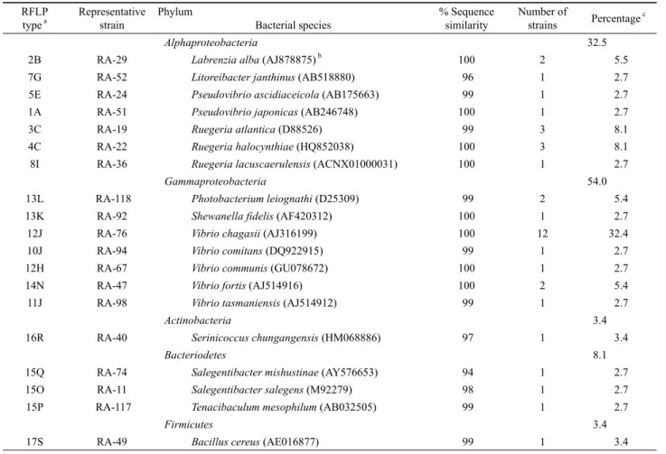
Alphaproteobacteria 32.5
2B RA-29 Labrenzia alba (AJ878875) b 100 2 5.5
7G RA-52 Litoreibacter janthinus (AB518880) 96 1 2.7
5E RA-24 Pseudovibrio ascidiaceicola (AB175663) 99 1 2.7
1A RA-51 Pseudovibrio japonicas (AB246748) 100 1 2.7
3C RA-19 Ruegeria atlantica (D88526) 99 3 8.1
4C RA-22 Ruegeria halocynthiae (HQ852038) 100 3 8.1
8I RA-36 Ruegeria lacuscaerulensis (ACNX01000031) 100 1 2.7
Gammaproteobacteria 54.0
13L RA-118 Photobacterium leiognathi (D25309) 99 2 5.4
13K RA-92 Shewanella fidelis (AF420312) 100 1 2.7
12J RA-76 Vibrio chagasii (AJ316199) 100 12 32.4
10J RA-94 Vibrio comitans (DQ922915) 99 1 2.7
12H RA-67 Vibrio communis (GU078672) 100 1 2.7
14N RA-47 Vibrio fortis (AJ514916) 100 2 5.4
11J RA-98 Vibrio tasmaniensis (AJ514912) 99 1 2.7
Actinobacteria 3.4
16R RA-40 Serinicoccus chungangensis (HM068886) 97 1 3.4
Bacteriodetes 8.1
15Q RA-74 Salegentibacter mishustinae (AY576653) 94 1 2.7
15O RA-11 Salegentibacter salegens (M92279) 98 1 2.7
15P RA-117 Tenacibaculum mesophilum (AB032505) 99 1 2.7
Firmicutes 3.4
17S RA-49 Bacillus cereus (AE016877) 99 1 3.4
a Each combination of number and alphabet represents RFLP types of the restriction enzymes: HaeIII and MspI
b Number in parenthesis presents accession number
c The number represents percentage of phylum and species in total bacterial community
Table 1. RFLP fingerprinting types and relative abundance of major groups in cultivable bacterial community of the marine sponge, A. simplex (RA)
AGG CAG CAG-3′)와 518r (5′-ATT ACC GCG GCT GCT GG-3′)의 프라이머쌍을 이용하여 16S rRNA 유전자의 V3 영역 을 증폭하였다. PCR 반응은 GeneAmp PCR system 2700 thermal cycler를 이용하였으며 PCR 혼합물의 조성은 16S rDNA의 PCR 증폭의 경우와 동일하게 하였다. PCR 조건은 94℃에서 40초간 변성, 65℃ (1회 당 0.5℃ touch down)에서 40 초간 냉각, 72℃에서 1분간 신장, 이 과정을 30회 반복 수행한 후 최종적으로 72℃에서 10분간 신장시켰다. 증폭된 DNA의 확 인을 위해 PCR 반응액 2 μl를 취하여 2% 아가로스젤을 이용하 여 Mupid-ex로 100 V에서 25분간 1× TAE 완충용액에서 전기 영동 하였다. 전기영동 후 EtBr로 염색하여 Gel Logic 200을 이 용, 자외선 조사로 확인하였다. 증폭된 DNA의 크기를 확인하기 위한 marker로는 100 bp ladder (Intron, Korea)를 사용하였다.
DGGE 전기영동
DGGE는 Bio-Rad Dcode system (Bio-Rad, USA)을 이용하 여 수행하였다. 증폭된 PCR 산물은 bis acrylamide (Bio-Rad)를 포함한 8% polyacrylamide를 이용하여 30%에서 70%의 농도 구배 조성(7 M urea, 40% formamide)으로 전기영동을 수행하
였다. Polyacrylamide 젤은 크기 20×13 (W×H cm), 두께 1 mm 로 작성하여 PCR 시료는 2× loading dye와 혼합하여 40 μl의 시 료를 loading하였다. 1× TAE 완충용액을 사용하여 60℃, 30 V 에서 1시간 안정화시킨 후 60 V로 전압을 올려 15시간 전기영동 을 수행하였다. 전기영동 후 DGGE 젤은 EtBr로 1시간 염색하 여 Gel Logic 200을 이용, 자외선 조사로 확인하였다.
DGGE 밴드의 동정
DGGE 젤 상에서 분리된 밴드 중, 각 밴드를 젤에서 잘라내어 Gel Extraction kit (QIAGEN, Germany)를 이용하여 DNA를 회 수하였다. 염기서열 분석을 위하여 밴드로부터 회수된 DNA를 주형으로 사용하였다. 프라이머는 GC가 제거된 341f와 518r을 이용하여 위와 동일한 조건으로 PCR을 수행하였다. PCR 증폭 산물은 Wizard SV Gel and PCR Clean-Up System (Promega, USA)을 이용하여 정제한 후 염기서열 분석(Macrogen)을 의뢰 하였다.
계통학적 분석 및 계통수 작성
RFLP 타입의 패턴 분석을 위해 FPQuestTM (Bio-Rad, Belgium)
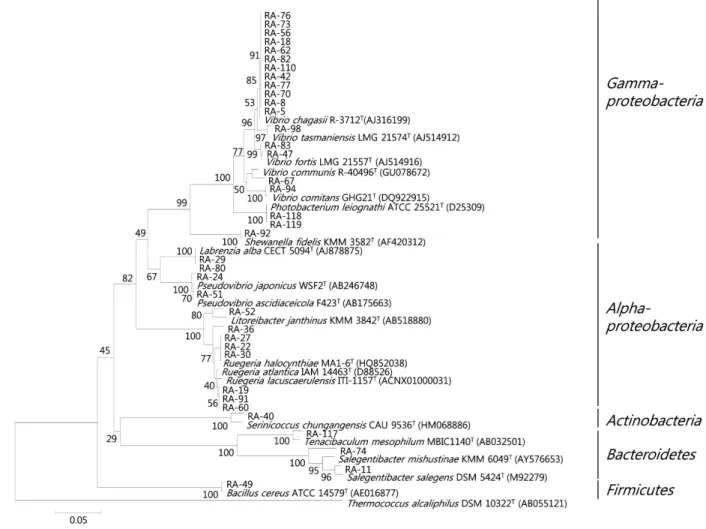
Fig. 1. Dendrogram showing the 16S rDNA-RFLP profiles and the relationship among bacterial strains isolated from the marine sponge, A. simplex.
software를 이용하였다. RFLP 및 DGGE 결과로 얻어진 염기서 열은 NCBI (the National Center for Biotechnology Information) 에 등록된 염기서열을 대상으로 Blast search를 수행하였다. 각 염기서열의 alignment는 CLUSTAL W (Thompson et al., 1994) 를 이용하여 정렬하였고 Molecular Evolutionary Genetics Analysis (MEGA) software version 4.0 을 이용하여(Tamura et al., 2007) neighbor-joining 방법(Saitou and Nei, 1987)에 의해 진화거리 를 계산하고 계통수를 추론하였다. 1,000회 반복 bootstrap 분석 에 의해 계통수를 확인하였다.
결 과
해면 공생세균의 분리 및 RFLP 분석
제주도 무릉아치에서 채집된 A. simplex 해면의 배양 가능한
공생세균 군집구조를 16S rDNA의 PCR-RFLP 방법에 의해 조 사하기 위하여 무작위로 선별된 총 120균주에 대하여 두 종류의 제한 효소를 이용하여 16S rDNA의 RFLP 타입을 분석하였다.
RFLP 타입은 각각 HaeIII를 이용한 경우 17개, MspI의 경우 19 개의 타입이 관찰되었다. 두 효소를 이용한 RFLP 타입을 조합 하여 총 24개의 서로 다른 RFLP 타입이 구분되었다(Table 1 and Fig. 1).
DGGE 밴드 양상
비배양법에 기초한 16S rDNA의 PCR-DGGE를 수행하여 A.
simplex의 해면 공생세균의 계통학적 다양성을 조사하였다. 해 면 시료로부터 추출한 유전체 DNA를 주형으로 16S rDNA 유전 자의 V3영역을 증폭하여 194 bp의 예상된 크기의 PCR 산물을 얻었다. A. simplex의 DGGE 밴드 패턴 분석 결과, 총 12개 이상
DGGEband Closest relative Accession
no. % Sequence
similarity Phylum
DA-5 Marivita sp. CZ-AX3 JX306767 98 Alphaproteobacteria
DA-2 Uncultured beta proteobacterium clone EC10 DQ889936 90 Betaproteobacteria DA-10 Uncultured gamma proteobacterium clone A44 FJ529272 94 Gammaproteobacteria
DA-8 Uncultured delta proteobacterium clone XC2A01 JN596694 100 Deltaproteobacteria
DA-11 Uncultured delta proteobacterium XC2A01 JN596694 100 Deltaproteobacteria
DA-4 Uncultured actinobacterium clone OTU115 JQ217275 98 Actinobacteria
DA-7 Uncultured actinobacterium clone Petrosia64 JN392390 100 Actinobacteria
DA-12 Uncultured actinobacterium clone ACT-E234 JN113055 99 Actinobacteria
DA-1 Uncultured Chloroflexi bacterium clone W04IS4E04 EF629722 99 Chloroflexi DA-6 Uncultured Chloroflexi bacterium clone XD2022 JN596747 97 Chloroflexi
DA-9 Uncultured Chloroflexi bacterium clone 289R EU819048 99 Chloroflexi
DA-3 Uncultured Nitrospira sp. clone XD2H12 HQ270376 91 Nitrospira
Table 2. Phylogenetic affiliation of re-amplified denaturing gradient gel electrophoresis (DGGE) bands derived from the sponge, A. simplex (DA)
Fig. 2. DGGE banding patterns of amplified 16S rDNA obtained from A. simplex (DA); DA1 and DA2 present the DGGE profiles from independent experiments using the same sponge sample.
의 밴드를 확인할 수 있었다(Fig. 2). 이 해면 종의 DGGE 밴드 가 나타내는 세균 다양성을 파악하기 위하여 16S rDNA의 부분 염기서열을 분석하였다.
염기서열 분석 및 계통학적 분석
16S rDNA-RFLP 분석에 의해 24개의 타입으로 나뉜, A.
simplex에서 분리된 세균들에 대해 각 RFLP 타입 별로 1–2개의
분리 균주를 선별하여 부분 염기서열(500 bp 이상)을 분석하였 다. 염기서열이 분석된 총 37개의 분리 균주는 모두 기존에 보고 된 세균 종과 94% 이상의 유사도를 나타내었다(Table 1). 염기 서열이 분석된 24개의 RFLP 타입은 19개의 세균 종을 나타내었 으며 이 결과를 근거로 처음 RFLP 분석에 쓰여진 120균주들을 분석하고 이들의 분석 결과에 근거하여 주요 공생세균 그룹에 대한 계통학적 다양성을 분석하였다(Fig. 3). 16S rDNA의 RFLP 분석에 의해 A. simplex 해면의 공생세균은 Alphaproteobacteria, Gammaproteobacteria, Actinobacteria, Bacteroidetes, Firmicutes, 5개의 문에 속하는 것으로 나타났다(Figs. 1 and 3).
DGGE 밴드 패턴으로부터 세균군집의 다양성을 분석하기 위 해 확인된 DGGE 밴드에서 DNA를 추출하여 염기서열을 분석 하였다. 결과, 12개의 밴드에 대한 염기서열을 확인할 수 있었으 며, 결정된 서열들은 모두 알려진 서열들과 90%에서 100%의 상 동성(similarity)을 나타내었다(Table 2). DGGE 밴드 패턴은 다 양하였으며, 밴드로부터 밝혀진 대부분의 서열들은 배양되지 않 은 세균(uncultured bacteria)들과 높은 상동성을 나타내었다.
DGGE 밴드에서 DA-1, DA-6과 DA-9는 uncultured Chloroflexi (EF629722, JN596747, EU819048)와 97%에서 99%의 상동성을 나타내었으며, DA-2는 uncultured Betaproteobacteria (DQ889936) 와 90%, DA-3는 uncultured Nitrospira (HQ270376)와 91% 상동 성을 나타내었다. DA-4, DA-7과 DA-12는 uncultured Actinobacteria (JQ217275, JN392390, JN113055)와 98%에서 100%의 상동성 을 나타내었으며, DA-5는 Marivita sp. (JX306767)와 98%, DA-8과 DA-11은 uncultured Deltaproteobacteria (JN596694)와 100% 상동성을 보였으며, 밴드 DA-10은 uncultured Gammaproteobacteria (FJ529272)와 94% 상동성을 나타내었다.
DGGE 밴드의 염기서열 분석에 근거하여 계통수를 작성한 결과 (Fig. 4), A. simplex의 주요 공생세균 그룹은 Alphaproteobacteria, Betaproteobacteria, Gammaproteobacteria, Deltaproteobacteria, Actinobacteria, Chloroflexi 그리고 Nitrospira의 7개의 문으로 나타났다.
Fig. 3. Phylogenetic relationship were determined by the 16S rDNA sequences of the isolated bacterial strains from the marine sponge, A.
simplex (RA). Numbers above branches indicate bootstrap values of neighbor-joining analysis (>50%) from 1,000 replicates. The scale bar represents 0.05 substitution per nucleotide position.
고 찰
제주도에 서식하는 해양 해면 A. simplex의 공생세균의 다양 성을 알아보기 위하여 배양에 의한 PCR-RFLP와 비배양 분자 지문법인 DGGE 분석을 수행하였다.
PCR-RFLP 분석에 의해 A. simplex의 배양 가능한 공생세균 은 총 5개의 문에 포함되며 그 중 Gammaproteobacteria가 54%로 가장 우점하는 세균 군집이었다. Alphaproteobacteria의 경우는 32.5%로 나타나 Gammaproteobacteria에 이어 우점하는 세균 군집이었다(Table 1). Alphaproteobacteria와 Gammaproteobacteria 를 포함한 Proteobacteria 문은 총 86.5%를 차지하여 A. simplex 에서 가장 우점하는 세균 문임을 알 수 있었다. 이는 Li 등(2007) 에 의해 남중국해에 서식하는 4종의 해면, Stelletta tenuis, Halichondria rugosa, Dysidea avara, Craniella australiensis의 혼합배양에 의한 공생세균 군집 분석에서 Alphaproteobacteria와 Gammaproteobacteria가 가장 우점하는 세균 군집으로 보고된 바 이와 일치하는 결과였으며, 일반적으로 Alphaproteobacteria
와 Gammaproteobacteria는 해면 공생세균 군집에서 가장 우점 하는 세균 군집으로 알려져 있다(Webster et al., 2004). 가장 많 은 분포를 보인 세균 속은 Gammaproteobacteria의 Vibrio (45.9%)와 Alphaproteobacteria의 Ruegeria (19%)였다. Vibrio 종 중 V. Chagasii는 32.4% 차지하는 단일 우점종으로 넙치 애 벌레로부터 처음 분리되어 분리원이 해양환경인 것으로 알려져 있다(Thompson et al., 2003). Actinobacteria와 Firmicutes는 각 각 3.4%를 차지하여 A. simplex 내에서 가장 적은 분포를 나타내 었다. Actinobacteria의 경우 Serinococcus 속, Firmicutes의 경우 Bacillus가 주요 속으로 구성되었다. RA-74 균주는 Salegentibacter mishustinae와 94% 상동성을 나타내어 신종 후보 균주임을 알 수 있었다(Table 1).
배양 가능한 공생세균 군집의 경우 5개의 문이 발견된데 비해 비배양에 기초한 DGGE법에 의한 경우 7개의 문이 나타나 공생 세균 군집 구조에 있어 방법에 따른 차이를 나타내었다(Figs. 3 and 4).
PCR-RFLP와 DGGE 방법에 의해 A. simplex 해면의 전체 공생세
Fig. 4. Phylogenetic tree from analysis of 16S rDNA sequences of DGGE bands from A. simplex (DA). Numbers above branches indicate bootstrap values of neighbor-joining analysis (>50%) from 1,000 replicates. The scale bar represents 0.05 substitution per nucleotide position.
균의 다양성 분석 결과, Alphaproteobacteria, Gammaproteobacteria, Actinobacteria의 그룹이 공통으로 존재하는 세균 문이었다. 제 주도 무릉연안에서 서식하는 종류가 다른 해면, Spirastrella abata에 대한 PCR-RFLP와 DGGE 방법에 의한 공생세균 다양 성에 관한 연구에서도 Alphaproteobacteria, Gammaproteobacteria, Actinobacteria는 공통적으로 나타나는 공생세균 군집으로 밝혀 졌으며(Jeong et al., 2010), 그 외 해외의 해양 해면 종에 관한 연 구에서도 이들 세균 문은 해면 종에 널리 분포하는 세균 문으로 보 고된 바 있다(Zhang et al., 2006; Mohamed et al., 2008; Kennedy et al., 2009). 한편, Bacteroidetes와 Firmicutes는 RFLP 방법에 의해서만 발견되었으며 Betaproteobacteria, Chloroflexi, Deltaproteobacteria, Nitrospira는 비배양 DGGE에 의해서만 발견되어 방법에 따른 공생세균 다양성에 차이를 나타내었다.
해양 해면에서 널리 분포하는 것으로 알려져 있는 Chloroflexi, Nitrospira는 일반적으로 비배양 군집분석의 경우 높은 빈도로 검출되나 배양법에 의한 경우 발견 빈도가 매우 낮다(Jeong et al., 2010; Erwin et al., 2011). 이는 배양배지의 조성, 배양 조건 등 분리 방법의 차이에 기인한 것으로 생각되며, 따라서 다양한 배지를 이용하여 분리효율을 극대화 하고자 하는 노력이 배양에 의한 해면 공생세균 다양성에 관한 연구에서 이루어지고 있다 (Park et al., 2002; Sipkema et al., 2011). Haliclona sp. 해면의 배양 가능한 공생세균을 분리하기 위해 한천평판배양, 액체배 양, floating filter 배양, 3가지의 서로 다른 방법을 이용한 경우, 배양 방법에 따라 세균 다양성이 현저히 다르다는 것을 보고하였다 (Sipkema et al., 2011). 또한 해면 공생세균의 분리에 있어 다양
한 분리 방법을 적용했을 경우 세균의 우점 그룹은 역시 Alphaproteobacteria (89%)였으나, 구성하는 속에 있어서는 일 반적인 배양법에 의해서는 잘 분리되지 않는 Planctomycetes, Verrucomicrobia, Delataproteobacteria의 분리 빈도가 높다는 것을 보고하였다(Sipkema et al., 2011). Hymeniacidon perleve 해면으로부터 방선균을 분리하여 16S rRNA gene-RFLP에 의 해 다양성을 조사한 연구에서도 배양조건에 따라 방선균의 다양 성은 매우 달라지며, 특히 해양세균의 배양에 흔히 사용되는 MA 배지를 사용한 경우 세균의 다양성은 매우 제한적이라는 것 을 보고하고 있다(Zhang et al., 2006). Jackson 등(2012)에 의한 연구에 따르면 배양에 의한 경우, 분리한 200개의 균주가 4개의 문에 속한 반면 pyrosequencing에 의해 다양성을 조사한 경우 10개의 문이 발견되었다. 본 연구 결과에서도 배양에 의한 RFLP 방법의 경우 비배양 DGGE에 비해 다양성이 낮은 것으로 나타나 배양법에 의한 경우 다양성이 매우 저평가 되는 것 (Jackson et al., 2012)을 알 수 있었다. 그러나 배양에 의하지 않 는 DGGE 방법 역시 증폭된 단편들이 서로 엉겨 잘 분리되지 않 거나 젤의 농도 구배에 따라 검출되는 세균 그룹이 다르거나 또 한 매우 짧은 염기서열(약 150 bp)로 인해 세균 다양성의 분석에 는 한계가 있다.
배양에 의한 RFLP와 비배양 DGGE에 의해 다양성을 분석하 는 경우, 흔히 RFLP에 의한 경우보다 DGGE에 의한 경우 약간 더 많은 세균 문이 발견되며, 비배양 DGGE에 의한 경우 배양되 지 않는(uncultured) 세균 그룹이 흔히 발견되지만 배양 없이 total DNA를 추출하여 다양성을 분석하는 DGGE임에도 배양법
에 의해 검출되는 세균 그룹이 발견되지 않는 경우도 매우 흔하 여(Zhang et al., 2006; Jeong et al., 2010), 이 두 가지 방법은 보 완적으로 사용되어야 할 것으로 생각된다.
결론적으로 RFLP와 DGGE의 서로 다른 두 가지 방법에 의해 A. simplex의 공생세균 다양성을 분석한 결과, Alphaproteobacteria, Betaproteobacteria, Deltaproteobacteria, Gammaproteobacteria, Actinobacteria, Bacteroidetes, Chloroflexi, Firmicutes 그리고 Nitrospira, 총 9개의 문이 존재하는 것으로 나타났다. 서로 다른 두 실험 방법을 적용한 경우, Alphaproteobacteria, Gammaproteobacteria, Actinobacteria가 공통 세균 그룹으로 나타났으나 방법에 따라 각각 서로 다른 다양한 세균문을 나타내는 것으로 관찰되었다.
따라서 연구 방법에 따라 검출되는 세균 다양성의 차이를 고려 하여 해면 공생세균의 군집 분석이 이루어져야 할 것으로 사료 된다.
적 요
해양 해면 Asteropus simplex를 제주도에서 채집하여 배양에 의한 RFLP와 비배양에 의한 DGGE 분석 방법에 의해 세균군집 구조를 조사하였다. 16S rDNA-RFLP 분석을 위해 변형된 Zobell 배지와 MA를 이용하여 120균주를 선별하고 제한효소, HaeIII와 MspI을 사용하여 각각의 다른 RFLP 패턴으로 구분하 였다. RFLP 패턴으로부터 유래한 16S rDNA 염기서열 분석 결과, 알려진 세균 종과 94% 이상의 유사도를 나타내었으며 Alphaproteobacteria, Gammaproteobacteria, Actinobacteria, Bacteroidetes, Firmicutes, 5개의 문이 관찰되었다. 그 중 Gammaproteobacteria가 우점하였다. 같은 해면, A. simplex의 DGGE 분석을 위해 total genomic DNA로부터 16S rDNA를 증 폭하여 DGGE fingerprinting을 수행한 결과 12개의 서로 다른 밴드가 관찰되었다. 각 밴드의 16S rDNA 염기서열은 알려진 세 균의 염기서열과 90% 이상의 유사성을 나타내었으며 대부분의 염기서열은 uncultured bacteria에 속하였다. DGGE 분석으로부 터 미생물의 군집은 Alphaproteobacteria, Betaproteobacteria, Gammaproteobacteria, Deltaproteobacteria, Actinobacteria, Chloroflexi, Nitrospira, 7개의 문으로 나타났다. RFLP와 DGGE 방법에 의해 Alphaproteobacteria, Gammaproteobacteria, Actinobacteria가 공통적으로 발견되었으나 전체적인 공생세균 의 군집구조는 분석방법에 따른 차이를 나타내었다. 배양에 의 한 방법보다 비배양 방법에서 더 다양한 세균군집구조를 나타내 었다.
감사의 말
이 논문은 2012년 한남대학교 학술연구조성비 지원에 의해 수행되었으며 이에 감사 드립니다.
참고문헌
Bergman, O., Haber, M., Mayzel, B., Anderson, M.A., Shpigel, M., Hill,
R.T., and Ilan, M. 2011. Marine-based cultivation of Diacarnus sponges and the bacterial community composition of wild and maricultured sponges and their larvae. Mar. Biotechnol. 13, 1169–
1182.
Botstein, D., White, R.L., Skolnick, M., and Davis, R.W. 1980.
Construction of a genetic linkage map in man using restriction fragment length polymorphisms. Am. J. Hum. Genet. 32, 314–331.
Erwin, P.M., Olson, J.B., and Thacker, R.W. 2011. Phylogenetic diversity, host-specificity and community profiling of sponge-associated bacteria in the Northern Gulf of Mexico. PLoS.
6, e26806. doi:10.1371/journal.pone.0026806.
Fischer, S.G. and Lerman, L.S. 1983. DNA fragments differing by single base-pair substitutions are separated in denaturing gradient gels:
correspondence with melting theory. Proc. Natl. Acad. Sci. USA 80, 1579–1583.
Hentschel, U., Fieseler, L., Wehrl, M., Gernert, C., Steinert, M., Hacker, J., and Horn, M. 2003. Microbial diversity of marine sponges. Prog.
Mol. Subcell. Biol. 37, 59–88.
Jackson, S.A., Kennedy, J., Morrissey, J.P., O’Gara, F., and Dobson, A.W. 2012. Pyrosequencing reveals diverse and distinct sponge-specific microbial communities in sponges from a single geographical location in Irish waters. Microb. Ecol. 64, 105–116.
Jeong, E.J., Im, C.S., and Park, J.S. 2010. A comparison of bacterial diversity associated with the sponge Spirastrella abata depending on RFLP and DGGE. Kor. J. Microbiol. 46, 366–374.
Kennedy, J., Baker, P., Piper, C., Cotter, P.D., Walsh, M., Mooij, M.J., Bourke, M.B., Rea, M.C., O’Connor, P.M., Ross, R.P., and et al. 2009.
Isolation and analysis of bacteria with antimicrobial activities from the marine sponge Haliclona simulans collected from Irish waters.
Mar. Biotechnol. 11, 384–396.
Li, Z., He, L., and Miao, X. 2007. Cultivable bacterial community from south china sea sponge as revealed by DGGE fingerprinting and 16S rDNA phylogenetic analysis. Curr. Microbiol. 55, 465–472.
Mohamed, N.M., Rao, V., Hamann, M.T., Kelly, M., and Hill, R.T. 2008.
Monitoring bacterial diversity of the marine sponge Ircinia strobilina upon transfer into aquaculture. Appl. Environ. Microbiol. 74, 4133–
4143.
Murayama, S., Nakao, Y., and Matsunaga, S. 2008. Asteropterin, an inhibitor of cathepsin B, from the marine sponge Asteropus simplex.
Tetrahedron Lett. 49, 4186–4188.
Park, S.H., Kwon, K.K., Lee, D.S., and Lee, H.K. 2002. Morphological diversity of marine microorganisms on different media. J. Microbiol.
40, 161–165.
Saitou, N. and Nei, M. 1987. The neighbor-joining method: a new method for reconstructing phylogenetic trees. Mol. Biol. Evol. 4, 406–425.
Sipkema, D., Schippers, K., Maalcke, W.J., Yang, Y., Salim, S., and Blanch, H.W. 2011. Multiple approaches to enhance the cultivability of bacteria associated with the marine sponge Haliclona (gellius) sp.
Appl. Environ. Microbiol. 77, 2130–2140.
Tamura, K., Dudley, J., Nei, M., and Kumar, S. 2007. MEGA4: molecular evolutionary genetics analysis (MEGA) software version 4.0. Mol.
Biol. Evol. 24, 1596–1599.
Taylor, M.W., Hill, R., and Hentschel, U. 2011. Meeting report: 1st international symposium on sponge microbiology. Mar. Biotechnol.
13, 1057–1061.
Thomas, T.R., Kavlekar, D.P., and LokaBharathi, P.A. 2010. Marine drugs from sponge-microbe association-a review. Mar. Drugs 8, 1417 –1468.
Thompson, F.L., Thompson, C.C., Li, Y., Gomez-Gil, B., Vandenberghe,
J., Hoste, B., and Swings, J. 2003. Vibrio kanaloae sp. nov., Vibrio pomeroyi sp. nov. and Vibrio chagasii sp. nov., from sea water and marine animals. Int. J. Syst. Evol. Microbiol. 53, 753–759.
Thompson, J.D., Higgins, D.G., and Gibson, T.J. 1994. CLUSTAL W:
improving the sensitivity of progressive multiple sequence alignment through sequence weighting, position-specific gap penalties and weight matrix choice. Nucleic Acids Res. 22, 4673–4680.
Webster, N.S., Cobb, R.E., Soo, R., Anthony, S.L., Battershill, C.N., Whalan, S., and Evans-Illidge, E. 2011. Bacterial community
dynamics in the marine sponge Rhopaloeides odorabile under in situ and ex situ cultivation. Mar. Biotechnol. 13, 296–304.
Webster, N.S., Negri, A.P., Munro, M.M., and Battershill, C.N. 2004.
Diverse microbial communities inhabit Antarctic sponges. Environ.
Microbiol. 6, 288–300.
Zhang, H., Lee, Y.K., Zhang, W., and Lee, H.K. 2006. Culturable actinobacteria from the marine sponge Hymeniacidon perleve:
isolation and phylogenetic diversity by 16S rRNA gene-RFLP analysis. Antonie van Leeuwenhoek 90, 159–169.