A large ulcerated exophytic mass involving the stom- ach is the classic CT finding of a gastrointestinal stromal tumor (GIST) (1). An exophytic adenocarcinoma of the colon is very rare with only a few reported cases (2, 3).
To the best of our knowledge, the CT appearance of colon cancer which simulated the classic appearance of a GIST has only been reported once in the world’s litera- ture (2). Here we present another such similar case.
Case Report
A 62-year-old male patient was admitted to the hospi-
tal complaining of dizziness. The patient’s hemoglobin level was 10.1 g/dL (13-17 g/dL) and a negative occult blood stool test. In addition, the patient’s serum carci- noembryonic antigen was 1.3 ng/mL (0-5 ng/mL). A gas- trofiberscopy was performed, which demonstrated a fungating mass with exudates in the posterior wall of the gastric upper body (Fig. 1A). A biopsy of the fungat- ing mass with the gastrofiberscopy revealed no evidence of malignancy, chronic active gastritis with intestinal metaplasia or ulcer with necrosis. A colonofiberscopy demonstrated an ulcerated lobulated mass in the distal transverse colon (Fig. 1B). A biopsy of the mass along with a colonofiberscopy revealed an ulcer with inflam- matory debris, chronic active inflammation and no evi- dence of malignancy. A contrast-enhanced multidetec- tor-row CT (Somatom Sensation 64; Siemens, Erlangen, Germany) revealed an approximately 13.8×7.3 cm lob- ulated heterogeneous enhancing mass with a smooth margin in the left upper abdomen located between the
J Korean Soc Radiol 2009;60:267-270
─ 267 ─
Exophytic Colon Cancer: Resemblance to a Gastrointestinal Stromal Tumor of the Stomach:
A Case Report
1Chul Hi Park, M.D., Ha Na Kim, M.D., Sung Su Byun, M.D., Seung Yeon Ha, M.D.2
1Department of Radiology, Gachon University, Gil Medical Center
2Department of Pathology, Gachon University, Gil Medical Center Received November 27, 2008 ; Accepted January 8, 2009
Address reprint requests to : Ha Na Kim, M.D., Department of Radiology, Gachon University, Gil Medical Center, 1198 Guwol-dong, Namdong-gu, Inchon, 405-760, Republic of Korea.
Tel. 82-32-460-3060 Fax. 82-32-460-3065 E-mail: [email protected]
An exophytic adenocarcinoma of the colon is very rare with only a few reports to date. To the best of our knowledge, the CT appearance of colon cancer, which simulat- ed the classic appearance of a GIST has only been reported once in the world’s litera- ture. We recently evaluated a patient with a large lobulated mass involving the stom- ach, pancreas and colon. The CT appearance of the case was consistent with the diag- nosis of an exophytic gastric GIST. However, at surgery, the patient was found to have a large ulcerated carcinoma of the colon near the splenic flexure that had invaded the stomach and pancreas. We report a case of an exophytic adenocarcinoma of the colon that resembled the classic appearance of a gastrointestinal stromal tumor of the stom- ach.
Index words :Colon neoplasms
Gastrointestinal stromal tumors Tomography, X-ray computed
stomach and splenic flexure of the colon (Figs. 1C-F).
The CT appearance was most suggestive of an exophytic gastric GIST. Moreover, the tail portion of the pancreas was invaded by the mass. The proximal colon was not dilated and no definite evidence of lymphadenopathy was observed.
The patient underwent surgical treatment, which in- cluded a wedge resection of the stomach, segmental re- section of the transverse colon, distal pancreatectomy and splenectomy.
The final pathology findings were a moderately differ- entiated adenocarcinoma of the colon which invaded the stomach and pancreas, and no evidence of lymph
node metastasis (Fig. 1G).
Discussion
An exophytic mass of the gastrointestinal tract is char- acteristic of a gastric GIST. To date, only two cases have been reported with bulky exophytic colon cancer resem- bling leiomyosarcoma upon a CT scan (2). However, in these two cases, the CT revealed a gastrocolic fistula.
The site of the fistula revealed that the propensity for the left portion of the transverse colon to be involved may be related to its anatomical relationship; the distal three-fifths of the transverse colon lies immediately be-
Chul Hi Park, et al: Exophytic Colon Cancer: Resemblance to a Gastrointestinal Stromal Tumor of the Stomach
─ 268 ─
A B
C D
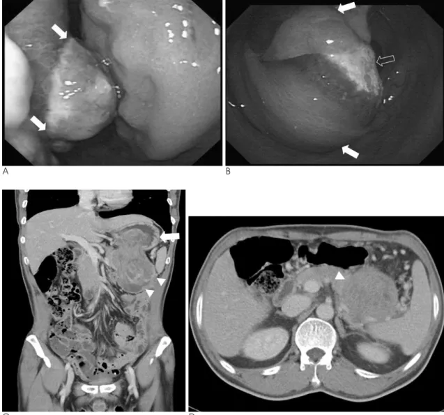
Fig. 1. A 62-year-old man presenting with dizziness.
A. A gastrofiberscopy shows a large lobulated mass with exudates (white arrows) in the posterior wall of the gastric upper body.
B. A colonofiberscopy shows a protruding mass (white arrows) with ulceration (open arrow) in the distal transeverse colon.
C. An enhanced CT scan of the abdomen shows a 13.8×7.3 cm lobulated heterogeneous enhancing mass with a smooth margin in the left upper abdomen located between the stomach (white arrow) and splenic flexure of the colon (white arrow heads) on the coronal view. The colonic wall is thickened and shows a wide angle between the mass.
D. The tail portion of the pancreas is compressed by the mass (white arrow head).
low the greater curvature of the stomach. In addition, the greater omentum between them is very short (4-6).
Because a gastrocolic fistula has not been reported to oc- cur with a GIST, Lee et al. (2) suggested that a bulky mass between the stomach and colon, with a gastrocolic fistula, should first suggest an adenocarcinoma of the stomach or colon. However, in our case, the fistula for- mation of the mass between the colon and stomach was not resolved on the CT or pathologic findings. In addi- tion, the colon located proximal to the mass was not di- lated and showed no distinct evidence of periregional lymphadenopathy.
In many cases of gastric GIST, the bulk of the tumor will be in an extragastric location, which makes it diffi- cult to determine the origin of the tumor from the gas- tric wall on CT images (7). Levy et al. (7) suggested that the tumor may be attached to the gastric wall by a thin pedicle and that careful evaluation of the gastric wall in these cases may reveal subtle wall thickening that will
help establish the stomach as the origin of the mass. In our case, we believe that the mass originated from the colon because of the lack of definite gastric wall thicken- ing around the exophytic growing mass on CT in addi- tion to the wide angle between the mass and the colon and the colonic wall thickening. Also, we think that the differential diagnosis between a GIST and an exophytic colon cancer is important when making a decision of the tumor’s stage and origin.
An exophytic sigmoid colon cancer resembling an ovarian tumor was previously reported and the author cited the difficulty of determining the origin of the tu- mors that presented as a pelvic mass, however the ex- amination of the relationship of the venous anatomy such as the inferior mesenteric vein, the gonadal vein and the mass on the CT, provided useful information.
However, in our case, the large exophytic mass com- pressed the surrounding vessels, including the splenic vein and multiple omental collateral vessels. Therefore,
J Korean Soc Radiol 2009;60:267-270
─ 269 ─
E F
G
Fig. 1. E. The heterogeneous enhancing mass (open arrow head) is also protruding into the lumen of the splenic flexure in the colon (white arrow heads).
F. The gross findings reveal a lobulated tumor (white arrows) be- tween the colon (two asterisks) and stomach (white arrow head) by the surgical wedge resection of the stomach, segmental re- section of the distal transeverse colon and the splenectomy (as- terisk).
G. The microscopic findings reveal that the tumor shows gland formation (open arrow) with severe necrosis (white arrow) (H &
E, ×100).
the examination of the relationship between the venous anatomy and the mass on the CT did not provide useful information with regard to the origin of the mass.
The differential diagnosis of a gastric GIST includes other mesenchymal neoplasms such as true leiomy- omas, leiomyosarcomas, schwannomas, neurofibromas, and neuroendocrine neoplasms. Because all of these neoplasms develop in the gastric wall, their imaging fea- tures may be similar to those of a GIST (7).
Here we have reported a rare case of exophytic colon cancer resembling a gastric GIST and, although this case was rare, the differential diagnosis of a gastric GIST should include the possibility of an exophytic colon can- cer.
References
1. Scatarige JG, Fishman EK, Jones B, Cameron JL, Sanders RC, Siegelman SS. Gastric leiomyosarcoma: CT observations. J Comput Assist Tomogr 1985;9:320-327
2. Lee WJ, Horton KM, Fishman EK. Gastrocolic fistula due to ade- nocarcinoma of the colon: simulation of primary gastric leiomyosarcoma on CT. Clin Imaging 1999;23:295-297
3. Okamoto D, Asayama Y, Yoshimitsu K, Irie H, Aibe H, Utsunomiya T, et al. Exophytic colon cancer mimicking an ovari- an tumor: the value of evaluation of the venous anatomy on MD- CT. CMIG Extra 2005;Cases 29:1-4
4. Smith DL, Dockerty MB, Black BM. Gastrocolic fistulas of malig- nant origin. Surg Gynecol Obstet 1972;134:829-832
5. Theony RH, Hodgson JR, Scudamore HH. The roentgenologic di- agnosis of gastrocolic and gastrojejunocolic fistulas. Am J Roentgenol Radium Ther Nucl Med 1960;83:876-881
6. MacMahon CE, Lund P. Gastrocolic fistula of malignant origin. A consideration of its nature and report of five cases. Am J Surg 1963;106:333-347
7. Levy AD, Remotti HE, Thompson WM, Sobin LH, Miettinen M.
Gastrointestinal stromal tumors: radiologic features with patholo- bic correlation. Radiographics 2003;23:283-304
Chul Hi Park, et al: Exophytic Colon Cancer: Resemblance to a Gastrointestinal Stromal Tumor of the Stomach
─ 270 ─
대한영상의학회지 2009;60:267-270
위에서 발생한 위장관 간질종양과 유사한 모습을 보이는 외장성 대장암: 1예 보고1
1가천의과대학교 의학전문대학원 영상의학교실
2가천의과대학교 의학전문대학원 해부병리학교실
박 철 희∙김 하 나∙변 성 수∙하 승 연2
대장의 외장성 선암은 매우 드물게 보고되고 있다. 저자들이 아는 한 위장관 간질종양과 유사한 모습을 보이는 대 장암의 전산화 단층촬영 소견은 단 한번 보고 되었다. 저자들은 최근 위, 췌장 그리고 대장을 침범한 커다란 종괴가 있는 환자를 검사하였다. 이 환자의 컴퓨터 단층 촬영 소견은 위에서 기원한 외장성 간질종양의 모습이었으나 수술 상 대장에서 기원한 커다란 궤양성 암이 위와 대장을 침범한 것으로 확인되었다. 저자들은 위에서 발생한 위장관 간 질종양의 전형적인 모습과 유사한 대장에서 발생한 외장성 선암 1예를 경험하여 보고하고자 한다.