Case Report
Mesenteric Pseudocyst of the Small Bowel in Gastric Cancer Patient: A Case Report
Sang Eok Lee, In Seok Choi, Won Jun Choi, Dae Sung Yoon, Ju Ik Moon,Yu Mi Ra, Hyun Sik Min, Yong Seok Kim1, Sun Moon Kim1, Jang Sihn Sohn2, and Bong Soo Lee3
Department of Surgery, 1Division of Gastroenterology and Hepatology, 2Depatrment of Pathology, Konyang University College of Medicine, 3Sokpyunhan Internal Medicine Clinic, Daejeon, Korea
Mesenteric pseudocyst is rare. This term is used to describe the abdominal cystic mass, without the origin of abdominal organ. We presented a case of mesenteric pseudocyst of the small bowel in a 70-year-old man. Esophago-gastro-duodenoscopy showed a 3.5 cm sized excavated lesion on the posterior wall of angle. Endocopic biopsy confirmed a histologic diagnosis of the poorly differentiated adenocarcinoma, which includes the signet ring cell component. Abdominal computed tomography scan showed a focal mucosal en- hancement in the posterior wall of angle of the stomach, a 2.4 cm sized enhancing mass on the distal small bowel loop, without distant metastases or ascites in rectal shelf, and multiple gallbladder stones. The patient underwent subtotal gastrectomy with gastroduodenos- tomy, segmental resection of the small bowel, and cholecystectomy. The final pathological diagnosis was mesenteric pseudocyst. This is the first case report describing incidentally detected mesenteric pseudocyst of the small bowel in gastric cancer patients.
Key Words: Mesenteric cyst; Stomach neoplasms; Pseudocyst
J Gastric Cancer 2012;12(1):43-45 http://dx.doi.org/10.5230/jgc.2012.12.1.43
Correspondence to: Won Jun Choi
Department of Surgery, Konyang University Hospital, 685, Gasuwon- dong, Seo-gu, Daejeon 320-718, Korea
Tel: +82-42-600-8956, Fax: +82-42-543-8956 E-mail: [email protected]
Received February 2, 2012 Revised March 7, 2012 Accepted March 7, 2012
Copyrights © 2012 by The Korean Gastric Cancer Association www.jgc-online.org
This is an open-access article distributed under the terms of the Creative Commons Attribution Non-Commercial License (http://creativecommons.org/
licenses/by-nc/3.0) which permits unrestricted noncommercial use, distribution, and reproduction in any medium, provided the original work is properly cited.
Introduction
Mesenteric pseudocyst is a term used to describe abdominal cystic mass without the origin of abdominal organ.(1) This has been classified according to embryologic, ehiologic, histologic, and ther data, causing considerable confusion. It was considered the term mesenteric cyst as merely descriptive, and apply a histologic classification such as lymphangioma, pseudocyst, enteric duplication cyst, enteric cyst, and mesothelial cyst.(2) We presented a case of mesenteric pseudocyst of the small bowel in a 70-year-old man.
Case Report
A 70-year-old man was referred to our hospital for operation of gastric cancer with a 1-month history of progressively worsen- ing epigastric and intermittent peri-umbilical discomfort. He had no specific previous medical or surgical history including cancer.
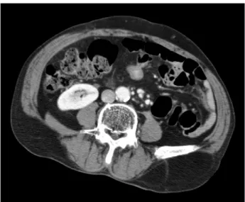
On physical examination, no tenderness or palpable mass were identified. Esophago-gastro-duodenoscopy (EGD) showed a 3.5 cm sized excavated lesion on the posterior wall of angle. Endocopic biopsy confirmed a histologic diagnosis of poorly differentiated adenocarcinoma including signet ring cell component. Endoscopic ultrasonography revealed invasion of caner to the proper muscle layer. Abdominal computed tomography (CT) scan showed a focal mucosal enhancement in posterior wall of angle of stomach, a 2.4 cm sized enhancing mass on distal small bowel loop without distant metastases or ascites in rectovesical pouch, and multiple gallbladder stones (Fig. 1). These physical, laboratory, and radiological findings prompted us to diagnose early gastric cancer, and gastrointestinal stromal tumor of small bowel. A pheripheral blood count showed
Lee SE, et al.
44
no leukocytosis or anemia. Laboratory testing revealed alfa-feto- protein level of 2.88 (normal range, 0 to 9 ng/ml), carcino-em- bryonic antigen level of 1.45 ng/ml (normal range, 0 to 5 ng/ml), carbohydrate antigen (CA) 19-9 level of 6.5 U/ml (normal range, 1 to 35 U/ml), and CA 72-4 level of 4.8 U/ml (normal range, 0 to 4 U/ml). Other laboratory test results were within normal limit.
The patient underwent subtotal gastrectomy with gastroduode- nostomy, segmental resection of small bowel, and cholecystectomy.
Mesenteric mass was adhered severely with greater omentum at the mesenteric side of small bowel, and mesenteric fat tissues. It looked like having connection with small bowel lumen. Small bowel, mesentery, and mesenteric mass were resected en-bloc methods, and end to end anastomosis was performed. After fixation of the surgical specimen, macroscopic examination revealed a uni-locular cyst measuring 3×3×2 cm in size. The mass contained yellow
gelatinous materials with 1mm sized wall diameter. Pathological examination revealed 3 cm sized fibrous cystic wall without endo- thelial or epithelial lining and foam cell collection (Fig. 2, 3). The final pathological diagnosis was mesenteric pseudocyst. Pathologic stage of gastric cancer was T1bN1M0 (6th International Union Against Cancer TNM staging system); invasion to submusosa, me- tastases to 4 perigastric lymph nodes out of 16 retrieved nodes, and negative resection margin. Cholecystectomy specimen was reported as chronic cholecystitis with multiple gallbladder stones.
Discussion
Mesenteric pseudocysts are very rare intraabdominal mass with an incidence of about 1 case per 100,000 hospital admissions.(3) Ros et al.(2) first used the term “pseudocyst” in the classification of mesenteric cyst. Mesenteric pseudocyst could be located in the small bowel, large bowel mesentery and even retroperitoneum.(1,4) Most reports were pseudocyst of large bowel or retroperitoneum.(1) Although most mesenteric pseudocysts are asymptomatic, symp- tomatic mesenteric cysts could be associated with cyst size, cyst lo- cation, and complications, including infection, rupture, hemorrhage, and intestinal obstruction.(5) In our patient, there was no specific symptom associated with mesenteric pseudocyst except for inter- mittent vague periumbilical discomfort. If EGD and CT scan were not performed in this patient presenting non-specific abdominal pain, the diagnosis of mesenteric pseudocyst would be delayed.
To the best of our knowledge, this is the first case report de- scribing incidentally detected mesenteric pseudocyst of small bowel in gastric cancer patients. When clinician performed staging work up for gastric cancer, should be aware the possibility of associated intraabdominal lesions.
Fig. 2. Fibrous cystic wall without specific endothelial or epithelial lin- ing epithelium. Cystic content shows cholesterol cleft (H&E, ×12.5).
Fig. 3. Fibrous cystic wall and focal foam cell collection (H&E, ×40).
Fig. 1. Preoperative computed tomography.
Mesenteric Pseudocyst in Gastric Cancer Patient
45
References
1. Gallego JC, González JM, Fernández-Virgós A, del Castillo M.
Retrorectal mesenteric cyst (non-pancreatic pseudocyst) in adult. Eur J Radiol 1996;23:135-137.
2. Ros PR, Olmsted WW, Moser RP Jr, Dachman AH, Hjermstad BH, Sobin LH. Mesenteric and omental cysts: histologic clas- sification with imaging correlation. Radiology 1987;164:327- 332.
3. Kurtz RJ, Heimann TM, Holt J, Beck AR. Mesenteric and ret- roperitoneal cysts. Ann Surg 1986;203:109-112.
4. Iida T, Suenaga M, Takeuchi Y, Kobayashi T, Tobinaga J, Sakai M, et al. Mesenteric pseudocyst of the sigmoid colon. J Gastro- enterol 2003;38:1081-1085.
5. Fan HL, Chen TW, Hong ZJ, Hsieh CB, Chan DC, Chen CJ, et al. Volvulus of small intestine: rare complication of mesenteric pseudocyst. Z Gastroenterol 2009;47:1208-1210.