111
개의 결장 간질종양에 대한 영상의학 및 면역조직화학 진단 1예
최지혜1·김현욱1·이혜경1·김준영2·윤정희2,*
1해마루 이차진료동물병원, 2서울대학교 수의과대학 BK21 수의과학연구인력양성사업단 (게재승인: 2008년 1월 17일)
Medical imaging and immunohistochemical diagnosis of gastrointestinal stromal tumor originated from colon in a dog
Jihye Choi
1, Hyunwook Kim
1, Haekyung Lee
1, Junyoung Kim
2, Junghee Yoon
2,*
1
Haemaru Referral Animal Hospital, Seongnam 463-050, Korea
2
College of Veterinary Medicine and BK21 Program for Veterinary Science, Seoul National University, Seoul 151-742, Korea
(Accepted: January 17, 2008)
Abstract : Gastrointestinal stromal tumor (GIST) is one of the mesenchymal tumors originated from gastrointestinal submucosa. A 10 year-old, male, mixed breed dog with persistent diarrhea, anorexia and lethargy was referred to Haemaru Animal Referral Hospital. Large mass originated from the transverse colon was observed and large amount of ascites and free gas were found on abdominal radiography and ultrasonography. The ascites was septic exudate mixed with bacteria that consisted with intestinal perforation.
There was no metastatic lesion. This mass was tentatively diagnosed as adenocarcinoma, leiomyosarcoma (LMS) and lymphosarcoma and surgical resection and histilogical examination were planned. However, according to owner’s request, the patient was euthanized and then the necropsy was performed. About 10 cm sized mass originated from the cecum, ascending colon and transverse colon was adhered to surrounding mesentery and the perforation and large amount of ascites were observed. GIST was suspected on histopathologic examination and confirmed according to CD 117 expression in immunohistochemistry. GIST, derived from interstitial cells of Cajal, can be distinguished from LMS and leiomyoma (LM) on the basis of expression of CD117 (KIT) immunohistochemically. GIST has a different biological behavior and clinical course compared with LMS and LM, therefore definite diagnosis for GIST using immunohistochemistry is clinically important to predict the precise prognosis of the patient.
Keywords : dog, gastrointestinal stromal tumor, mesenchymal, immunohistochemistry, CD117
서 론
원발성위장관종양은상피세포유래
,
신경내분비유래
,
조혈세포유래,
중간엽(mesenchymal)
유래로분류할수있다
[14].
이중개에서가장흔히발생하는원발성 위장관 종양은 점막층에서 기인하는 선암종
(adenocarcinoma)
으로70%
정도에서림프절,
간,
폐,
대 망으로전이된다.
중간엽유래종양은점막하층에서발생하며
,
평활근종양,
신경성분화를보이는종양,
위장관간질 종양
(gastrointestinal stromal tumor; GIST)
으로분류된다
[1].
종양발생시조직표본을hematoxylin
과eosin
으로염색한뒤광학현미경으로검사하여진단했던과거에는평활근과유사한모양을보이는비림프계중
간엽종양은모두평활근종
(Leiomyoma; LM)
이나평활근육종
(Leiomyosarcoma; LMS)
으로진단되어왔다[8].
하지만평활근종양으로분류되었던이종양을최근고
*Corresponding author: Junghee Yoon
College of Veterinary Medicine and BK21 Program for Veterinary Science, Seoul National University, Seoul 151-742, Korea [Tel: +82-2-880-1265, Fax: +82-2-880-8662, E-mail: [email protected]]
112 최지혜·김현욱·이혜경·김준영·윤정희
유의
kit
단백질을 면역조직화학법(immunohistoche-
mistry; IH)
으로재분류한결과다수가GIST
로확인되었다
[1, 10, 13]. GIST
는 점막하층의 카잘 간질 세포(Interstitil cell of Cajal)
에서발생하는육종으로, kit
라고하는신호전달단백질이변형되어발생하는것으로알
려져있다
[5, 9, 11, 12]. GIST
는과거평활근종양으로생각했으나
, IH
검사혹은전자현미경적분화양상에따라평활근분화
(myogenic)
를보이는종양,
신경분화(neurogenic)
를보이는종양,
평활근과신경분화를동시에보이는종양
(mixed),
특별한분화를보이지않는종양
(anaplastic)
등다양한분화과정을보이는것으로알려졌다
[4, 8]. GIST
는다른원발성위장관종양처럼주로평균
10
살이상의노령견에서발생하며,
품종소인은없는것으로보고되었다
[8].
종양발생시체중저하,
구토
,
식욕부진,
설사등다양한소화기증상을나타내 며,
위와소장에서더흔히발생하는평활근종양과는달리
,
주로소장과대장에서관찰되었다[8, 13, 14].
또 한조직검사상대장에서발생한종양은소장종양에비 해더악성지표를보였고,
장천공과복막염을동반한경우가많았다
. GIST
와LMS, LM
은조직학적검사상평활근성상을보여감별이매우어려우나
, GIST
는평활근유래종양과는행동특성과예후등이다르므로추 가적인
IH
검사를통한감별이필요하다.
본증례는결장에서발생한종괴로인해결장이폐색 되고이차적으로파열되어심한복막염이발생하였다
.
감별진단목록으로원발성위장관종양중선암종
,
평활근유래종양
,
림프육종등을의심하였으나,
조직학적검사상평활근성상종양으로분류되었고
IH
검사를통해
GIST
로확진되었다.
본보고를통해결장종양에대한진단영상과
GIST
의확진을위해반드시실시해야하는
IH
검사에대해고찰하고자한다.
증 례
설사와구토
,
기력저하,
식욕감소를보이는11
살의 수컷잡종견이10
일간대증치료에증상이개선되지않아진료의뢰되었다
.
내원당시설사,
식욕절폐,
기력 저하,
점막창백,
복부팽만이관찰되었다.
신체검사상 복부중앙부에종괴가촉진되어,
혈액검사와방사선검사를 실시하였다
.
혈액 검사상백혈구증가증과band cell
증가(14%),
빈혈(PCV=23%),
저알부민혈증(1.2 g/dl,
정상범위
; 2.2-3.9 g/dl), ALKP
증가(507 µ/l,
정상범위;
23-212 µ/l)
가확인되었다.
복부방사선검사상복수에의해복강내대비도가감소되고
,
중하복부에서연부조직 종괴와방광내결석이관찰되었다(Fig. 1).
외측상에서종괴의앞쪽과뒤쪽에
,
그리고간의뒤쪽변연과유문부사이에가스음영이관찰되어 복강내유리가스
(free
gas)
가존재하는것을확인할수있었고,
종괴에의해내림결장이등쪽으로변위되어있었다
.
복수와유리가 스로인해종괴의변연이불분명하고주변장기와의경 계가명확하게구별되지않았으나,
종괴의위치와주변장기가변위된방향을바탕으로결장이나소장에서유
Fig. 1. Abdominal radiographs of the dog. A mass is identified in middle abdomen and abdominal detail is decreased
due to ascites in lateral (A) and ventrodorsal view (B). Descending colon (dc) is displaced dorsally by a mass (m) in
lateral view and the lumen is interrupted (long arrow) steeply. Abnormal free gases (short arrow) are observed as linear
shape caudal to the liver (l) and the gastric pylorus (p) and the concurrent ascites and abdominal free gas suggest the
rupture of alimentary tract. Additionally, abnormal round shape gas bubbles were observed in mid-abdomen. Incidentally,
a calculus is found in urinary bladder (ub).
래한종양혹은장간막림프절에서유래한종양사이에 감별이필요하였고
,
복강내에복수와유리가스가함께확인되어이차적인장관파열이의심되었다
.
복부초음 파검사상다량의복수가고에코로영상화되어초음파유도하에
700 cc
정도를제거하였다.
복강내유리가스로인해초음파영상이좋지않았으나
,
중하복부에서주 변조직과경계가불분명하고변연이울퉁불퉁한종괴 가관찰되었고,
종괴내에국소적으로가스가포함된부분이있어저에코와고에코가혼재된양상으로영상화
되었다
(Fig. 2).
종괴의앞쪽으로정상적인장분절이주행하다가 종괴로 연결되었으나
,
종괴 내에 장벽의layering
이전혀관찰되지않았고장분절의내강으로추정할만한선상의고에코음영도관찰되지않았다
.
하지 만,
정상적인구조를가진내림결장은좌측복부에서관찰되었으나오름결장과가로결장은영상화되지않 았다
.
커다란종괴로인해좌측신장과비장은복부좌 측으로변위되고복막염에의해장간막이심하게비후 되어있었다.
다른장기에서종양의전이소견은보이지 않았다.
종괴가발생한부위와파열부위를좀더명확 하게평가하여수술가능성여부를판단하기위해가스 트로그라핀(gastrografin; Schering, Spain) 20 ml
를주입 하여결장조영술을실시하였다(Fig. 3).
직장과내림결 장은조영제로충진되었으나가로결장부분은내강이 좁아지면서앞쪽부분은더이상조영제가채워지지않아가로결장의폐색이 확인되었다
.
조영제가찬내림 결장은종괴에 의해등쪽으로변위되어있었으나,
이부위에서조영제의유출소견은확인되지않았다
.
초음파유도하에천자한복수는삼출물
(exudates)
로세포학적검사결과다수의간균과구균
,
세균을탐식하고있는염증세포가관찰되었다
.
이상의결과를바탕으로가 로결장유래종괴에의해장폐색이발생하고결국파 열되어복막염으로진행한것으로진단하였으며,
종괴가둥근고립병변
(solitary lesion)
형태로확인되어선암종
(adenocarcinoma),
평활근육종(leiomyosarcoma),
림프육종
(lymphosarcoma)
등을감별항목으로설정하였다.
폐로의전이여부를평가하기위해실시한흉부방사선검 사에서특이적인이상은보이지않았다
.
수술을통해파 열된결장을제거하고문합술을실시할것을추천하였 으나,
환자상태가좋지않아보호자의요청으로안락사 하였다.
부검시복강전체에심한악취와농성복수가확인되
었고복부중앙장간막에심하게유착된직경
10 cm
크기의검붉은종괴가확인되었다
(Fig. 4).
유착된장간막 은비후되고충혈되어있었으며다수의림프절이종대 되어있었다.
유착된장간막을조심스럽게제거하자종 괴의파열된부분이확인되었고응고된혈괴가부착되 어있었다.
종괴는가로결장과오름결장,
맹장에서기인한것으로확인되었고
,
이들장기는정상적인장벽을Fig. 2. Abdominal ultrasonography of the dog. A hypoechoic mass with indistinct and irregular border containing anechoic
fluid (arrow) is observed in middle abdomen (A) and multifocal hyperechoic gases (long arrows) with acoustic shadowing
(short arrows) are identified within the parenchyma of a mass (B, C). A focal mass (m) with relatively distinct border
containing anechoic fluid (arrowheads) and hyperehoic lesions (long arrows). This part of the mass is suspected as cecum
during postmortem examination. Large amount of echogenic ascites and normal small intestinal loops (s) are observed (E).
114 최지혜·김현욱·이혜경·김준영·윤정희
소실하고내강이폐색되어있었다
.
그외소장과내림 결장등다른장기에는육안검사상특이적인이상소견은 발견되지 않았다
.
종괴를 적출하여 조직 검사(Antech Diagnostics, USA)
를의뢰하여다음과같은결Fig. 3. Contrast colon study with Gastrografin of the dog. The descending colon (dc) is filled with contrast medium and interrupted (long arrow). This lesion represents the obstruction of descending colon. However, there is no leakage of contrast medium. A soft tissue density mass is identified in middle abdomen (m). Lots of free gases (short arrows) are observed and a part of the mass is delineated by free gas (arrowheads).
Fig. 4. Postmortem examination of the dog. A pale descending colon (dc) is connected with a dark red mass (m). The
perforation (arrow) due to necrosis is observed (A). After pulling back the mass, ascending colon (ac) and cecum (c) are
identified to be involved into the mass (B). The surface of the mass is irregular and messy and it is measured about 10 cm.
과를얻었다
(Fig. 5).
경계가불분명하고capsule
에싸여있지않은종괴는방향성없이배열된세포로구성되어
있었고 세포는 세밀한 섬유혈관 기질
(fibrovascular
stroma)
로지지되어작은크기의나선형과비월주사된다발
(interlacing bundles)
로관찰되었다.
방추형의 세포내에는호염성의응집된염색질
(chromatin)
을가지는길다란핵이하나관찰되었고
,
세포질의양은적거나중등도로확인되어육종
(sarcoma)
의특징을보였다.
유사분열은고배율에서 시야당
0-2
개정도관찰되어,
이러한 형태학적인특징을바탕으로grade 1
의GIST
로잠정진단하였다
.
하지만,
이러한형태학적인특징은평활근육 종,
섬유육종,
신경다발막육종(nerve sheath sarcoma),
간엽세포종
(mesenchymoma)
에서도유사하게관찰되므로,
확진을 위해
c-kit
에대한IH
검사를 추가하였고,
종양세포가
c-kit
에대해면역양성반응을보여다른육종은배제하고
GIST
로최종진단하였다.
고 찰
점막하층에서발생하는중간엽종양은평활근종양
,
신경성분화를보이는종양
,
그리고GIST
로구분된다[1]. GIST
는위장관의연동운동을조절하는네트워크를형성하는것으로알려진카잘간질세포에서유래하며
,
LM, LMS
및신경집종등과구별되는고유의IH
검사상특징을갖는다
[1, 6, 8].
평활근성상을보이는대부분의위장관중간엽종양은현미경검사를통해
LM
이나
LMS
로진단되어 왔다[8, 15].
하지만,
최근에는CD117
발현이GIST
와평활근유래종양의감별에유용하다고밝혀지면서
,
사람에서조직검사를통해평활근종양으로 진단되었던 많은중간엽 종양이
CD117
(KIT)
검사를통해GIST
로재분류되고있어그중요성이높아지고 있다
[1].
개에서도소장과맹장에서조직검사를통해
LM
과LMS
로진단된표본을IH
검사로평활근유래종양과
GIST
로재분류한다양한보고에서소장과맹장모두에서평활근유래종양보다
GIST
의발생률이높은것을확인할수있었다
[7, 10, 13].
GIST
는c-kit
유전자돌연변이와연관이있다고알려져있다
[1, 7].
이는tyrosine kinase
수용체를만드는유 전자로세포분열이필요한경우외부에서신호가오면kit
단백질이활성화되고이에따라세포가분열하게되 는데,
이유전자에돌연변이가생겨kit
단백질이변형 되면외부신호가없어도단백질이활성화되어 암세포 가지속적으로자라나도록신호를보내고이로인해세포분열이촉진되어
GIST
가발생하게된다[5, 9, 11, 12].
GIST
는다른원발성위장관종양처럼주로평균11
살의노령견에서발생하며
,
체중저하,
구토,
식욕부진,
설사등비특이적인소화기증상을보이며무증상으로 진행하기도한다
[5, 8, 14]. GIST
는암수에서동일한비율로보고되었고
,
본증례도품종,
연령,
성별,
임상증 상으로는특별한감별진단리스트를세우기어려웠다[5].
평활근유래종양이위와소장에서다발하는 것과는달리
, GIST
는주로소장과대장에서보고되었고,
본증례역시맹장
,
오름결장,
가로결장등대장에서유래하였다
[13].
조직검사상GIST
가대장에서유래한경우소장유래종양에비해악성지표가높고
,
본증례와 같이장이천공되고복막염을동반한경우가많다고보고되어있다
[10, 13].
본증례는악성도의조직학적기준을따라종양의유사분열수
,
세포밀도,
이형성,
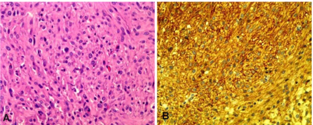
괴사Fig. 5. Microscopically, the mass is composed of cells arranged haphazardly and in interlacing bundles and small whorls
supported by a fine fibrovascular stroma. The cells are spindle to elongate with scant to moderate cytoplasm and a single
elongate nucleus with basophilic aggregated chromatin. Mitoses are 0-2/hpf (A, H & E,
×400). This neoplastic cells are
immunoreactive for c-kit. (B, immunohistochemical stain,
×400).
116 최지혜·김현욱·이혜경·김준영·윤정희
및출혈유무등으로분류했을때
grade 1
정도의비교적낮은악성도를보였다
[1].
하지만, GIST
의악성도는조직학적소견만으로판단하기어렵고임상및다양한 예후인자들을종합적인판단하여결정해야한다는이 전연구
[1]
처럼,
본증례도조직학적분류와는달리종괴의파급으로인해맹장
,
오름결장,
가로결장의벽구 조가완전히소실되고내강이폐색되는등종양의행동 은매우공격적인양상을보여결과적으로환자의예후 는좋지않았다.
GIST
는평활근유래종양과는달리주로혈관을통해전파되므로주변림프절로의전이가적다
.
본증례에서도종양의크기가
10 cm
정도로크고장내강의폐색과파열이발생할정도로진행하였으나
,
체표림프절이나주변장간막림프절로의전이가확인되지않았고다 른장기에서도이상소견은없었다
.
하지만, GIST
로진 단된21
마리중29%
가간이나복강으로전이된보고도있어
, GIST
가의심되는경우에도주변장기로의전이여부를평가해야할것이다
[5].
또한,
위장관종양이발생한
42
마리의개에서GIST
와LMS
의특성을비교한결과평균생존기간이각각
11.6
과7.8
개월로나타났고
,
특히수술전후기간을고려하면각각37.4
와7.8
개월로비교적큰차이를보이는등
, GIST
는평활근유래종양과는생물학적특성이다르다
[3, 8, 13].
따라서,
GIST
와LMS, LM
은조직학적검사상평활근성상을보여감별이매우어려우나
,
추가적인IH
검사를통한감별이반드시필요하다
[1].
위장관종양이의심되는경우방사선검사
,
초음파검사
,
내시경, CT
검사등을통해종괴여부를확인하고,
조직학적검사상방추세포가확인되고 육종의특성을
보이면주로
LMS
와GIST
의감별이필요하다. GIST
는고유의
kit
단백질을IH
검사를통해확인하며, CD117
의발현만으로도
LMS
와명확한구별이가능하다[2, 5].
다른위장관종양은주변림프절을포함한광범위한
절제술이필요한것과는달리
, GIST
는주변림프절로의전이가없어수술적으로종양부분만을절제한다
[3].
사람에서는
GIST
에서특이적으로발현하는kit
단백질길항제를사용하여
GIST
를억제하는연구가이루어지고있다
[6].
본증례는종양의파열로인해보호자가치료를거부하여이러한치료방법을적용하지못하였으나
,
위장관종양특히대장유래종양이발생한경우조직
검사와
IH
검사를통해GIST
로진단된경우수술적절제와
kit
길항제를적용해보는것이추천된다.
결 론
GIST
는원발성위장관질환중중간엽에서유래하는종양으로그발생빈도가비교적높은편이다
.
평활근유래종양과형태학적으로유사하지만
,
혈액을통해전이되고주로소장이나대장에서호발하며치료방법과 예후등다양한특성이서로달라이들종양간의감별 이필수적이다
.
방사선검사와초음파검사등진단영 상을통해위장관종양이의심되는경우조직학적검사 를실시하고,
평활근유래종양의특성을보이는 경우IH
검사를통해GIST
에대한확진이필요하다.
참고문헌
1.
강동욱,
김주헌,
김동훈,
김경희,
박미자,
강대영.
위장관간질종양에서