INTRODUCTION
Probiotics are non-pathogenic microorganisms that confer a health benefit on the host1 by improving the balance of the in- testinal microflora and possibly by augmenting host defenses.
Several studies have characterized the ability of probiotic strains to alter cytokine production in the gut and associated lymphoid tissue, demonstrating immunomodulatory effects on some al- lergic diseases.2-4 Successful probiotic treatments in animal mod- els of asthma have recently been described,5,6 and we have re- ported that oral Lactobacillus rhamnosus (Lcr35) treatment pri- or to allergic sensitization can attenuate airway inflammation and hyperreactivity in a mouse model of allergic airway inflam- mation.7 However, the beneficial effects of probiotics on the de- velopment of allergic asthma and allergic rhinitis in human clin- ical trials remains controversial.8,9 Despite conflicting results, it is generally thought that the immunomodulatory effect of pro- biotics on allergic diseases is a promising area of research.10,11
A number of studies have suggested that the immunosup- pressive cytokines interleukin-10 (IL-10) and transforming
Asthma Prevention by Lactobacillus Rhamnosus in a Mouse Model is Associated With CD4 + CD25 + Foxp3 + T Cells
Seong-Ok Jang,
1Ha-Jung Kim,
1Young-Joon Kim,
1Mi-Jin Kang,
1Ji-Won Kwon,
2Ju-Hee Seo,
2Hyung Young Kim,
2Byoung-Ju Kim,
3Jinho Yu,
2Soo-Jong Hong
2*
1Asan Institute for Life Sciences, University of Ulsan College of Medicine, Seoul, Korea
2Department of Pediatrics, Childhood Asthma Atopy Center, Asan Medical Center, University of Ulsan College of Medicine, Seoul, Korea
3Department of Pediatrics, Inje University Haeundae Paik Hospital, Busan, Korea
growth factor (TGF)-β along with regulatory T (Treg) cells are the main mechanisms by which probiotics suppress allergic in- flammation.2,5,12 Nevertheless, there have been few mechanistic studies of probiotic activity in allergic disease. In the present study, we aimed to confirm the involvement of Treg cells in the protective effect of Lcr35 in a mouse model of allergic asthma.
MATERIALS AND METHODS Mice
Female BALB/c mice weighing 20-25 g (4 weeks old) were purchased from Orient Bio (Orient Bio Inc., Seongnam, Korea) Allergy Asthma Immunol Res. 2012 May;4(3):150-156.
http://dx.doi.org/10.4168/aair.2012.4.3.150 pISSN 2092-7355 • eISSN 2092-7363
Purpose: Probiotic bacteria can induce immune regulation or immune tolerance in allergic diseases. The underlying mechanisms have been recent- ly investigated, but are still unclear. The aim of this study was to evaluate the protective effects of the probiotic Lactobacillus rhamnosus (Lcr35) in a mouse model of asthma and to identify its mechanism of action. Methods: Lcr35 was administered daily by the oral route at a dosage of 1×10⁹ CFU/mouse in BALB/c mice for 7 days before the first sensitization. Clinical parameters and regulatory T (Treg) cells were examined. The role of CD4+CD25+Foxp3+ Treg cells was analyzed using a Treg cell-depleting anti-CD25 monoclonal antibody (mAb). Results: Airway hyperresponsive- ness, total IgE production, pulmonary eosinophilic inflammation, and splenic lymphocyte proliferation were suppressed after Lcr35 treatment. Th1 (IFN-γ) and Th2 (IL-4, IL-5, and IL-13) cytokines in the serum were suppressed, and the percentage of CD4+CD25+Foxp3+ Treg cells in the spleen was significantly increased in the Lcr35 treatment group. Anti-CD25 mAb administration abolished the protective effects of Lcr35, indicating that CD4+ CD25+Foxp3+ Treg cells are essential in mediating the activity of Lcr35. Conclusions: Oral administration of Lcr35 attenuated the features of aller- gic asthma in a mouse model and induced immune regulation by a CD4+CD25+Foxp3+ Treg cell-mediated mechanism.
Key Words: Asthma; probiotics; mice; T-lymphocytes, Regulatory
This is an Open Access article distributed under the terms of the Creative Commons Attribution Non-Commercial License (http://creativecommons.org/licenses/by-nc/3.0/) which permits unrestricted non-commercial use, distribution, and reproduction in any medium, provided the original work is properly cited.
Correspondence to: Soo-Jong Hong, MD, PhD, Department of Pediatrics, Childhood Asthma Atopy Center, Asan Medical Center, University of Ulsan College of Medicine, Pungnap 2-dong, Songpa-gu, Seoul 138-736, Korea.
Tel: +82-2-3010-3379; Fax: +82-2-473-3725; E-mail: sjhong@amc.seoul.kr Received: June 22, 2011; Revised: September 26, 2011;
Accepted: November 1, 2011
•Seong-Ok Jang and Ha-Jung Kim contributed equally to this work.
•There are no financial or other issues that might lead to conflict of interest.
and treated in accordance with the guidelines of the Institu- tional Animal Care and Use Committee (IACUC) at Asan Medi- cal Center and Ulsan University College of Medicine.
Probiotic preparation
The Lcr35 used in this study was obtained from the Lyocen- tre® laboratory (Aurillac, France) and was cultured in sterile Lactobacillus MRS broth (Difco, Sparks, MD, USA) at 37°C for 24 hours. After cultivation, cells were collected by centrifuga- tion at 5,000×g for 10 minutes, washed twice with phosphate buffered saline (PBS), and lyophilized. The lyophilized powder was reconstituted with sterile saline before use.
Establishment of allergic asthma and probiotic treatment Six-week-old female BALB/c mice (N=7 per group) were sen- sitized by intraperitoneal (i.p.) administration of an ovalbumin (OVA; 10 µg, grade V; Sigma Chemical Co., St. Louis, MO, USA) and alum (2.25 mg; Imject®, Pierce, Rockford, IL, USA) mixture.
One week after the first sensitization, the mixture was adminis- tered a second time. Seven days later, the mice inhaled 1% OVA via an ultrasonic sprayer (Nescosonic UN-511; Alfresa, Osaka, Japan) for 30 minutes daily for three successive days (OVA chal- lenge). The mice received Lcr35 (1×10⁹ colony-forming units/
600 μL/mouse/day) orally from one week before primary sensi- tization to the endpoint of the study. Negative controls received only saline instead of OVA at both sensitizations and airway challenge. Positive controls received nothing more after OVA sensitization.
Clinical evaluations in vivo
Airway hyperresponsiveness (AHR) in response to inhaled methacholine (MeCh; Sigma Chemical Co.), administered 24 hours after OVA challenge, was measured in conscious, unre- strained mice using a barometric whole-body plethysmograph (Buxco; EMKA Technologies, Paris, France). Briefly, mice were placed in a whole-body chamber, and basal readings were ob- tained for 3 minutes and averaged. Aerosolized saline followed by 5-50 mg/mL MeCh were inhaled for 3 minutes after each MeCh inhalation.
Treg cells were depleted using anti-CD25 monoclonal anti- body (mAb). Briefly, mice received 250 μg of rat anti-mouse CD25 mAb (clone PC61; eBioscience, San Diego, CA, USA) i.p.
in 400 μL of normal saline one day before 1% OVA challenge.
Control mice were injected with 250 μg of rat IgG1 (Sigma Chemical Co.).
Bronchoalveolar lavage (BAL) fluid analysis
After measurement of AHR, the mice were anesthetized by i.p.
administration of ketamine-xylazine, and the trachea was im- mediately exposed. The airways were lavaged through a trache- al cannula, two times with 1-mL aliquots of pyrogen-free saline warmed to 37°C. The recovered lavage fluid was pooled, and
the cells were collected by centrifugation (5,000 rpm, 4°C, 5 minutes) and resuspended in 100 mL of cold PBS. The cells were stained with trypan blue to determine viability, and total nucleated cells were counted using a hemocytometer.
For differential BAL cell counts, cytospin preparations were made and stained with Diff-Quik (Sysmex, Takatsukadai, Ja- pan). After the samples were coded, all cytospin preparations were evaluated by one observer using an oil immersion micro- scope (magnification, ×1,000). At least 200 cells were counted per preparation, and the absolute number of each cell type was calculated.
Serum Ig analysis
Serum was obtained from blood taken during exsanguination of the mice after airway measurement, and 100 μL (1/10 dilu- tion in carbonate-bicarbonate buffer) were added to each well of a 96-well plate. An IgE-specific enzyme linked immunosor- bent assay (ELISA) was used to quantitate total IgE in the se- rum, using matching antibody pairs (eBioscience) according to the manufacturer’s instructions. For the ELISA, 96-well plates were first coated overnight with rat anti-mouse IgE (10 μL in 100 μL of PBS; PharMingen, San Diego, CA, USA), rat anti- mouse IgG1 (20 μg in 100 μL of PBS; PharMingen), or rat anti- mouse IgG2a (20 μg in 100 μL of PBS; PharMingen). The remain- ing binding sites were blocked, and the plates were incubated with 100 μL of diluted serum (1:5 for IgE, 1:10 for IgG1 or IgG2a).
After the plate was washed, each of the following was sequen- tially added, incubated, and removed by washing: OVA (1 μg/
100 μL), peroxidase-labeled rabbit anti-OVA Ig (240 ng/100 μL, PharMingen), and 3,3,5,5-tetramethylbenzidine solution (Sig- ma Chemical Co.). The optical density was measured at 450 nm, and the Ig level was determined relative to that of a refer- ence pool of serum from OVA-sensitized BALB/c mice (assigned a value of 100 experimental units/mL). Determinations were performed in duplicate.
Cytokine assays
Commercial preparations of paired antibodies and protein standards for measurements of mouse IL-4, IL-5, IL-13, and IFN-γ (eBioscience) in sera were used to develop ELISAs ac- cording to the manufacturer’s instructions. Determinations were performed in duplicate.
Lymphocyte proliferation assay
After BAL, the mouse spleen was resected. Mouse splenocytes were separated on a Histopaque (Sigma Chemical Co.) gradient, and the collected cells were washed with PBS. RBCs were lysed by gently mixing the cells with 3.6 mL of 0.24% NaCl for 20 sec- onds, followed by the quick addition of 0.3 mL of 8.7% NaCl and further dilution with PBS. The pellet was suspended in Is- cove’s Modified Dulbecco’s Medium (IMDM), and stored over- night at 4°C. The next morning, the cells were centrifuged at
4°C, suspended in cold PBS, stained with trypan blue, and counted using a hemocytometer.
Splenic T cells were cultured in IMDM supplemented with 25 mM HEPES, 10% (v/v) heat inactivated fetal bovine serum (FBS), 60 mg/L (100 U/mL) penicillin, 100 mg/L streptomycin, and 0.29 g/L L-glutamine. Splenic T cells were adjusted to 1×10⁵ cells/200 μL/well, transferred to 96-well plates, and incubated at 37°C in a humidified 5% CO2 incubator for 72 hours. The cells were stimulated with OVA treatment (100 μg/mL) for 72 hours.
At 12 hours before the end of the incubation, 1 μCi of [3H]-thy- midine was added to each well. The cells were harvested onto a glass microfiber filter (Simport, Beloeil, Canada), and radioac- tivity was measured in a liquid scintillation counter. The incor- poration during the last 12 hours of culture (counts per minute) was used as an index of proliferation. All cultures were tested in triplicate.
Flow cytometry
Mouse Treg cells were collected from the spleen and analyzed for CD4+CD25+Foxp3+ expression using a mouse Treg cell stain- ing kit containing FITC-labeled anti-CD4, APC-labeled anti- CD25, and PE-labeled anti-Foxp3 (eBioscience) according to the manufacturer’s instructions. Briefly, prepared cells (1×10⁶) were washed by centrifugation with cold PBS, resuspended in 1 mL of fixation/permeabilization solution, and incubated in the dark at 4°C for 30-60 minutes. The cells were washed once with 2 mL of permeabilization buffer, collected by centrifugation, re- suspended in 20 mL of blocking agent with 2% (2 mL) normal rat serum in permeabilization buffer, and incubated at 4°C for 15 minutes. Next, 20 mL of fluorochrome-conjugated antibody or isotype control in permeabilization buffer were added, fol- lowed by incubation in the dark at 4°C for 30 minutes. Finally, the cells were washed with 2 mL of permeabilization buffer, re- suspended in flow cytometry buffer (PBS with 2% FBS), and analyzed by flow cytometry using a FACSCalibur with CellQuest software (BD Biosciences, Mountain View, CA, USA). Determi- nations were performed in duplicate.
Lung histopathology
For the histological evaluation of lung tissue, the left lung of each mouse was embedded in paraffin, sectioned to a thickness of 5 μm, and stained with hematoxylin and eosin (H&E) to as- sess eosinophilic infiltration. Inflammation was scored by two independent, blinded investigators. The degree of peribronchi- al and perivascular inflammation was evaluated on a subjec- tive scale of 0-3, as described elsewhere.13 Cellular infiltration in five randomly selected fields was assessed under a Zeiss Axio- phot microscope (magnification, ×100; Carl Zeiss, Inc., Thorn- wood, NY, USA).
Statistical analysis
We analyzed the association between groups and positive
controls using the Mann-Whitney test. Data are expressed as means±standard error. All statistical analyses were performed using SPSS ver. 18.0 for Windows (SPSS Inc., Chicago, IL, USA).
A P value<0.05 indicated significance.
RESULTS
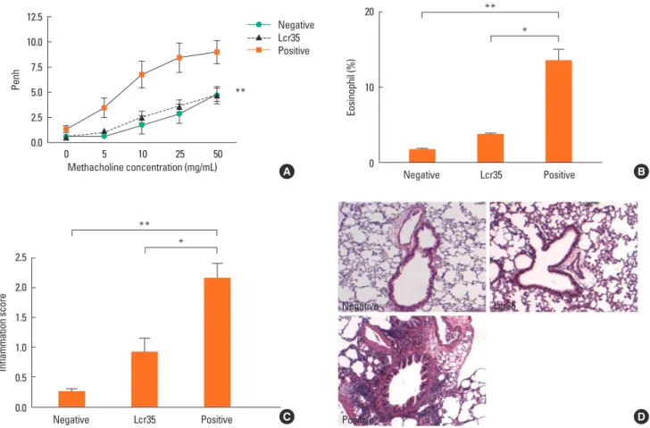
Effect of Lcr35 treatment on maximal Penh and airway eosinophilia
Lcr35 administration to allergic mice significantly suppressed AHR, to the level of the negative control group at the maximum MeCh dose (negative control: 4.72±2.19; Lcr35-treated: 4.75±
1.56; positive control: 9.00±2.87; P<0.01) (Fig. 1A).
Lcr35-treated mice exhibited a significant suppression of BAL fluid total cell count compared with positive controls (Lcr35- treated: 4.77×10⁵ cells/mL; positive control: 8.29×10⁵ cells/mL;
P<0.05). Oral administration of Lcr35 significantly reduced the number of eosinophils in BAL fluid compared with positive controls (Lcr35-treated: 3.79±0.29%; positive control: 13.60±
3.00%; P<0.05) (Fig. 1B).
Lung inflammation after OVA inhalation was significantly re- duced by oral administration of Lcr35 (Lcr35-treated: 2.16±
0.55; positive control: 0.92±0.52; P<0.05) (Fig. 1C). Examina- tion of the lung tissue of positive control mice revealed peri- bronchial and perivascular cellular infiltrates; Lcr35 treatment produced a marked decrease in both cellular infiltration and inflammatory changes, as determined by histopathology (Fig.
1D). Thus, Lcr35 treatment inhibited allergen-induced pulmo- nary inflammation, including the influx of eosinophils.
Mechanism of the effect of Lcr35 on allergic responses The administration of Lcr35 significantly reduced the total IgE level compared with that of the positive control, but had no sig-
Table. Systemic immune responses and cytokine levels of each group
Parameter Group Negative Lcr35 Positive
Systemic immunoglobulins
Total IgE (pg/mL) 4,041±229.3* 5,777±387.7† 7,427±381.1 OVA-specific IgE
(ng/mL) 4,232±170.0* 6,370±423.5 7,666±391.2
OVA-specific IgG1 (μg/mL)
3,978±457.7* 7,201±518.3 7,813±553.8 OVA-specific IgG2a
(μg/mL) 36.48±13.25* 68.74±3.11 75.90±3.83
Serum cytokines
IL-4 (pg/mL) 3.96±0.28* 4.68±0.52† 7.68±0.74
IL-5 (pg/mL) 42.60±5.67* 84.40±6.49† 151.0±14.29 IL-13 (pg/mL) 441.4±29.6* 957.5±35.6† 1,377.0±112.1 IFN-γ (pg/mL) 87.76±12.43† 176.00±5.30 191.60±21.19
*P<0.01; †P<0.05 (compared with the positive control).
Lcr35, Lactobacillus rhamnosus; OVA, ovalbumin.
nificant effect on OVA-specific IgE, IgG1, or IgG2a (Table). Se- rum IL-4, -5, and -13 levels were significantly suppressed by orally administered Lcr35. The IFN-γ level was also decreased in Lcr35-treated mice, but the difference was not significant (Table). These results suggest that Lcr35 treatment suppresses Th2-dependent cytokines. OVA-induced cell proliferation was significantly lower in Lcr35-treated mice than in positive con- trol mice (Fig. 2). Thus, Lcr35 effectively inhibited the OVA-spe- cific splenic T cell response.
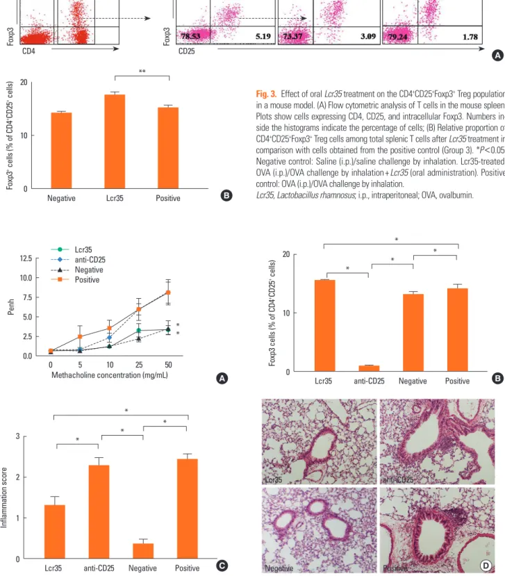
Lcr35 treatment led to a significant increase in CD4+CD25+ Foxp3+ Treg cells, compared with the percentage in positive control mice (Lcr35-treated: 17.60±0.49%; positive control:
15.20±0.44%; P<0.05) (Fig. 3B). This suggests that Lcr35-in- duced attenuation of allergic responses in the mouse model of asthma is associated with an increased CD4+CD25+Foxp3+ Treg cell population. To confirm the involvement of CD4+CD25+ Foxp3+ Treg cells, Treg cell depletion was achieved by anti- CD25 mAb treatment. Anti-CD25 mAb treatment before OVA challenge significantly increased AHR at the maximum MeCh dose, compared with AHR in Lcr35-treated mice (anti-CD25- treated: 8.16±1.63; Lcr35-treated: 3.30±0.57; P<0.05) (Fig. 4A).
Fig. 2. Effect of oral Lcr35 treatment on the suppression of OVA-specific splen- ic T cell proliferation in a mouse model. *P<0.05 and **P<0.01. Negative con- trol: Saline (i.p.)/saline challenge by inhalation. Lcr35-treated: OVA (i.p.)/OVA challenge by inhalation+Lcr35 (oral administration). Positive control: OVA (i.p.)/
OVA challenge by inhalation.
Lcr35, Lactobacillus rhamnosus; OVA, ovalbumin; i.p., intraperitoneal.
3[H]-thymidine incorporation (cpm) 7,000 6,000 5,000 4,000 3,000 2,000 1,000
0 Negative Lcr35 Positive
**
*
OVA (-) OVA (+)
Fig. 1. Effect of oral Lcr35 treatment on allergic asthma in a mouse model. (A) Airway hyperresponsiveness; (B) Eosinophil proportion in bronchoalveolar lavage flu- id; (C) Peribronchial and perivascular lung inflammation score; (D) Lung pathology. *P<0.05 and **P<0.01. Negative control: Saline (i.p.)/saline challenge by inhala- tion. Lcr35-treated: OVA (i.p.)/OVA challenge by inhalation+Lcr35 (oral administration). Positive control: OVA (i.p.)/OVA challenge by inhalation.
Lcr35, Lactobacillus rhamnosus; i.p., intraperitoneal; OVA, ovalbumin.
Eosinophil (%)
20
10
0 Negative Lcr35 Positive
*
**
Inflammation score
2.5 2.0 1.5 1.0 0.5
0.0 Negative Lcr35 Positive
*
**
A B
C D
Negative Lcr35
Positive
Penh
12.5 10.0 7.5 5.0 2.5
0.0 0 5 10 25 50
Methacholine concentration (mg/mL)
Negative Lcr35 Positive
**
Negative Lcr35 Positive
Foxp3+ cells (% of CD4+CD25+ cells) 20
10
0
**
Foxp3
CD4
R2
Foxp3
CD25
Negative Lcr35 Positive
Fig. 3. Effect of oral Lcr35 treatment on the CD4+CD25+Foxp3+ Treg population in a mouse model. (A) Flow cytometric analysis of T cells in the mouse spleen.
Plots show cells expressing CD4, CD25, and intracellular Foxp3. Numbers in- side the histograms indicate the percentage of cells; (B) Relative proportion of CD4+CD25+Foxp3+ Treg cells among total splenic T cells after Lcr35 treatment in comparison with cells obtained from the positive control (Group 3). *P<0.05.
Negative control: Saline (i.p.)/saline challenge by inhalation. Lcr35-treated:
OVA (i.p.)/OVA challenge by inhalation+Lcr35 (oral administration). Positive control: OVA (i.p.)/OVA challenge by inhalation.
Lcr35, Lactobacillus rhamnosus; i.p., intraperitoneal; OVA, ovalbumin.
A
B
Fig. 4. Effect of anti-CD25 mAb treatment on allergic asthma in a mouse model after oral Lcr35-treatment. (A) Airway hyperresponsiveness; (B) Relative proportion of CD4+CD25+Foxp3+ Treg cells among total splenic T cells; (C) Comparison of pulmonary inflammation scores; (D) Lung pathology (H&E stain; ×100). *P <0.05. Lcr35- treated: OVA (i.p.)/OVA challenge by inhalation+Lcr35 (oral administration). Anti-CD25: OVA (i.p.)/OVA challenge by inhalation+Lcr35 (oral administration)+anti-CD25 Ab (i.p.). Negative control: Saline (i.p.)/saline challenge by inhalation. Positive control: OVA (i.p.)/OVA challenge by inhalation.
Lcr35, Lactobacillus rhamnosus; OVA, ovalbumin; i.p., intraperitoneal.
Penh
12.5 10.0 7.5 5.0 2.5
0.0 0 5 10 25 50
Methacholine concentration (mg/mL) Lcr35
anti-CD25 Negative Positive
Foxp3 cells (% of CD4+CD25+ cells) 20
10
0 Lcr35 anti-CD25 Negative Positive
*
* *
*
Inflammation score
3
2
1
0 Lcr35 anti-CD25 Negative Positive
*
* *
*
A B
C
Lcr35 anti-CD25
Negative Positive
**
D
The isotype control rat IgG1 had no effect on AHR (data not shown). Anti-CD25 treatment also significantly decreased the population of CD4+CD25+Foxp3+ Treg cells in Lcr35-treated mice (anti-CD25 treated: 1.00±0.08%; Lcr35-treated: 15.53±
0.10%; P<0.05) (Fig. 4B). These results suggest that CD4+CD25+ Foxp3+ Treg cells are involved in the suppression of AHR medi- ated by oral administration of Lcr35.
The pulmonary inflammation score was also significantly higher in anti-CD25 mAb-treated Lcr35 mice compared with Lcr35-treated mice that did not receive anti-CD25 mAb (anti- CD25 treated: 2.28±0.19; Lcr35-treated 1.32±0.20; P<0.01) (Fig. 4C). The histopathology revealed enhanced eosinophilic inflammation in the lungs of anti-CD25-treated Lcr35 mice compared with anti-CD25 mAb-non-treated Lcr35 mice (Fig.
4D). The data indicate that administration of anti-CD25 mAb reverses the effect of oral Lcr35 treatment on lung inflamma- tion and that Treg cells mediate the effect of Lcr35.
DISCUSSION
This study examined the effect of oral Lcr35 on allergic asth- ma in a mouse model. The major finding was that Lcr35 sup- presses allergic parameters, including AHR, airway inflamma- tion, and total IgE, by regulating the activity of CD4+CD25+Foxp3+ Treg cells. In addition, these effects were reversed by adminis- tration of anti-CD25 mAb.
The effect of probiotics on Th1 cytokines is controversial, as probiotics have been shown to promote, inhibit, and have no effect on Th1 cytokine production.14-16 In our study, serum lev- els of Th2 cytokines were reduced by oral Lcr35 treatment, whereas Th1 cytokine levels were not significantly reduced. We did not determine cytokine levels in target organs or BAL fluid;
this is a limitation of this study.
To test our hypothesis, CD4+CD25+Foxp3+ Treg cell numbers in the spleen were determined. Treg cell numbers were signifi- cantly higher after Lcr35 treatment. Exacerbation readouts of asthma after Treg depletion demonstrated the importance of Treg cells in the protective effect of Lcr35. This suggests that Treg cells may be involved in the protection conferred by oral Lcr35 in the mouse model of asthma. However, other potential mechanisms such as immunosuppressive cytokines (IL-10 and TGF-β) were not examined. To our knowledge, this is the first study to identify the mechanism of action of probiotics using depletion of CD4+CD25+Foxp3+ Treg cells in a mouse model of asthma.
Some reports have suggested that the involvement of thymus- derived natural Treg cells expressing Foxp3 and CD25 in toler- ance to allergens is unlikely, and the expression of Foxp3 and CD25 on Treg cells remains controversial.10 Further studies are needed to confirm the mechanism underlying the protective effect of Lcr35.
Many studies have attempted to identify the mechanism of
action of probiotics in allergic diseases.2-4 Several have implicat- ed Treg cells in the activity of probiotics,2,17 but these studies did not investigate changes in the inflammatory response after Treg cell depletion. Our study is important in that it confirms previous hypotheses that Treg cells contribute to the protective effect of probiotics.
Oral administration of Lcr35 completely blocked OVA-specific proliferation of splenic T cells in the present study. This sug- gests that Lcr35 regulates the systemic immune response in OVA-sensitized and -challenged mice. Oral administration of probiotics induces regulatory dendritic cells, which in turn pro- mote the generation of CD4+Foxp3+ Treg cells in mesenteric lymph nodes.2 Dendritic cells can directly present antigens from commensal bacteria to mesenteric lymph nodes and in- teract with T and B cells to maintain a non-inflammatory im- mune response.3,4
In contrast to the massive eosinophilic infiltration in the peri- bronchial and perivascular areas of positive control mice, ani- mals treated with Lcr35 showed significantly less eosinophilic inflammation. This was coincident with decreased production of the Th2 cell-derived cytokines IL-4, IL-5, and IL-13 in the Lcr35 treatment group.
There is strong evidence implicating CD4+CD25+ Treg cells in controlling allergic diseases. For example, the transfer of CD4+ CD25+ Treg cells to sensitized mice reduced AHR, eosinophilic inflammation, and Th2 cytokine induction in the lung, in an IL- 10-dependent manner.18,19 It is widely believed that administra- tion of an anti-CD25 mAb results in the rapid and efficient de- pletion of CD4+CD25+ Treg cells, and this has been confirmed by secondary staining with a mAb directed against a different CD25 epitope.20,21 Previous studies have shown that the num- ber of CD4+CD25+ Treg cells was significantly decreased within 3 days of anti-CD25 mAb treatment in vivo, but had recovered to normal levels by day 10.22,23 Therefore, in this study, we ad- ministered anti-CD25 mAb one day before OVA challenge. As expected, the anti-allergic effects of Lcr35 disappeared, and the CD4+CD25+Foxp3+ Treg cell population was significantly re- duced. This demonstrates that the beneficial effect of Lcr35 on allergic asthma in the mice was mediated by CD4+CD25+Foxp3+ Treg cells.
In conclusion, we demonstrated that the suppression of aller- gic responses and immunomodulation by the probiotic Lcr35 in a mouse model of asthma was mediated by the activity of CD4+CD25+Foxp3+ Treg cells.
ACKNOWLEDGMENTS
This work was supported by Basic Science Research Program through the National Research Foundation of Korea (NRF) funded by the Ministry of Education, Science and Technology (MEST) (NRF-2007-313-E00268).
REFERENCES
1. Marteau P. Probiotics, prebiotics, synbiotics: ecological treatment for inflammatory bowel disease? Gut 2006;55:1692-3.
2. Kwon HK, Lee CG, So JS, Chae CS, Hwang JS, Sahoo A, Nam JH, Rhee JH, Hwang KC, Im SH. Generation of regulatory dendritic cells and CD4+Foxp3+ T cells by probiotics administration sup- presses immune disorders. Proc Natl Acad Sci U S A 2010;107:2159- 64.
3. Macpherson AJ, Uhr T. Induction of protective IgA by intestinal dendritic cells carrying commensal bacteria. Science 2004;303:
1662-5.
4. Huang FP, Platt N, Wykes M, Major JR, Powell TJ, Jenkins CD, MacPherson GG. A discrete subpopulation of dendritic cells trans- ports apoptotic intestinal epithelial cells to T cell areas of mesen- teric lymph nodes. J Exp Med 2000;191:435-44.
5. Feleszko W, Jaworska J, Rha RD, Steinhausen S, Avagyan A, Jaud- szus A, Ahrens B, Groneberg DA, Wahn U, Hamelmann E. Probiot- ic-induced suppression of allergic sensitization and airway inflam- mation is associated with an increase of T regulatory-dependent mechanisms in a murine model of asthma. Clin Exp Allergy 2007;
37:498-505.
6. Hong HJ, Kim E, Cho D, Kim TS. Differential suppression of heat- killed lactobacilli isolated from kimchi, a Korean traditional food, on airway hyper-responsiveness in mice. J Clin Immunol 2010;30:
449-58.
7. Yu J, Jang SO, Kim BJ, Song YH, Kwon JW, Kang MJ, Choi WA, Jung HD, Hong SJ. The effects of Lactobacillus rhamnosus on the pre- vention of asthma in a murine model. Allergy Asthma Immunol Res 2010;2:199-205.
8. Kalliomäki M, Salminen S, Arvilommi H, Kero P, Koskinen P, Iso- lauri E. Probiotics in primary prevention of atopic disease: a ran- domised placebo-controlled trial. Lancet 2001;357:1076-9.
9. Helin T, Haahtela S, Haahtela T. No effect of oral treatment with an intestinal bacterial strain, Lactobacillus rhamnosus (ATCC 53103), on birch-pollen allergy: a placebo-controlled double-blind study.
Allergy 2002;57:243-6.
10. Shida K, Takahashi R, Iwadate E, Takamizawa K, Yasui H, Sato T, Habu S, Hachimura S, Kaminogawa S. Lactobacillus casei strain Shirota suppresses serum immunoglobulin E and immunoglobu- lin G1 responses and systemic anaphylaxis in a food allergy model.
Clin Exp Allergy 2002;32:563-70.
11. Pochard P, Gosset P, Grangette C, Andre C, Tonnel AB, Pestel J, Mercenier A. Lactic acid bacteria inhibit Th2 cytokine production by mononuclear cells from allergic patients. J Allergy Clin Immu- nol 2002;110:617-23.
12. Smits HH, Engering A, van der Kleij D, de Jong EC, Schipper K, van
Capel TM, Zaat BA, Yazdanbakhsh M, Wierenga EA, van Kooyk Y, Kapsenberg ML. Selective probiotic bacteria induce IL-10-produc- ing regulatory T cells in vitro by modulating dendritic cell function through dendritic cell-specific intercellular adhesion molecule 3-grabbing nonintegrin. J Allergy Clin Immunol 2005;115:1260-7.
13. Tournoy KG, Kips JC, Schou C, Pauwels RA. Airway eosinophilia is not a requirement for allergen-induced airway hyperresponsive- ness. Clin Exp Allergy 2000;30:79-85.
14. Borchers AT, Selmi C, Meyers FJ, Keen CL, Gershwin ME. Probiot- ics and immunity. J Gastroenterol 2009;44:26-46.
15. Torii A, Torii S, Fujiwara S, Tanaka H, Inagaki N, Nagai H. Lactoba- cillus acidophilus strain L-92 regulates the production of Th1 cyto- kine as well as Th2 cytokines. Allergol Int 2007;56:293-301.
16. Segawa S, Nakakita Y, Takata Y, Wakita Y, Kaneko T, Kaneda H, Wa- tari J, Yasui H. Effect of oral administration of heat-killed Lactoba- cillus brevis SBC8803 on total and ovalbumin-specific immuno- globulin E production through the improvement of Th1/Th2 bal- ance. Int J Food Microbiol 2008;121:1-10.
17. Mastrangeli G, Corinti S, Butteroni C, Afferni C, Bonura A, Boiri- vant M, Colombo P, Di Felice G. Effects of live and inactivated VSL#3 probiotic preparations in the modulation of in vitro and in vivo allergen-induced Th2 responses. Int Arch Allergy Immunol 2009;150:133-43.
18. Kearley J, Barker JE, Robinson DS, Lloyd CM. Resolution of airway inflammation and hyperreactivity after in vivo transfer of CD4+
CD25+ regulatory T cells is interleukin 10 dependent. J Exp Med 2005;202:1539-47.
19. Kearley J, Robinson DS, Lloyd CM. CD4+CD25+ regulatory T cells reverse established allergic airway inflammation and prevent air- way remodeling. J Allergy Clin Immunol 2008;122:617-24.e6.
20. McHugh RS, Shevach EM. Cutting edge: depletion of CD4+CD25+
regulatory T cells is necessary, but not sufficient, for induction of organ-specific autoimmune disease. J Immunol 2002;168:5979-83.
21. Kohm AP, Williams JS, Miller SD. Cutting edge: ligation of the glu- cocorticoid-induced TNF receptor enhances autoreactive CD4+ T cell activation and experimental autoimmune encephalomyelitis. J Immunol 2004;172:4686-90.
22. Kohm AP, Williams JS, Bickford AL, McMahon JS, Chatenoud L, Bach JF, Bluestone JA, Miller SD. Treatment with nonmitogenic an- ti-CD3 monoclonal antibody induces CD4+ T cell unresponsive- ness and functional reversal of established experimental autoim- mune encephalomyelitis. J Immunol 2005;174:4525-34.
23. Kohm AP, McMahon JS, Podojil JR, Begolka WS, DeGutes M, Kasprowicz DJ, Ziegler SF, Miller SD. Cutting Edge: Anti-CD25 monoclonal antibody injection results in the functional inactiva- tion, not depletion, of CD4+CD25+ T regulatory cells. J Immunol 2006;176:3301-5.