Abstract (J. Kor. Oral Maxillofac. Surg. 2009;35:187-192)
Introduction
Primary marginal zone B-cell lymphoma was first reported by Sheibani et al, as a monocytoid B-cell lymphoma, and recently
1), WHO classified it as non-metastatic primary lym- phoma, which occur within the lymph node. Histopathologi- cally, infiltration of tumorous lymphocytes and monocytoid B- cells is observed around lymph nodes, blood vessels and lymph follicles.
Lymphoma in the oral cavity can occur primarily, and also, much more often, secondarily from the lymphoma which occurred in the other region. They are localized commonly in the palatine tonsil, which is part of the Waldeyer’s ring, an interesting lymphoid barrier between the lymph node and the lymphoid system associated to the mucosa. Waldeyer’s ring lymphomas constitute nearly 5-10% of malignant lymphoma.
It is rare in the jaw, vestibule, gingival mucosa and is very sel- dom in the tongue region
2). According to serial papers dealing with head and neck lymphomas, only 3% of the head & neck lymphoma can be considered as the primary marginal zone B- cell lymphoma
3,4).
In the past, lymphomas of the oral cavity as were classified
by ordinary classification for nodal lymphoma in other extra- nodal sites. Most of them were B-cell lymphomas or T cell lymphoma and rarely were Hodgkin’s lymphomas. The patho- logical characteristics of MALT lymphoma in the oral cavity was defined after Isaacson and Write (1983) et al reported a series of patients with lymphoma B gastrointestinal of low grade of malignancy. Since then, MALT lymphoma in the sali- vary gland in relation with the Sjo
¨gren’s disease was reported, however it occurred mostly within the Waldeyer’s ring and was reported to occur seldom in the tongue
5).
This case report presents a 71-year-old female with primary MALT lymphoma showing lymphocyte infiltration in the minor salivary gland in the lateral surface of the tongue.
Clinical case
A 71-year-old female patient visited the clinic with a chief complaint of a bilateral mass on the lateral surface of the tongue with the onset of about 20 days. She also complained of dry mouth with burning sensations. She didn’t show any sys- temic symptoms such as weight loss, nocturnal diaphoresis or fever. She also didn’t have any unusual past medical history or family history.
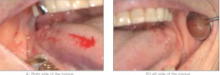
The lateral surface of the tongue showed some bilateral swelling, and the mass was soft, with no induration nor tender- ness to palpation. The surface of the lesion showed redness and seemed to adhere with adjacent tissues(Fig. 1). No specific systemic history was present and biopsy was carried out on her first visit due to malignancy suspicion.
이 백 수
경희대학교 치과대학 부속병원 구강악안면외과
130-701 서울특별시동대문구회기동1 경희대학교치과대학구강악안면외과학교실 Lee Baek-Soo
Dept. of Oral and Maxillofacial Surgery, KyungHee University Dental School, Hoegi 1, Dongdaemunku, Seoul, 130-701, Korea
TEL: 02-958-9440 E-mail: [email protected]
Low-grade mucosa-associated lymphoid tissue(MALT) marginal zone B-cell lymphoma of the tongue-A case report
Joo-Young Ohe, Baek-Soo Lee, Yeo-Gab Kim, Yong-dae Kwon, Byung-jun Choi, Young-Ran Kim Department of Oral & Maxillofacial Surgery, Kyung Hee University dental college, Seoul, Korea
Out of all oral malignant tumor, malignant lymphoma occurs in only 3.5%. Especially, most of the primary malignant lymphomas, which occur in the head & neck region are high-grade diffuse large B-cell lymphoma and mucosa-associated lymphoid tissue (MALT) marginal zone B-cell lym- phoma is very rare. In the head & neck region, malignant lymphoma is reported to occur in the thyroid, salivary gland, trachea, larynx, orbital lobe and the Waldeyer’s ring. Among the Waldeyer’s ring, palatal tonsil is reported to be the most common region, but, only 1 case report was published in Korea. Until now, there were no case reports of MALT lymphoma that occurred in the tongue. The purpose of this case report is to report and discuss on a case of MALT lymphoma of the tongue.
Key words: MALT lymphoma, Tongue
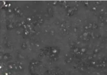
Histologically, central cell atypical lymphoid cells were observed. The size of the cells varied a lot and some cells showed Dutcher body(intranuclear inclusion) and plasmocytic differentiation, a typical histologic finding of MALT lym- phoma. Squamous epithelium entrapped by atypical lymphoid cells with formation of so-called lymphoepithelial lesion.
Some showed reactive germinal centers, well polarized and with starry-sky histiocytes(Fig. 2).
Immunohistologic test results displayed positive with the markers of CD20, CD43, CD79a, Bcl-2 etc, and negative result with the markers of CD3, Bcl-6, CD10, TDT(Fig. 3-6).
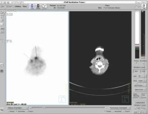
Then MRI of the craniocervical region and systemic PET CT scan were taken. In MRI, soft tissue mass formation was observed on the lingual and palatine tonsil. MRI showed
swelling of the tongue tonsil, which is suspected to be a lym- phoma, and showed a low intensity signal on the T2W1 image(Fig. 7). An enlarged LN at the right internal jugular chain(level III) was observed, but its size and shape did not give an impression of metastases and other salivary glands did not show any unusual findings. On the systemic PET CT scan, a slightly imbalanced intake of FDG was found on the base and ventral part of the tongue and was spreaded out inferiorly, showing a possibility of malignancy(Fig. 8). Systemically no unusual FDG intake was observed, and it was concluded that there were no signs of metastases. The lesion was completely excised and she received radiation therapy after transferring to a different hospital.
Fig. 1.Lateral surface of the tongue showed bilateral swelling with slightly redness & seemed to adhere with adjacent tissues.
Fig. 2.Neoplastic lymphoid cells infiltrate squamous epithelium resembling a lymphoepithelial lesion(H-E stain X100). Large sized lymphoma cells are diffusely infiltrated. Most of them have round nuclei, with vesicular chromatin and nucleoli. Some showed reactive germinal centers, well polarized and with starry-sky histiocytes(H-E stain X400).
A) Right side of the tongue B)Left side of the tongue
(H-E stain X100) (H-E stain X400)
Fig. 3. Immunohistochemical stain of CD20 shows diffuse positive reaction in the cell membrane (X200).
Fig. 4. Immunohistochemical stain of CD43 shows diffuse positive reaction in the cell membrane (X200).
Fig. 5. Immunohistochemical stain of CD79 shows diffuse positive reaction in the cell membrane (X200).
Fig. 6. Immunohistochemical stain of bcl-2 shows diffuse positive reaction in the cell membrane (X200).
Fig. 7.In MRI, soft tissue mass formation was observed on the lingual and palatine tonsil.
Fig. 8-2.Suggestive of metastastic lesion in suprahyoid area.
Fig. 8-1.A slightly imbalanced intake of FDG was found on the base of the tongue.
Discussion
Clinically, MALT lymphoma incline to enter an indolence phase. They tend to remain localized for years and if dissemi- nation occurs, they tend to recur in other extra-nodal sites. This tumor is classified as the B-cell type lymphoma of non- hodgkin lymphoma and is a lymphoepithelial lesion with monocytic B-cell proliferation. Unlike other low-grade B-cell lymphoma, they does not metastases remotely, because they are limited by lymphocyte homing phenomenon, and the growth of the tumor is affected greatly by exposure to anti- gens. Even when they progress higher grade, prognosis is bet- ter respect to the nodal counterpart, because they can be cured by surgical excision
6).
It is reported that chronic inflammation is closely related to the pathogenesis of MALT lymphoma. Due to chronic inflam- mation, antigen-stimulated lymphocyte is transformed malig- nantly, and it is continuously accumulated, and finally takes form a lymphoma
7).
It is hard to diagnose the lesion in the early stage because it shows localization of tumor, insidious growth and no special symptoms. Radiographic tests are useful in deciding on the boundaries of excision, but are not very useful in diagnosing
8).
Histologically, MALT lymphomas repeat the architecture of normal lymphoid follicles surrounded by B cells of medium size like centrocyte cells with the tendency to destroy epithelial structures and to colonise germinal centers. Generally, it is comprised of centrocyte-like cleaved cell, monocytoid B cell, small lymphocyte, diffuse heterogenous infiltration of plasma cell. Intranuclear inclusion(Dutcher body) may be seen in the plasma or lymphoplasmacytic cells. Lymphoepithelial lesions, representing infiltration of ductal and epithelial structurs by neoplastic B cells, are seen in both lymphoepithelial sialadeni- tis and marginal zone B cell lymphoma. An important early change is the formation of “halos”, comprised of monocytoid and centrocyte-like B cell surrounding epimyoepithelial islands(lymphoepithelial lesion)
9). A MALT lymphoma could be suspected and further immunohistological analysis confirms the B cell type of the tumor cells. Among the immunohistolog- ic tests CD3, CD5, CD20, CD43 etc. are tested. Especially CD5 negative result is very useful for differential diagnose is with CD5 positive chronic lymphocytic leukemia and node- based mantle cell lymphoma
10).
When diagnosed as MALT lymphoma, CT images of the neck, chest, abdominal, pelvis area is necessary because of possible distant metastasis and in some cases bone marrow biopsy may be necessary to define the stage of the disease. In
the case of extra-nodal craniocervical lymphoma, it is reported that metastasis in the bone marrow is found in 25% of the cas- es. Also when CT scan results are doubtful, upper gastro- intestinal microscopy is strongly suggested because gastroin- testinal MALT lymphoma can be found together sometimes
10).
Generally, there are several ways to treat MALT lymphoma, chemotherapy, radiation therapy, surgical method and combi- nation therapy. When among them only one therapy is given, chemotherapy is known to be the most effective single method.
Surgical method or radiation therapy is given when chemother- apy is not an option rejection symptoms
11).
According to Tsang et al., MALT lymphoma present in vari- ety of extranodal site with 70-90% of patients presenting with localized(stage IE-IIE) disease. Due to the radiation sensitivity characteristie of MALT lymphoma, a moderate dose local radi- ation treatment(30-35Gy) for these stage is efficacious.
Following IF RT for stage I and II MALT lymphomas, relapse of the disease occurs in 30-40% of patient after 10 years.
Localized relapse of MALT lymphoma usually involves a dif- ferent site and is frequently amenable to further local radiation therapy. Prognosis of MALT lymphoma, compared to other low grade B cell lymphoma, is reported to be relatively good.
Also, since the lesion tends to stay localized in one region for a long time, local therapy such as surgical excision and radiation therapy is possible in many cases showing a good prognosis
12). In this case, becase of long distance from our hospital, she received radiation therapy after transferring to a different hos- pital. And we confirmed that she was treated by radiothera- py(30 Gy), thus resulting in complete remission.
In conclusion, therapeutic strategies for this kind of disease are not standardized yet due to the small number of MALT lymphomas described in the head and neck region. Therefore, it remains to be established whether the currently applied ther- apeutic strategies of combined radiochemotherapy for advanced lesions and either radiation therapy or surgery for small lesions provide the best treatment of MALT lymphomas in this location.
Reference
1. Sheibani K, Sohn CC, Burke JS, Winberg CD, Wu AM, Rappaport H. Monocytoid B-cell lymphoma. A novel B-cell neo- plasm. Am J Pathol 1986;124:310-8.
2. Menarguez J, Mollejo M, Carrion R, Oliva H, Bellas C, Forteza J, et al. Waldeyer ring lymphomas. A clinicopathological study of 79 cases. Histopathology 1994;24:13-22.
3. Fukuda Y, Ishida T, Fujimoto M, Ueda T, Aozasa K. Malignant lymphoma of the oral cavity: clinicopathologic analysis of 20 cases. J Oral Pathol 1987;16:8-12.
4. Wolvius EB, van der Valk P, van der Wal JE, van Diest PJ, Huijgens PC, van der Waal I, et al. Primary extranodal non-
Hodgkin lymphoma of the oral cavity. An analysis of 34 cases.
Eur J Cancer B Oral Oncol 1994;30B:121-5.
5. Isaacson P, Wright DH. Malignant lymphoma of mucosa-associ- ated lymphoid tissue. A distinctive type of B-cell lymphoma.
Cancer 1983;52:1410-6.
6. Isaacson PG, Spencer J. The biology of low grade MALT lym- phoma. J Clin Pathol 1995;48:395-7.
7. Harris NL. Low-grade B-cell lymphoma of mucosa-associated lymphoid tissue and monocytoid B-cell lymphoma. Related enti- ties that are distinct from other low-grade B-cell lymphomas.
Arch Pathol Lab Med 1993;117:771-5.
8. Harris NL, Jaffe ES, Stein H, Banks PM, Chan JK, Cleary ML, et al. A revised European-American classification of lymphoid neo- plasms: a proposal from the International Lymphoma Study
Group. Blood 1994;84:1361-92.
9. Ellis GL. Lymphoid lesions of salivary glands: malignant and be- nign. Med Oral Patol Oral Cir Bucal 2007;12:E479-85.
10. Bhattacharyya N, Frankenthaler RA, Gomolin HI, Kadin ME, Lauretano AM. Clinical and pathologic characterization of mu- cosa-associated lymphoid tissue lymphoma of the head and neck.
Ann Otol Rhinol Laryngol 1998;107:801-6.
11. Yoshino T, Ichimura K, Mannami T, Takase S, Ohara N, Okada H, et al. Multiple organ mucosa-associated lymphoid tissue lym- phomas often involve the intestine. Cancer 2001;91:346-53.
12. Tsang RW, Gospodarowicz MK. Radiation therapy for localized low-grade non-Hodgkin’s lymphomas. Hematol Oncol 2005;
23:10-7.