Bilateral Conjunctival Mucosa-Associated Lymphoid Tissue Type Lymphoma in a Kidney Transplant Recipient
Eun-Young Ji, M.D.
1, Ji-Yeun Chang, M.D.
1, Chul Woo Yang, M.D.
1, Seok-Goo Cho, M.D.
2and Byung Ha Chung, M.D.
1Divisions of Nephrology
1, Hematology
2, Department of Internal Medicine, Seoul St. Mary’s Hospital, College of Medicine, The Catholic University of Korea, Seoul, Korea
Lymphoproliferative disorder in a posttransplant setting has emerged as a difficult problem in kidney transplantation (KT).
Lymphoma involving adnexa of the eye has rarely been reported due to scarcity of lymphoreticular tissue in the ocular area. This report presents a case of a 37-year-old KT recipient who was diagnosed with conjunctival mucosa-associated lymphoid tissue lymphoma with a chief complaint of seeing black spots. Unlike other post-transplant lymphoproliferative diseases associated with the Epstein-Barr virus (EBV) reactivation via immunosuppression, the lesion was not related to the virus. The patient received radiotherapy with concomitant conversion from the tacrolimus to the sirolimus. Overall, the results presented herein indicate lymphoma may be an important differential diagnosis when KT recipients complain of ocular discomfort.
Key Words: Kidney transplantation, Marginal zone B-cell lymphoma, Lymphoproliferative disorders
중심 단어: 신장이식, MALT 림프종, 림프증식성질환Received February 21, 2018 Revised March 31, 2018 Accepted April 23, 2018
Corresponding author: Byung Ha Chung
Division of Nephrology, Department of Internal Medicine, Seoul St.
Mary's Hospital, College of Medicine, The Catholic University of Korea, 222 Banpo-daero, Seocho-gu, Seoul 06591, Korea Tel: 82-2-2258-6066, Fax: 82-2-536-0323
E-mail: [email protected]
INTRODUCTION
Kidney transplantation (KT) is an emerging option for renal replacement therapy that improves the quality of life, as compared to dialysis. However, immunosuppressive ther- apy for the prevention of rejection can lead to lymphoproli- ferative disease after KT up to 1% to 20%(1). The extra- nodal marginal zone lymphoma of mucosa-associated lym- phoid tissue (MALT) type that belonged to the indolent B-cell lymphomas which is mainly an observed gastric mu- cosa and it is associated with the
Helicobacter pylori
in- fection(2-5), has been rarely reported as originating fromother sites. We herein report a case of a KT recipient who is diagnosed with extranodal marginal zone lymphoma in- volving both conjunctivae.
CASE REPORT
A 37-year-old woman with end-stage renal disease due to lupus nephritis received a living-donor KT after 5 years of peritoneal dialysis. The donor was her mother, and the human leukocyte antigen mismatch number was 2. She un- derwent induction therapy by using basiliximab, and there- after maintained immunosuppression with tacrolimus, myco- phenolic acid, and deflazacort. The trough level of tacroli- mus has been maintained between 4∼5 ng/mL. The allog- raft function was kept stable with an estimated glomerular filtration rate of 60 mL/min/1.73 m2, and there was no sur- gical or immunological complication except for urinary tract infection over a 4-year post-transplant period.
Three years and eight months after KT, the patient was admitted to the hospital because of a black spot in her left
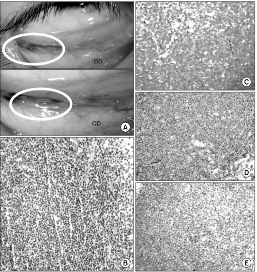
Fig. 1. Clinical appearance (A) and Immunohistochemical staining pro- perties (B-D). (A) Slit-lamp exam- ination showed a salmon color appea- rance of conjunctival mucosa-asso- ciated lymphoid tissue lymphoma.
(B) Monotonous atypical cells infil- trated of mucosa by small lympho- cytes (HE stain, ×400). (C) The specimen was negative to Epstein- Barr virus encoded RNA ( in situ hybridization, ×400). (D) Immuno- histochemistry was diffusely positive for CD20 (×400). (E) Microscopic finding revealed lymphoepithelial lesions (Pancytokeratin, ×400).
eye vision. There were no accompanying symptoms, such as loss of vision, eye pain, or inflammation signs. There was also no recent eye trauma or eye surgery. On admission, her blood pressure was 130/80 mmHg, heart rate was 90 beats/min, and body temperature was 36.7oC. She did not complain of any systemic symptom, such as weight loss, night sweat, or fatigue. There was no palpable mass, sub- cutaneous nodule, or organomegaly. The admission labo- ratory examination showed normal complete blood count (CBC), normal levels of lactate dehydrogenase and liver function tests, and minimally elevated erythrocyte sed- imentation rate. The tests for human immunodeficiency vi- rus and viral hepatitis A, B, and C were all negative.
Cytomegalovirus and Epstein-Barr virus (EBV) were not detected in the real-time polymerase chain reaction test. In the ophthalmic examination, multiple cystic nodules located
in both lower conjunctivae were found. The cystic nodules were biopsied and diagnosed with extranodal marginal zone lymphoma of MALT type (Fig. 1). During the staging workup for lymphoma, orbital magnetic resonance imaging and positron emission tomography computed tomography (PET-CT) showed hypertrophic changes in the lower con- junctivae of both eyes and tonsils (Fig. 2). The bone mar- row finding was normal, and the chromosome analysis of hematology/oncology showed no atypical clone. The stain for EBV was negative. The patient also underwent esoph- agogastroduodenoscopy with a negative result of
H. pylori
infection.The patient’s final diagnosis was extranodal marginal cell MALT lymphoma stage IE, and she was scheduled to re- ceive curative involved field radiation therapy on the con- junctivae with a dose of 25.2 Gy in 14 fractions. The pa-
Fig. 2. (A) Orbit magnetic resonance imaging showed no definite enhance- ment or mass in both orbit conjunc- tivae. (B) Coronal positron emission tomography computer tomography image showed intense fludeoxyglucose uptake in bilateral conjunctivae (white arrows) and both palatine tonsils.
tient’s tacrolimus was converted to 2 mg of sirolimus. At 6 months after the radiotherapy, the patient achieved a clin- ical complete remission without additional imaging studies.
A hematologist evaluated the treatment response by careful clinical judgment including CBC, serum chemistries, and lactate dehydrogenase. The patient tolerated the local radio- therapy without extraorbital relapse or late complications, including keratitis or cataract. We planned to assess her on the risk of relapse every 6 months for at least 5 years.
DISCUSSION
Post-transplant lymphoproliferative disease (PTLD) is one of the potentially fatal complications after KT that has been known to be a result of immunosuppressive ther- apy(6). PTLD is divided into four histologic categories by the World Health Organization (WHO) classification, name- ly, early hyperplastic lesions, polymorphic lesions, mono- morphic lesions, and classic Hodgkin-type lymphoma, and they are usually associated with EBV. However, MALT lymphoma, which is a lymphoproliferative disorder charac- terized by transformation from acquired marginal zone B-cell to malignant lymphocyte, is specifically excluded from the WHO category of PLTD(7-10). Recently, few post-transplantation MALT lymphomas have been reported, and they required differentiation from PTLD due to differ- ent management and prognosis(11).
It is generally known that MALT lymphoma most fre- quently develops in the stomach due to the
H. pylori
in-fection(12). Only rare nongastric MALT lymphomas with lung, salivary gland, small bowel, colon, or cutaneous in- volvement have been described in the post-transplant set- ting(13,14). Reports of post-transplant orbital and ocular lymphomas were rarely reported probably due to the scar- city of lymphoreticular tissue in these areas(15). Therefore, the present case was noteworthy to be reported in view of the extranodal marginal zone MALT lymphoma that oc- curred in the conjunctivae in the post-transplant setting.
The major etiology of PTLD is the detrimental effect of immunosuppressive agents on the immune control of EBV and 60%∼80% of PLTD was associated with the virus (16,17). However, the pathogenesis of it is still unclear and very complex due to the interplay of many different fac- tors, especially in EBV non-associated lymphoma(18). The patient of this case had a past infection of EBV, but there was no evidence of viral reactivation or invasion to the tissue. Therefore, the authors concluded that the present case was EBV non-associated lymphoma. In cases of EBV-related PTLD, the reduction of immunosuppression has been a mainstay of PTLD treatment(19). Rituximab, which is an anti-CD20 monoclonal antibody, is strongly sug- gested in a systemic disease(20). This case was an EBV-neg- ative lymphoma and the disease extent was limited to the eye; therefore, we decided that a local radiotherapy would be the treatment modality because radiotherapy is one of the competent options among the treatment modalities for orbital MALT lymphoma(21-25). Recent studies reported that a radiotherapy dose range of 25∼35 Gy achieved ex-
cellent survival rates for stage IEA orbital MALT lympho- ma(26). In addition, we changed tacrolimus to sirolimus for its anti-proliferative effect. Sirolimus is a macrolide anti- biotic with immunosuppressive properties, and it was shown in vitro to suppress the growth of a number of lines of B-cell lymphomas(27). The mechanism of the anti-pro- liferative effect of sirolimus is that the inhibition of inter- leukin-10 secretion induces apoptosis of the tumor cells.
Furthermore, the use of sirolimus instead of tacrolimus is safer than the reduction of immunosuppression in view of allograft rejection. Several cases reported a complete re- mission with good allograft function via sirolimus con- version without any chemotherapy(28,29). The patient of this case has maintained her allograft function without any sign of rejection during the treatment. PTLDs with EBV negativity have been known to have a poor prognosis with late onset(30). However, orbital MALT lymphomas have a good prognosis in comparison with other ocular adnexal lymphomas despite EBV negativity(31).
The patient achieved a complete remission after radio- therapy by careful clinical judgment. No additional radio- logic studies were performed other than CT simulation for involved field radiation therapy. According to previous re- ports, follow-up CT or PET is not routinely performed in lymphoma to evaluate response(32).
In conclusion, this report presents a case of conjunctival MALT lymphoma that produces visual discomfort in KT recipients. The patient received a lymphoma treatment with the conversion of the tacrolimus to sirolimus and a local ra- diotherapy without major complications or disease progression.
We suggest that the lymphoma should be suspected as a pos- sible cause of visual disturbance in KT recipients when oth- er common etiologies are ruled out.
ACKNOWLEDGEMENTS
This research was supported by a grant of the Korea Health Technology R&D Project through the Korea Health Industry Development Institute (KHIDI), funded by the Ministry of Health and Welfare, Republic of Korea (grant number: HC15C1129).