DOI : 10.3341/jkos.2008.49.4.601
한국인 정상안에서 빛간섭단층촬영으로 측정한 황반 두께와 내장된 정상값의 비교
윤상철․이대영․남동흔 가천의과학대학교 안과학교실
목적 : 한국인 정상안에서 황반부 망막두께를 OCT3 (version 4.0)로 측정한 뒤 이를 내장된 Normative data (ND)와 비교하여 그 차이를 알아보고자 하였다.
대상과 방법 : 전신 질환 및 안질환이 없고, 안저 검사상 정상이며, 교정시력이 0.8 이상인 만 20세에서 69세 사이의 성인 71명 111안을 대상으로 하였으며 대상군과 ND의 연령분포는 유사하도록 구성하였다. OCT3의 fast macular map을 이용하여 황반부 두께를 측정하였으며 이를 내장된 ND와 비교 분석 하였다.
결과 : ND와 대상군은 전반적으로 유사한 양상이였다. 그러나 ND에 비해 바깥쪽 원의 비측은 두꺼웠으며 이측은 얇 았다(p<.0001). 20, 30대 층에서는 안쪽 원의 상측, 비측과 바깥쪽 원의 비측이 두꺼웠다(p<.05). 바깥쪽 원의 비 측의 경우, 대상군의 황반부 소견은 25.2%에서 임상적으로 정상이지만 OCT 검사상 ND에 비해 두꺼운 것으로 나타 났다. 이것은 다른 영역들에 비해 높은 수치였다.
결론 : 한국인 정상안의 황반부 망막두께와 OCT3의 ND는 유사한 양상이나 일부분에서 차이가 있었고 임상에서 황반 부 두께를 측정하고 해석하는데 이 차이를 고려해야 할 것이다.
<한안지 49(4):601-610, 2008>
<접수일 : 2007년 5월 18일, 심사통과일 : 2007년 11월 15일>
통신저자 : 남 동 흔
인천시 남동구 구월동 1198 가천의과학대학교 길병원 안과
Tel: 032-460-3364, 3368, Fax: 032-460-3358 E-mail: [email protected]
* 본 논문의 요지는 2006년 대한안과학회 제96회 추계학술대회 에서 구연으로 발표되었음.
여러 망막질환들은 황반부에 병변을 야기하며 이로 인해 황반부에 부종, 위축 또는 열공 등의 변화가 생기 며 시력저하가 유발된다. 부종을 유발하는 주된 망막질 환으로는 당뇨망막병증, 혈관폐색질환, 중심 장액맥락 망막병증 등이 있으며 연령관련황반변성과 같은 퇴행성 질환은 위축성 변화를 유발하기도 한다.1-3 이러한 질환 들에 있어서 황반부 두께를 측정하는 것은 치료전에 병 변의 위치 및 정도를 파악하고 치료 후 효과를 판정하 는 데 중요하다.4 망막두께 변화를 확인하는 방법으로 는 여러가지가 있는데 세극등 검사, 안저입체 사진촬 영, 형광안저촬영 등을 사용하여 왔다. 하지만 안저검 사의 경우 검사자의 주관이 많이 개입되며 형광안저촬 영은 형광누출의 확인에는 유용하지만 망막두께 변화를
객관적으로 측정할 수 없다는 단점이 있다.5,6 이러한 단점을 보완하기 위해 개발된 장비로는 망막두께분석기 (Retinal thickness analyzer, RTA)와 빛간섭단 층촬영기(Optical coherence tomography, OCT) 가 있다. RTA의 경우 비교적 높은 해상도로 50 µm 까 지 망막두께 측정이 가능해 졌으나 안축장의 길이와 굴 절력에 의해 측정값에 오차가 발생하는 문제점이 있었 다.7 OCT의 경우 다른 검사들에 비해 민감도가 매우 높고 정확한 정량적인 측정이 가능하기에 망막두께 측 정에 널리 쓰이고 있다.8,9
최근 OCT3, version 4.0에는 Normative data 가 내장되어 있어 검사결과를 이와 비교해보도록 되어있 다. 이 Normative data는 미국의 6개 multicenter study를 통해 얻어진 것으로 안질환이 없는 정상인 328 scans으로 산출하였다. 하지만 인구구성이 대부 분 백인 또는 히스패닉계였으며 아시아계는 3%에 불과 하여 한국인을 대상으로 임상적으로 적용할 시에 황반 두께의 인종적 차이가 있을 가능성을 배제할 수 없 다.10 따라서 본 연구에서는 OCT3를 이용하여 한국 인 정상안의 망막두께를 산출하였으며 이를 OCT3 (version 4.0)에 내장된 Normative data와 비교해 보았다.
Age (y) Present study Normative data 20~29 27 eyes (24%) 58 eyes (18%) 30~39 20 eyes (18%) 45 eyes (14%) 40~49 25 eyes (23%) 74 eyes (23%) 50~59 22 eyes (20%) 71 eyes (21%) 60~ 17 eyes (15%) 80 eyes (24%) Average age (y) 44±13.8 47±15.8 Test of homogeneity: p-value=0.18.
Table 1. Subject distribution by age
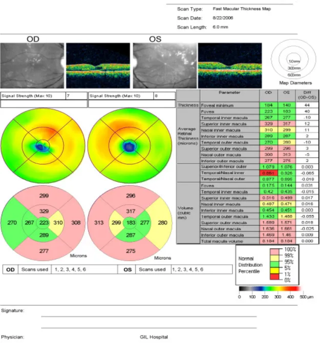
Figure 1. Sample OCT3 display of diabetic macular edema patient with normative data. It displays the macular normative database information in the form of clinically useful fast macular map and chart. Colored bands demonstrate the normal distribution of macular thickness measurements.
대상과 방법
2006년 6월부터 2006년 9월까지 본원 안과에 내원 한 정상 성인군 중 특별한 전신질환 및 안과적 질환이 없고, 안과 수술의 기왕력이 없으며 최대교정시력이 한 천석씨 시력표로 0.8 이상이고, 안압은 정상범위 이내 이며, 굴절이상은 -3.0 diopter에서 +3.0 diopter이 내이고 안저소견이 정상인 20세에서 69세까지의 71명 111안을 대상으로 하였다. 대조군인 Normative data의 대상환자들의 선정 기준은 ETDRS 시력표상
Present data (µm) Normative data (µm) p-value
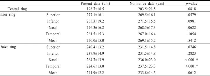
Central ring 198.7±16.5 203.5±21.5 .0818
Inner ring Superior 277.1±16.1 269.5±16.1 .0579
Inferior 265.3±19.2 271.5±15.5 .0981
Nasal 276.3±16.2 268.5±17.3 .0622
Temporal 261.5±15.3 267.0±16.4 .1054
Mean 270.0±15.0 269.1±15.2 .5412
Outer ring Superior 240.4±13.2 231.5±14.8 .0746
Inferior 237.9±14.9 231.5±14.8 .2823
Nasal 264.7±13.9 236.0±23.0 <.0001*
Temporal 224.6±13.0 237.5±23.3 <.0001*
Mean 241.9±12.2 233.8±14.5 .0612
* Statistical significance was calculated by goodness of fit test (p<.05).
Table 2. Comparison between macular thickness of healthy Korean and normative data of OCT3 0.6 이상의 최대교정시력과 21 mmHg 이하 의 안압
을 가진 18세 이상의 성인으로 하였으며 평균 구면렌즈 대응치(spherical equivalent)는 -0.54±1.9D 였 다. 그리고 안내수술의 과거력 및 안과적 기저질환과 당뇨 과거력이 있는 경우는 대상에서 제외하였다.10 동 질성 검정으로 통한 본 연구와 Normative data의 연 령분포를 비교해 보았을 때 통계학적으로 유사한 것으로 나타났다(Test of homogeneity, p=.18)(Table 1).
측정도구로 Zeiss사의 OCT3, version 4.0 (Zeiss- Humphrey, Dublin, CA)를 사용하였다. 황반 두께 의 측정은 단일 검사자에 의해 시행 되었으며, 비산동 상태에서 피검자의 황반부를 고정시키고 안구의 이동을 억제하기 위해 검사받는 눈으로 고정하는 내측주시 (internal fixation) 방법을 사용하였다. 중심소와를 지나는 지나는 6 mm 길이 6개의 선형스캔이 30° 간격 으로 방사상 주사를 하는 fast macular map 을 사용 하여 측정하였으며 이는 중심와를 중심으로 6 mm 직 경의 황반두께지도(macular thickness map)를 만 들게 되며 normative data를 측정한 방법도 이와 동 일하였다. 황반두께지도는 중심 1 mm 이내의 중심원 (central ring)과 3 mm 안쪽원(inner ring), 6 mm 바깥쪽원(outer ring)로 구성되며 안쪽원과 바 깥쪽원은 상측, 비측, 하측, 이측 4분역으로 나누워져 9개의 구역이 되며 µm단위로 측정된 값은 Retinal thickness analysis system을 통해 분석된다. 이를 통해 황반부의 이상부위를 알 수 있으며 옆에 도표를 통해 수치적으로도 파악할 수 있도록 하고 좌우 차이도 같이 제시된다. 그리고 이러한 데이터 값은 색깔로 구 분되어 0~1 percentile, 1~5 percentile, 5~95 percentile, 95~99 percentile, 99~100 percentile 이 각각의 색으로 표시된다(Fig. 1).
OCT3의 재현성 여부를 확인하기 위해 정상인 20 인을 동일한 검사자가 30분 간격으로 총 5회 반복하 여 검사한 후 측정치의 변이계수(Coefficient of variation: 측정치의 표준편차/측정치의 평균 ×100) 를 산출하였다.
결 과
대상군 71 명 111안 중 남자는 58안, 여자는 53안 이 였으며 평균 구면렌즈대응치(spherical equivalent) 는 -0.19±0.32D 이었다. 변이계수는 1 mm 중심원에 서는 1.94%, 안쪽원과 outer ring 총 8개 분획에서 도 2% 미만의 변이계수를 보여 재현성이 뛰어난 결과 를 보였다.
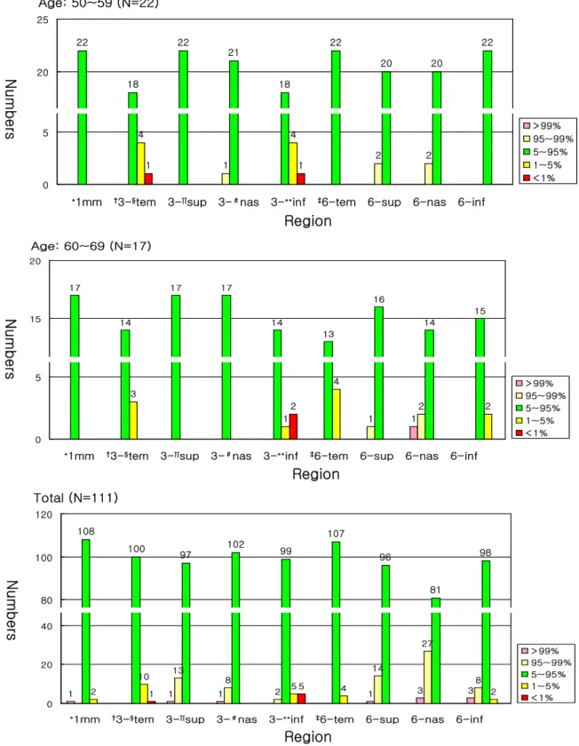
대상군의 망막 두께의 평균치는 1 mm 중심원은 198.7±16.5 µm 이었고 안쪽원은 270.05±16.7 µm, 바깥쪽원은 241.9±13.75 µm로 측정되었다. 안쪽원의 경우 이측이 가장 얇으며 상측이 가장 두꺼웠다. 바깥 쪽원은 이측이 가장 얇으며 하측, 상측, 비측 순으로 두 꺼웠다. 이를 OCT3에 내장된 Normative data와 비 교해 보았을 때 전반적으로 유사한 양상이나 바깥쪽 원 의 비측은 통계적으로 유의하게 두꺼웠으며 이측은 얇 았다(Z-test, p<.0001)(Table 2). 황반부 소견은 정 상이나 OCT3 결과상 95 percentile 이상으로 표시 되는 비율이 제일 높은 곳은 바깥쪽원의 비측이고 전체 대상군 중 25.2%가 이에 해당하였다(Fig. 2). 중심원 과 안쪽원, 바깥쪽원의 각 구획별 평균값을 보면 안쪽 원이 가장 두껍고 바깥쪽원, 중심원 순이었다. 연령별 변 화를 보면 모든 영역에서 30대 이후로 연령증가에 따라 점차 얇아지는 양상 이였으며 남녀간 비교에선 유의한 차이는 보이지 않았다(Z-test, p=.82)(Fig. 3, 4).
25 26
24 25 26 27
23
22 22
20 25 30
Age: 20~29 (N=27)
1
3 2
4 5 5
1 1 1
0 5 10
> 99%
95~99%
5~95%
1~5%
< 1%
*1mm †3-§tem 3-∏sup 3-#nas 3-**inf ‡6-tem 6-sup 6-nas 6-inf
1 1 1
2
3 8
4
1
4
12
2
20 20
11
15
17
20
15
6
15
1 1 1
0 5 10 15 20 25
> 99%
95~99%
5~95%
1~5%
< 1%
*1mm †3-§tem 3-∏sup 3-#nas 3-**inf ‡6-tem 6-sup 6-nas 6-inf Age: 30~39 (N=20)
24
22 23 24 24 25
22
19
24
20 25 30
Age: 40~49 (N=25)
2
1 1
3
6
1 1
3
1 1
0 5
> 99%
95~99%
5~95%
1~5%
< 1%
*1mm †3-§tem 3-∏sup 3-#nas 3-**inf ‡6-tem 6-sup 6-nas 6-inf
Numbers
Region
Region
Region
NumbersNumbers
22
18
22
21
18
22
20 20
22
20 25
Age: 50~59 (N=22)
1
2 2
4 4
1 1
0
5 > 99%
95~99%
5~95%
1~5%
< 1%
*1mm †3-§tem 3-∏sup 3-#nas 3-**inf ‡6-tem 6-sup 6-nas 6-inf
Age: 60~69 (N=17)
17
14
17 17
14
13
16
14
15 15
20
1 1
2 3
1
4
2 2
0
5 > 99%
95~99%
5~95%
1~5%
< 1%
*1mm †3-§tem 3-∏sup 3-#nas 3-**inf ‡6-tem 6-sup 6-nas 6-inf
Total (N=111)
108
100 97
102 99
107
96
81
98
80 100 120
*1mm †3-§tem 3-∏sup 3-#nas 3-**inf ‡6-tem 6-sup 6-nas 6-inf
1 1 1 1 3 3
13
8
2
14
27
8 2
10
5 4 2
1 5
0 20 40
> 99%
95~99%
5~95%
1~5%
< 1%
Figure 2. An analysis of the spread of macular thickness by OCT3 normative data in healthy Korean. At first the population was divided in age categories, and then five sub‐groups were made according to color‐code of the normative data.
Color-code of the normative data was divided in following percentile categories: more than 99% as very thick, 95~99% as thick, 5~95% as normal, 1~ 5% as thin, and less than 1% as very thin. Number of eyes was presented in each macular area of each sub-group. Some cases represented abnormal, even though patients’ macula was normal clinically.
* 1 mm=1 mm central ring; †3=3 mm inner ring; ‡6=6 mm outer ring; §tem=temporal; ∏sup=superior; #nas=nasal; ** inf=inferior.
NumbersNumbersNumbers
Region
Region
Region
Figure 4. Comparison of healthy Korean macular thickness between male and female.
* 1 mm=1 mm central ring; †3=3 mm inner ring; ‡6=6 mm outer ring; §tem=temporal; ∏ sup=superior; #nas=nasal; ** inf=inferior.
Figure 3. Comparison of healthy Korean macular thickness among the age groups.
고 찰
황반부는 여러 망막질환에 있어 그 두께의 변화가 나 타나며 이러한 황반부의 변화를 정량적으로 평가하는 것은 시력예후, 치료시기 판정, 치료효과 평가에 중요 한 관건이 되었으므로 여러 연구가 시도되어 왔다. 객 관적이고 정량적 측정이 가능하도록 개발된 검사로는 RTA와 OCT가 있다. RTA는 세극등 현미경과 유사한
방식으로 빛을 투사하여 산란되는 빛을 안저카메라로 분석하여 하나의 스캔에서 여러 개의 광학적 횡단영상 을 얻어내는 진단 기기이다. 망막두께를 정량화할 수 있다는 장점이 있으나 안축장과 굴절률에 따라 측정 오 차가 발생할 수도 있으며 망막내 많은 경성 삼출물이 있는 경우 반사광의 방해를 받아 정확한 상을 얻기 힘 들고 매질의 혼탁이 있는 경우도 사용하기 어려운 점이 있다.7
Macular thickness
Age
Region
Macular thickness
Source No. of eyes studied Type of OCT device Protocol No. of scans
Macular Thickness (1 mm diameter)
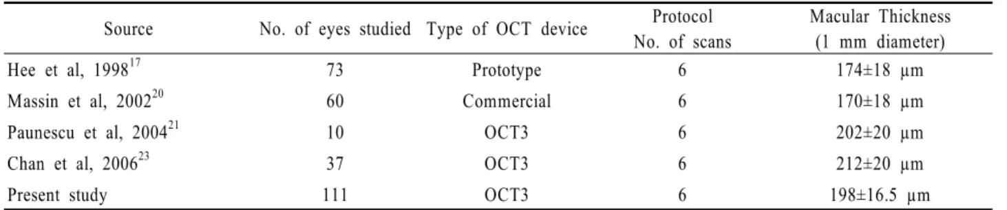
Hee et al, 199817 73 Prototype 6 174±18 µm
Massin et al, 200220 60 Commercial 6 170±18 µm
Paunescu et al, 200421 10 OCT3 6 202±20 µm
Chan et al, 200623 37 OCT3 6 212±20 µm
Present study 111 OCT3 6 198±16.5 µm
Table 3. Macular thickness measurements in healthy eyes reported in previous studies using OCT
Source No. of eyes studied Type of OCT device Protocol No. of scans
Macular Thickness (1 mm diameter)
Kang et al, 200422 112 OCT3 6 208±18 µm
Jung et al, 200413 120 OCT3 6 193.4±14.6 µm
Lee et al, 200514 104 OCT3 6 190.5±13.9 µm
Present study 111 OCT3 6 198.7±16.5 µm
Table 4. Macular thickness measurements in healthy korean eyes reported in previous studies using OCT OCT는 비접촉성, 비침습성 영상기술로 초음파 B-
scan과 유사하나 음파 대신 적외선 계열의 광선을 사 용하며 안조직에서 반사되어 나오는 반향반사지연시간 (echo delay time)을 통해 망막의 두께를 정량적으 로 측정할 수 있는 진단도구이며 약 10 um의 고해상도 단층상을 얻을 수 있다. 또한 OCT는 신뢰도가 매우 높 은 진단기구로 알려져 있다. 일반적으로 변이계수가 10% 미만일 때 신뢰할 수 있는 검사로 인정되며,11,12 본 연구에서는 모든 영역에서 2% 미만의 변이계수를 보였고 OCT를 통한 국내외 여러 연구에서도 대부분 2% 내외의 높은 재현성을 보여 진단기구로서 신뢰도가 매우 높음을 입증하였다.13,14
하지만 광학적 방법을 통한 검사이므로 유리체 출혈, 심한 백내장과 같은 매체의 혼탁이 심한 경우 빛의 투 사나 반사가 간섭받을 수 있어 망막의 측정이 제한 될 수 있으며 직접 검사자가 조준 광선을 주고 영상을 눈 으로 확인하며 시행하므로 어느 정도 환자의 주시상실 로 인한 오차를 RTA에 비해 줄일 수 있다고는 하나, 환자의 협조도가 현저히 떨어지는 경우 정확한 중심와 두께의 측정이 아닌 중심와 주변의 망막두께가 측정될 수 있는 가능성이 있다.15,16 따라서 이러한 제한점을 보 완하기 위해 OCT3에는 retinal map 기능이 추가되 어 있다.11,17
OCT3의 software 4.0에는 기존의 retinal map 에 부가적으로 normative data가 제시되었다.
Normative data의 목적은 기존의 정상안으로 도출 한 황반두께와 환자의 황반두께를 정량적으로 비교함으 로써 진단 및 치료 효과 판정에 도움을 주고자 하는 것 이다. Normative data와 비교한 환자의 황반두께
는 원 그래프와 표로 제시되며 해당하는 백분지수 (percentile, %ile)에 따라 각각 다른 색깔로 표시된 다. 이 Normative data는 미국의 6개 multicenter study를 통해 얻어진 것으로 기저 안질환 및 전신질환 이 없는 328 scans으로 산출한 것이고 대상군의 나이 는 18세에서 85세 까지 였으며 평균나이는 47±15.8세 이고 남녀간의 차이는 거의 없었으며(M:F=47:52) 좌우안의 구성비는 동일하였다. 대상군의 인종별 구성 을 보면 주로 백인과 히스패닉계열이 주를 이루었는데 그 비율은 63%, 24%로 매우 높았으며 아시아계와 흑 인비율은 3%, 8%에 불과하였다.
정상 황반두께를 측정하기 위해서 다양한 방법으로 여러 연구들이 시행되어왔다. 먼저 Hogan et al18은 병리조직학적 검사를 통해 중심와 두께가 130 µm라고 하였다. Asrani et al19은 RTA를 통해 망막두께 정상 치를 산출 하였는데 중심와의 두께는 167 µm, 후극부 의 평균두께는 229 µm라 하였다. OCT를 이용한 여러 연구들이 있었는데 Hee et al17은 중심와 두께가 174±18 µm라 하였으며 Massin et al20은 170±18 µm, Paunescu et al21은 202±20 µm 라고 보고하였 다. 상기 측정값들은 검사 방법에 따른 차이가 존재한 다고 보여지며 OCT라는 동일한 검사방법으로도 검사 자 마다 약간의 차이를 보이는 것은 OCT 기종의 차이, 검사자들의 측정 오차, 대상군의 인종구성비 차이 등으 로 생각된다(Table 3). 한국인을 대상으로 OCT3로 측정한 중심와 두께는 Kang et al22은 208±18 µm, Jung et al13은 193.4±14.6 µm, Lee and Lee14은 190.5±13.9 µm 라고 하였으며 본 연구는 198.7±16.5 µm 였다(Table 4). 대체적으로 한국인에서 OCT3로
측정한 평균 중심와 두께는 190~200 µm로 생각되며 일부 연구에서 차이가 있는 것은 측정오차 및 대상군 범위의 차이로 인한 표본편중으로 생각된다.
인종간 황반부 두께 차이에 대해서는 기존의 여러 연 구들이 있었으나 인종간의 차이가 있을 것이라는 가정 을 하고 있을 뿐 아직까지 원인을 명확히 밝히지 못하 고 있으며 각 인종별 황반부 두께 평균값에 대한 정보 도 부족한 상태이다. Asrani et al19은 RTA를 사용하 여 흑인과 백인간 황반부 두께의 차이에 대해 보고하였 는데 흑인 여성과 백인 여성간에는 약 17 µm 정도 백 인여성이 두꺼웠으며 통계적으로 유의한 차이를 보였지 만 흑인 남성과 백인 남성간에는 그 차이가 9 µm에 불 과하며 유의한 차이를 보이지 않았다. Tewari et al
24은 인도인 정상안에서 OCT3를 통해 산출한 황반부 두께를 다른 인종을 대상으로 한 기존의 연구와 비교해 볼 때 차이가 있다고 하였고 이러한 원인 중 하나로 인 종간의 차이를 들었다. Huynh et al25는 6세 아이들 을 대상으로 한 연구에서 동아시아계 아이들과 백인아 이들의 황반부 두께를 비교해 볼 때 통계적으로 유의한 차이가 있음을 보았으며 이러한 차이의 원인을 인종간 의 황반부 두께의 차이일 것이라고 가정하였다.
한국인을 대상으로 한 본 연구와 백인과 히스패닉계 가 주를 이룬 normative data와 중심와 두께를 비교 해 보았을 때 차이가 4.8±5 µm 이며 통계적으로 차이가 없었고 한국인을 대상으로 한 다른 연구들과 normative data를 비교했을 시에도 통계적인 차이는 보이지 않았 다. 하지만 각 구역별 두께를 비교해 보면 바깥쪽 원의 비측이 통계적으로 유의하게 두꺼웠으며 바깥쪽 원의 이측은 얇았다. 한국인을 대상으로 한 Jung et al13 의 연구 결과도 본 연구와 유사하였으며 따라서 normative data에 비해 바깥쪽 원의 비측이 두껍고 이측이 얇은 것은 한국인과 normative data의 인종 간 차이라고 생각된다. 각 인종별 황반부 두께 차이에 대해서는 더 많은 연구가 이루어져야 할 것이며 인종별 정상값 산출이 필요할 것으로 보인다. 이러한 추가적인 연구를 통해 RNFL normative data가 나이에 따른 보정기능이 있는 것처럼10 각 인종별로 normative data를 만들어 대상에 따라 각각 따로 적용하거나 인 종별 차이를 보정하도록 개선한다면 임상적으로 정확한 normative data가 될 것으로 생각된다.
본 연구를 통해 OCT3로 황반부 두꼐 측정시 한국인 황반부 두께의 기준을 제시하였으며 OCT3에 내장된 normative data와의 비교를 통해 한국인 황반부의 인종적 차이를 밝혔다. 따라서 임상에서 황반부 두께를 normavite data와 비교하여 해석할 때 이러한 인종 적 차이를 고려하여야 할 것으로 생각한다.
참고문헌
1) Ferris FL III, Patz A. Macular edema. A complication of diabetic retinopathy. Surv Ophthalmol 1984;28:S452-61.
2) Nussenblatt RB, Kaufman SC, Palestine AG, et al. Macular thickening and visual acuity. Measurement in patients with cystoid macular edema. Ophthalmology 1987;94:1134-9.
3) Hee MR, Puliafito CA, Wong C, et al. Quantitative assessment of macular edema with optical coherence tomography. Arch Ophthalmol 1995;113:1019-29.
4) Early Treatment Diabetic Retinopathy Study Research Group.
Grading diabetic retinopathy from stereoscopic color fundus photographs‐an extension of the modified Airlie House classification. ETDRS report number 10. Ophthalmology 1991;98:786-806.
5) Zeimer RC, Mori MT, Khoobehi B. Feasibility test of a new method to measure retinal thickness noninvasively. Invest Ophthalmol Vis Sci 1989;30:2099-105.
6) Gieser JP, Rusin MM, Mori M, et al. Clinical assessment of the macula by retinal topography and thickness mapping. Am J Ophthalmol 1997;124:648-60.
7) Polito A, Shah SM, Haller JA, et al. Comparison between retinal thickness analyzer and optical coherence tomography for assessment of foveal thickness in eyes with macular disease. Am J Ophthalmol 2002;134:240-51.
8) Puliafito CA, Hee MR, Schuman JS, Fujimoto JG. Optical Coherence Tomography of Ocular disease. Thorofare, NJ:
Slack, 1996;369-74.
9) Muscat S, Parks S, Kemp E, Keating D. Repeatability and reproducibility of macular thickness measurements with the Humphrey system. Invest Ophthalmol Vis Sci 2002;43:490-5.
10) Budenz DL, Anderson DR, Varma R, et al. Determinants of normal retinal nerve fiber layer thickness measured by Stratus OCT. Ophthalmology 2007;114:1046-52.
11) Massin P, Vicaut E, Haouchine B, et al. Reproducibility of retinal mapping using optical coherence tomography. Arch Ophthalmol 2001;119:1135-42.
12) Shrout PE, Fleiss JL. Intraclass correlations: uses in assessing rater reliability. Pshychol Bull 1979:428-9.
13) Jung HJ, Hyun JH, Kim YI, Yun IH. Normal macular thickness measured with macular mapping of OCT3. J Korean Ophthalmol Soc 2004;45:962-8.
14) Lee SM, Lee JH. Assessment of foveal thickness in normal Korean using retinal thickness analyzer and optical coherence tomography. J Korean Ophthalmol Soc 2005;46:1630-6.
15) Kholodnykh AI, Petrova IY, Larin KV, et al. Precision of measurement of tissue optical properties with optical coherence tomography. Appl Opt 2003;42:3027-37.
16) Huang D, Swanson EA, Lin CP, et al. Optical coherence tomography. Science 1991;254:1178-81.
17) Hee MR, Puliafito CA, Duker JS, et al. Topography of diabetic macular edema with optical coherence tomography.
Ophthalmology 1998;105:360-70.
18) Hogan MJ, Alvarado JA, Weddell JE. Histology of the Human Eye. Philadelphia: WB Saunders Company, 1971:492.
19) Asrani S, Zou S, d'Anna S, et al. Noninvasive mapping of the normal retinal thickness at the posterior pole. Ophthalmology 1999;106:269-73.
20) Massin P, Erginay A, Haouchine B, et al. Retinal thickness in healthy and diabetic subjects measured using optical coherence tomography mapping software. Eur J Ophthalmol 2002;12:102-8.
21) Paunescu LA, Schuman JS, Price LL, et al. Reproducibility of nerve fiber thickness, macular thickness, and optic nerve head measurements using Stratus OCT. Invest Ophthalmol Vis Sci 2004;45:1716-24.
22) Kang JH, Kim SA, Song WG, Yoon HS. Macular thickness changes with age in normal subjects measured by optical coherence tomography. J Korean Ophthalmol Soc 2004;45:592-8.
23) Chan A, Duker JS, Ko TJ, et al. Normal thickness measurements in healthy eyes using STRATUS optical coherence tomography. Arch Ophthalmol 2006;124:193-8.
24) Tewari HK, Wagh VB, Sony P, et al. Macular thickness evaluation using the optical coherence tomography in normal Indian eyes. Indian J Ophthalmol 2004;52:199-204.
25) Huynh SC, Wang XY, Rochtchina E, et al. Distribution of macular thickness by optical coherence tomography: Findings from a population‐based study of 6‐year‐old children.
Invest Ophthalmol Vis Sci 2006;47:2351-7.
=ABSTRACT=
Macular Thickness in Healthy Korean Eyes Using OCT3 Comparing with Normative Data
Sang Chul Yoon, M.D., Dae Yeong Lee, M.D., Dong Heun Nam, M.D., Ph.D.
Department of Ophthalmology, Gachon University Gil Medical Center, Incheon, Korea
Purpose: To determine whether there are significant differences between the retinal thicknesses of the macular area in eyes of healthy Koreans compared with normative data using OCT3 (version 4.0).
Methods: The study was performed in 111 eyes of 71 adults between the ages of 20 and 69 with corrected vision of 0.8 or greater and no systemic or ocular disease. The subject group was designed to have a similar age distribution to that of normative data. An OCT3 fast macular map was used to measure macular thickness.
The results were further analyzed and compared with the normative data.
Results: The overall results from the subject group were similar to the normative data. However, the nasal parts of the outer ring were thicker than the normative data and the temporal parts of the outer ring were thinner (p<.0001). When each age group was compared, the thickness of the superior part of the inner ring and the nasal part of the inner and outer rings were thicker than the normative data in the age groups of twenties and thirties (p<.05). For the nasal parts of outer ring, the macula were thicker by OCT examination than the normative data in 25.2% of subjects, although they were clinically normal.
Conclusions: Macular thickness in healthy Koreans was generally similar to the normative data of OCT3.
However, a slight difference may exist and must be considered when measuring and interpreting macular thickness measurements.
J Korean Ophthalmol Soc 49(4):601-610, 2008
Key Words: Macular thickness, Normative data, Optical coherence tomography
Address reprint requests to Dong Heun Nam, M.D., Ph.D.
Department of Ophthalmology, Gachon University Gil Medical Center
#1198 Kuwol-dong, Namdong-gu, Incheon 405-760, Korea
Tel: 82-32-460-3364, Fax: 82-32-460-3358, E-mail: [email protected]