J Korean Ophthalmol Soc 49(8):1275-1282, 2008 DOI : 10.3341/jkos.2008.49.8.1275
망막정맥폐쇄 및 당뇨병에 의한 황반부종 환자에서 유리체강내 베바시주맙 주입술의 단기효과
김종연․권의용․이동욱․조남천 전북대학교 의학전문대학원 안과학교실
목적: 망막정맥폐쇄 및 당뇨병에 의한 황반부종 환자에서 유리체강내 Bevacizumab (Avastin®) 주입술의 단기효과 에 대해 알아보고자 하였다.
대상과 방법: 망막정맥폐쇄 및 당뇨병으로 인한 황반부종의 소견을 보인 51명 59안을 대상으로 유리체강내로 Bevacizumab
을 주입하였으며 술 후 1, 3, 6개월째에 최대교정시력, 안압검사, 세극등현미경검사, 형광안저촬영, 빛간섭단층촬영을 시행하여 술 전과 비교하였다.
결과: 술 전 평균최대교정시력은 1.06±0.53였으며, 중심황반 두께는 479.6±160.4 µm이었다. 시술 후 평균 최대 교정시력은 1개월째 0.90±0.52, 3개월째 0.80±0.39, 6개월째 0.78±0.39이었으며 황반 두께는 1개월째 316.9±86.7 µm, 3개월째 281.1±67.4 µm, 6개월째 278.4±64.6 µm이었으며 통계학적으로 의의가 있었다. 2안에서 일시적인 안압이 상승이 발생한 것 이외에 합병증 등은 발생하지 않았다. 황반부종의 원인별로 결과간에 차이는 없었다.
결론: Bevacizumab의 유리체강내 주입술은 치명적인 합병증이 잘 생기지 않는 비교적 간단한 시술로 망막정맥폐쇄 및 당뇨병으로 인한 황반부종 환자에서 중심황반 두께 감소와 시력회복을 기대할 수 있는 치료로 유용하게 사용될 수 있을 것으로 사료된다.
<한안지 49(8):1275-1282, 2008>
<접수일 : 2008년 1월 3일, 심사통과일 : 2008년 4월 23일>
통신저자 : 권 의 용
전라북도 전주시 덕진구 금암동 634-18 전북대학교병원 안과
Tel: 063-250-1965, Fax: 063-250-1960 E-mail: [email protected]
* 본 논문의 요지는 2007년 대한안과학회 제98회 추계학술대회 에서 포스터로 발표되었음.
* 본 논문은 전북대학교병원에서 연구비의 일부를 2007년에 보조 받았음.
황반부종은 황반중심에서 반지름이 1 유두직경(1,500 µm)인 원 안에서 두꺼워진 망막이나 경성삼출물이 관 찰될 때를 말하는데 모세혈관 내피세포층, 즉 내측혈액 -망막장벽의 파괴로 체액과 혈장성분이 누출되어 황반 에 고임으로써 일어나며, 간혹 망막이나 망막혈관에 대 한 유리체견인에 의해 누출이 일어나는 경우도 있다.
이런 누출은 미세혈관류나 손상받은 모세혈관, 망막내 미세혈관이상으로부터 발생하는데, 새어나온 체액은 망막 전 층에 고이지만 특히 축삭들이 느슨하게 얽혀있 는 외망상층(outer plexiform layer)이나 내과립층
(inner nuclear layer)에 많이 고이게 된다. 황반부종 은 당뇨망막병증, 망막정맥폐쇄, 맥락막신생혈관, 인공 수정체안, 염증성 안질환, 혈관성 종양 등에서 발생하며 안저검사에서 경성삼출물, 형광안저혈관조영에서 혈관 누출 그리고 빛간섭단층촬영에서 두꺼워진 망막 등의 관 찰로 진단된다. 이는 당뇨망막병증이나 망막분지정맥폐 쇄에서 시력저하의 가장 큰 원인 중 하나로 알려져 있으 며 황반부종을 감소시키기 위한 방법으로 격자레이저 광 응고술, 유리체절제술, 유리체강내 스테로이드 주입술 (Intravitreal triamcinolone acetonide injection) 등이 알려져 있다.1-6
최근에는 신생혈관의 발생을 억제하는 항 혈관내피 성장인자 주입술이 새롭게 각광받고 있다. 혈관신생은 각종세포와 인자, 세포외기질 간의 상호작용에 의해 이 루어지는 복잡한 과정으로 상처치유와 같은 과정 이외 에서는 생체 내에서는 엄격히 조절되고 있으며 조절되 지 않은 혈관신생은 여러 가지 병리현상을 일으키게 된 다. 혈관신생유도물질 중 가장 중요한 역할을 하는 혈관 내피성장인자(vascular endothelial growth factor, VEGF)는 헤파린에 결합된 당단백으로서 강력한 혈관 생성, 유사분열 촉진물질이자 혈관내피세포에 대해 높
은 혈관투과성을 보인다. 주로 망막색소상피에서 주로 분비되고 맥락막모세혈관, 망막모세혈관, 망막색소상피 에 주로 수용체가 분포하며 신생혈관질환에서 높은 농 도로 검출되지만 정상 안구조직에서도 분비되고 전방수 보다 유리체에 더 많이 분포하며 혈관생성과 혈관투과 성을 증가시키는 기능이 있다.7,8 망막내피세포에는 다 량의 VEGF 수용체가 있고 허혈은 망막주위세포 (retinal pericytes), 망막내피세포(retinal endo- thelial cells), 망막색소상피세포(retinal pigment epithelial cells) 내의 mRNA의 VEGF 양을 늘리 며 이로 인해 증가된 VEGF는 황반 부종을 야기시킨
다.9,10 그리하여 이와 같은 VEGF의 기능을 억제하여
신생혈관생성을 막기 위한 항 혈관내피성장인자 주입술 이 허혈을 유발하는 당뇨망막병증과 망막정맥폐쇄로 인 한 황반 부종의 새로운 치료로 시도되고 있다.11,12 이에 사용되는 대표적인 항 혈관내피성장인자 항체중 하나인 Bevacizumab (Avastin®, Genentech Inc., San Francisco, CA)은 혈관내피성장인자에 결합하는 인 간화 단클론항체(humanized monoclonal antibody) 로 항암제로 사용되었으며 2004년 2월 직장암의 치료 로 FDA 승인이 되었다.13-15 최근에 Bevacizumab 주입술이 망막정맥폐쇄로 인한 황반부종과 증식성 당뇨 망막병증으로 인한 혈관 투과성 감소에 효과가 있다고
하였다.16,17 그러나 국내에는 아직까지 항 혈관내피성
장인자 주입술의 효과에 대한 보고가 없다.
이에 저자들은 망막정맥폐쇄 및 당뇨병에 의한 황반 부종 환자에서 Bevacizumab (Avastin®) 유리체강 내 주입술의 시력 및 해부학적인 단기효과에 대해 알아 보고자 하였다.
대상과 방법
2007년 1월 1일부터 2007년 12월 1일까지 본원 안 과에서 안저검사상 당뇨망막병증과 망막정맥폐쇄로 인 한 황반부종으로 진단받은 환자 51명 59안을 대상으로 후향적인 연구를 시행하였다. 빛간섭단층촬영상 황반부 가 300 µm 이상 증가되고 안저검사상 황반중심에서 1/2 유두지름의 원안에 1 유두 크기 이상으로 망막이 두꺼워지거나, 낭포성 변화 혹은 확산 부종이 있으며, 형 광안저촬영상 황반 중심 1/2 유두 지름 내에 형광 누출 을 보이는 경우를 대상으로 하였다. 녹내장이 있거나 시 술전 6개월 이내에 안과 수술 병력이 있는 환자들과 시 력저하를 유발할 수 있는 다른 질병을 가지고 있는 환자 들은 대상에서 제외하였다. 시술 전에 최대교정시력, 안 압검사, 세극등현미경검사, 안저검사, 형광안저촬영을 시행하였으며, 빛간섭단층촬영(Optical coherence
tomography, OCT)을 이용하여 황반부 두께를 측정 하였다. 59안 중 남자는 32안, 여자는 27안이었으며 평균 연령은 59.0세(범위:30~79세)이었고 평균 추적 관찰기간은 7.3개월(범위:6~8개월)이었다. 1차 주입 후 시력호전이나 황반부종의 감소를 보였던 환자에서 다시 술 전의 상태로 시력감소나 황반부종 증가를 보인 환자중 재주입술을 원하는 환자에 대하여 2차 주입술을 시행하였다. 시술방법은 환자를 천장을 보고 눕게하고 0.5% Proparacaine (Alcaine®, Alcon)으로 점안 마취를 시행하고 Povidone iodine으로 안검소독을 한 후 소독포를 덮은 뒤 소독된 개검기를 삽입하였다. 유 수정체안에서는 각막변연부에서 3.5 mm, 위수정체안 과 무수정체안에서는 각막윤부에서 2.5 mm 떨어진 상 측 부위의 평면부에 26 gauge 주사침을 이용하여 1.25 mg 또는 2.5 mg (0.05 또는 0.1 ml) Bevacizumab (Avastin®)을 유리체강내로 주입하였다. 술 후 2주일 간 1일 4회 Moxifloxacin (Vigamox®, Alcon)을 점안하도록 하였다.
시술 후 1개월, 3개월, 6개월째 외래경과 관찰하면 서 세극등현미경검사, 안저검사, 빛간섭단층촬영, 형광 안저촬영검사를 시행하였으며 주입 전과 주입 후 최대 교정시력과 황반부 두께 변화를 관찰하였다.
최대교정시력은 LogMAR (logarithm of the minimum angle of resolution)시력으로 전환하였 으며 통계적인 분석은 SPSS V.15.0를 사용하였으며 P-value가 0.05 미만인 경우를 통계학적으로 의의가 있는 것으로 정의하였다.
결 과
대상환자 51명의 평균연령은 59.0세(범위:30~79 세)였으며 남자가 26명(51.0%), 여자가 25명(49.0%) 이었다. 황반부종의 원인 질환으로는 당뇨망막병증 30 안(50.8%), 그 중 증식당뇨망막병증 17안(28.8%), 비증식성당뇨망막병증 13안(22.0%)이었으며, 망막분 지정맥폐쇄 12안(20.3%), 망막중심정맥폐쇄 17안 (28.8%)이었다. 황반부종의 진단부터 시술까지의 기 간은 평균 42일(범위: 5~58일)이었고, 시술 전 평균 최대교정시력은 1.06±0.53 (LogMAR)였으며, 평균 황반 두께는 479.6±160.4 µm이었고 시술 후 평균 관 찰기간은 7.3개월(범위:6~8개월)이었다(Table 1, 2).
술 후 1개월째의 평균 시력은 0.90±0.52, 3개월째 0.80±0.39, 6개월째 0.78±0.39로 각각 술 전보다 통 계학적으로 유의성 있게 향상되었다(p<0.05). 2회 이 상의 시술을 받은 안은 8안(13.56%)이었으며, 시술 후 시력변화가 없거나 시력이 감소한 경우는 59안 중 5
Variable Data Age (years)
Mean±SD* 59.0±7.2
Range 30-79
Total (eyes, %)
Male 32 (54.2%)
Female 27 (45.8%)
Unerlying disease (%)
Diabetic retinopathy, Proliferative 17 (28.8%)
Diabetic retinopathy, Non-Proliferative 13 (22.0%)
CRVO† 17 (28.8%)
BRVO‡ 12 (20.3%)
Follow up period (months)
Mean±SD* 7.3±0.31
Range 6-8
Preoperative BCVA§ (LogMAR#, mean±SD*) 1.06±0.53
Preoperative central macular thickness (µm, mean±SD*) 479.6±160.4
* SD=Standard deviation; †CRVO=Central retinal vein occlusion; ‡BRVO=Branched retinal vein occlusion; §BCVA=Best corrected visual acuity; #LogMAR=Logarithm of the minimum angle of resolution.
Table 1. Patients’ characteristics
DME RVO BRVO CRVO
Age (years) 57.9 61.5 65.4 57.5
Total (eyes) 30 29 12 17
Male 16 16 4 12
Female 14 13 8 5
Mean follow up time(months) 7.1 7.4 7.5 7.3
Preoperative BCVA*
(LogMAR†,mean±SD§)/
CMT‡(µm, mean±SD§)
1.06±0.53 / 480.7±153.2
1.07±0.55 / 478.4±170.2
0.62±0.41/
406.6±96.9
1.38±0.4 / 529.2±193.9
* BCVA=best corrected visual acuity; †LogMAR=logarithm of the minimum angle of resolution; ‡CMT=central macular thickness; § SD=standard deviation.
Table 2. The numbers of eyes, mean age, mean follow-up time, BCVA*, central macular thickness before intravitreal bevacizumab injection
안(8.47%)이었다. 황반 두께는 1개월째 316.9±86.7 µm, 3개월째 281.1±67.4 µm, 6개월째 278.4±64.6 µm로 모두 술 전보다 통계학적으로 유의하게 감소하였 다(p<0.05).
전체 59안 중 2안(3.4%)에서 일시적인 안압이 상승 하였으나 국소 점안 안압하강제로 호전되었고 5안 (8.47%)에서 결막하 출혈이 발생하였으나 1주일이내 에 사라졌으며 이외에 백내장, 망막박리, 유리체출혈, 안구내염 등 시력에 치명적으로 영향을 주는 합병증과 전신적 합병증은 발생하지 않았다.
황반부종의 원인 질환별로 당뇨망막병증의 경우 시 술 전 1.06±0.53, 1개월째 0.88±0.47, 3개월째 0.81±0.40, 6개월째 0.77±0.33로 시력호전을 보였으
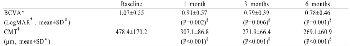
며 황반부 두께는 시술 전 480.7±153.2 µm, 1개월째 326.5±86.9 µm, 3개월째 290±68.2 µm, 6개월째 287.4±67.7 µm로 술 전에 비해 감소하였다(p<0.05, Table 3). 망막정맥폐쇄의 경우 시술 전 1.07±0.55, 1개월 째 0.91±0.57, 3개월째 0.79±0.39, 6개월째 0.78±0.46로 술전에 비해 시력호전을 보였으며 황 반부 두께는 시술 전 478.4±170.2 µm, 1개월째 307.1±86.8 µm, 3개월째 271.9±66.4 µm, 6개월째 269.1±60.9 µm로 술 전에 비해 감소하였다(p<0.05, Table 4).
황반부종의 원인별로 1, 3, 6개월에 시력 호전 정도 는 통계학적으로 차이를 보이지 않았으며(Fig. 1, 각각 p=0.56, p=0.74, p=0.49, 독립적 t-test) 1, 3, 6
Baseline 1 month 3 months 6 months BCVA*
(LogMAR†, mean±SD#)
1.06±0.53 0.88±0.47 (P=0.006)§
0.81±0.40 (P<0.001)§
0.77±0.33 (P<0.001)§ CMT‡
(µm, mean±SD#)
480.7±153.2 326.5±86.9 (P<0.001)§
290±68.2 (P<0.001)§
287.4±67.7 (P<0.001)§
* BCVA=best corrected visual acuity; †LogMAR=logarithm of the minimum angle of resolution; ‡CMT=central macular thickness; § Paired t-test; #SD=standard deviation.
Table 3. Changes in visual acuity and central retinal thickness in macular edema caused by diabetic macular edema(DME)
Baseline 1 month 3 months 6 months
BCVA*
(LogMAR†, mean±SD#)
1.07±0.55 0.91±0.57 (P=0.002)§
0.79±0.39 (P=0.006)§
0.78±0.46 (P=0.001)§ CMT‡
(µm, mean±SD#)
478.4±170.2 307.1±86.8 (P<0.001)§
271.9±66.4 (P<0.001)§
269.1±60.9 (P<0.001)§
* BCVA=best corrected visual acuity; †LogMAR=logarithm of the minimum angle of resolution; ‡CMT=central macular thickness; § Paired t-test; #SD=standard deviation.
Table 4. Changes in visual acuity and central retinal thickness in macular edema caused by retinal vein occlusion (RVO)
0.3 0.5 0.7 0.9 1.1 1.3 1.5 1.7
Preop 1 month 3 months 6 months
LogMAR
DME ME with RVO
Figure 1. Changes in LogMAR after intravitreal bevacizumab injections, according to macular edema etiology; DME=diabetic macular edema; ME=macular edema; RVO=retinal vein occlusion.
200 250 300 350 400 450 500 550 600 650 700
Preop 1 month 3 months 6 months
Central retinal thickness (㎛)
DME ME with RVO
Figure 2. Changes of central macular thickness with optical coherence tomography (OCT) after intravitreal bevacizumab injections.
DME=diabetic macular edema; ME=macular edema; RVO=
retinal vein occlusion.
-0.3 -0.2 -0.1 0 0.1 0.2 0.3 0.4
-40 -20 0 20 40 60 80
Macular thickness decrease (%)
Visual acuity increase
Figure 3. The relationship between visual acuity increase and macular thickness decrease (%) at 6 months after intravitreal bevacizumab injections.
개월에 황반 두께 감소도 의의있는 차이가 없었다(Fig. 2, 각각 p=0.39, p=0.30, p=0.28, 독립적 t-test).
또한 6개월째에 황반부 두께의 감소와 시력 호전 사 이에는 통계학적으로 의의있는 상관관계가 없었다 (Fig. 3, p=0.34, 회귀분석).
고 찰
황반부종은 모든 당뇨병환자의 약 10%에서 발생하 며 시력저하의 중요한 원인으로 알려져 있으며, 혈관내 피성장인자(vascular endothelial growth factor, VEGF), 인슐린양성장인자(insulin-like growth factor, IGF-I) 등 인자의 증가와 내측혈액-망막장벽의 파괴에 의한 혈관 투과성 증가로 발생한다.18 ETDRS (Early Treatment Diabetic Retinopathy Study)
에서 국소레이저 치료로 임상적으로 유의한 황반부종에 서 시력 감소 위험을 반으로 줄일 수 있음을 발표한 후 레이저광응고술이 주된 치료방법으로 적용되어져 왔다.
그러나 그 대상이 제한적이고 미만성 당뇨황반부종의 경우에는 예후가 나쁘며, 황반중심을 침범한 경우에는 특별한 치료방법이 없는 실정이다. 최근에는 혈관투과 성에 관여하는 프로스타글란딘(Prostaglandin)이나 혈관내피성장인자등이 황반부종을 일으키는 것으로 밝 혀져 안내 스테로이드 주입술이 황반부종을 감소시키고 시력호전에 효과적이라는 보고가 있었다.19,20 한편 최 근 안내 트리암시놀론 아세토나이드 주입후에 안압상 승, 안내염, 백내장, 망막박리 등의 문제점들이 보고되 면서 주의를 요하고 있다.21-24
망막분지정맥폐쇄는 당뇨망막병증과 함께 망막혈관 질환 중 가장 흔히 볼 수 있는 질환으로 고혈압, 심혈관 계 질환과 관련성이 높다. 망막분지정맥폐쇄는 60대의 노인층에서 호발하고 성별이 차이가 없으며 시력감퇴를 보이는 황반부종이나 황반비관류와 같은 합병증이 없는 경우라면 치료는 필요 없지만 황반부종이 발생시 예후 는 불량해진다. BVOS (Branch Vein Occlusion Study)에서는 황반부종으로 시력이 저하되었을 때, 몇 가지 기준을 충족하는 경우 아르곤레이저 광응고술 로 시력감소를 줄일 수 있다고 보고하였다.2 이는 망막 을 얇게 하여 맥락막 혈관으로부터 망막 내층으로 영양 공급이 가능하게 함으로써 누출되는 망막혈관들이 자가 조절로 수축하고 부종이 감소되는 효과를 기대하는 것 이다.25 스테로이드는 phospholipase 효소를 차단하 여 arachidonic acid pathway를 방해함으로써 prostaglandin의 생성을 억제하며 또한 혈관내피성 장인자의 생성도 감소시켜 황반부종의 환자에서 시력 호전의 효과가 있다고 보고하였다.5
망막중심정맥폐쇄는 50대 이상의 고령의 성인에서 영구적인 시력저하를 일으키는 대표적인 망막질환의 하 나로서 아직까지 이 질환의 발병기전이나 치료방법에 대해서는 뚜렷하게 알려지지 않았다. 또한 신생혈관이 나 황반부종 등의 합병증을 초래하여 이차적으로 시력 저하를 일으키기도 하며, 질환의 예후 또한 예측이 어 려워 환자의 관리가 어렵다. 아직까지 효과가 입증된 치료방법은 없으며, 이차적인 시력감소의 원인인 망막 부종이나 신생혈관 발생에 의한 합병증을 줄이기 위하 여 격자 레이저 광응고술이나 범망막광응고술이 시도 되고 있으나 중간 매체의 혼탁이 있는 환자에서는 시행 이 어렵고 또한 성공률도 낮았으며, 대부분의 환자에서 의미 있는 시력의 호전은 보이지 않았다. 여러 저자들 에 의하여 망막중심정맥폐쇄 환자에서 고에너지의 아르 곤 레이저와 Nd:YAG 레이저를 경우에 따라 병합하여
사용하여 사상판 부위에서의 폐쇄부위를 우회하는 망막 맥락막 곁순환의 형성을 유도하여 치료하려 하였지만 약간의 시력 호전을 보이는 경우가 있었으나 대부분 병 의 경과에는 영향을 주지 못하였다.26-28
최근에는 혈관내피성장인자 주입술을 통한 신생혈관 의 발생을 억제하여 황반부종을 감소시키려는 방법이 시도되고 있다. 혈관신생은 각종세포외 인자, 세포외기 질 간의 상호작용에 의해 이루어지는 복잡한 과정으로 상처치유와 같은 과정 이외에서는 생체 내에서는 엄격 히 조절되고 있으며 조절되지 않은 혈관신생은 여러 가 지 병리현상을 일으키게 된다.29 혈관신생유도물질 중 가장 중요한 역할을 하는 혈관내피성장인자(VEGF)는 1948년 Michaelson30이 허혈성 망막에서 분비되는 화학적 혈관생성인자가 망막 신생혈관의 생성을 유발한 다고 보고하였고 처음 factor X라고 명명하였다.
망막내피세포에는 다량의 VEGF 수용체가 있고 허 혈은 망막주위세포(retinal pericytes), 망막내피세 포(retinal endothelial cells), 망막색소상피세포 (retinal pigment epithelial cells) 내의 mRNA 의 VEGF 양을 늘린다. 혈류량의 감소(망막중심정맥 폐쇄증, 경동맥 폐쇄증), 망막 모세혈관 감소(당뇨병성 망막증, 방사선 망막증), 말초혈관 미형성 또는 협착 (미숙아 망막증, 겸상적혈구증), 망막색소상피로부터 맥락막 혈류공급 분리(망막박리) 등이 모두 상대적인 망막 허혈을 유발한다. 이러한 허혈은 망막주위세포, 망막내피세포, 망막색소상피세포 이외의 가능한 모든 세포내의 VEGF의 합성과 분비를 촉진하며 망막, 맥락 막, 각막, 홍채 등에 혈관신생을 일으킨다.31 이러한 결 과들은 VEGF의 기능을 억제하여 신생혈관생성으로 인해 발생한 합병증을 막기 위해 항 혈관내피성장인자 주입술을 시도할 수 있게 하였다.
이와 같이 혈관내피성장인자의 조절을 통해 치료하려 는 대표적인 항 혈관내피성장인자 항체(Anti-VEGF antibody)인 Bevacizumab(Avastin®)은 유전자 재조합 인간 단일클론항체(recombinant humanized monoclonal antibody)로서 선택적으로 혈관내피성 장인자와 결합하여 억제함으로써 VEGFR1과 VEGFR2 를 통한 활성화를 차단하여 결과적으로 혈관신생을 억 제하는 역할을 한다. 2004년 2월 FDA 승인을 받아 현재 전이성 대장암에 대해 5-fluorouracil을 사용하 는 화학치료법과 함께 사용되고 있으며 전이성 대장암 환자를 상대로 한 연구에서 5-FU/LV와 병용 투여하 였을 때 반응률, 무진행 생존기간 및 생존기간이 유의 하게 높은 것으로 알려져 있다.32
이와 같은 특성 때문에 유리체강내 Bevacizumab 주입술을 근시성 맥락막신생혈관, 특발성 맥락막신생혈
관, 결절맥락막혈관병증, 나이관련황반변성의 맥락막신 생혈관, 당뇨망막병증의 망막신생혈관, 망막정맥폐쇄의 황반부종등의 치료로 시도되고 있다.16,17,33-43
Rosenfeld et al41은 망막중심정맥폐쇄로 반복적인 황반부종이 발생하여 이전에 유리체강내 트리암시놀론 아세토나이드 주입술을 시행받은 환자에서 유리체강내 Bevacizumab 주입술후 빛간섭단층촬영의 변화에 대 해 보고하였으며 Iturralde et al42은 이전에 망막중 심정맥폐쇄로 황반부종이 발생한 환자 중 유리체강내 트 리암시놀론 아세토나이드 주입술 후 호전되지 않은 16 안에서 황반부종의 감소와 시력 호전이 보였다고 하였 다. Spandau et al43은 비허혈성 망막중심정맥폐쇄로 인한 황반부종 환자에서 유리체강내 Bevacizumab 주입술 후 유의한 효과를 볼 수 있었다고 하였으며 Arevalo JF et al44은 당뇨로 인한 황반부종 환자에 서 유리체강내 Bevacizumab 주입술후 시력의 개선 과 중심망막두께의 감소를 보였다고 하였다.
본 연구에서도 당뇨와 망막정맥폐쇄에 따른 황반 부 종 환자에서 시술 후 시력의 호전과 함께 황반부의 두 께 감소인 해부학적인 호전도 관찰할 수 있었다.
Pieramici et al45은 나이관련황반변성의 맥락막신 생혈관 치료를 위한 반복적인 Bevacizumab 주입술 후 전부포도막염이 발생한 경우를 보고하였고, Meyer et al46은 Bevacizumab 주입술 후 급성 망막색소상 피 파열이 발생한 2예를 보고하였다.
본 연구에서는 전체 59안 중 2안(3.39%)에서 일시 적인 안압이 상승이 상승하였으나 국소점안 안압하강제 로 호전되었고 이외에 백내장, 망막박리, 유리체출혈, 안구내염 등 시력에 치명적으로 영향을 주는 안과적합 병증과 출혈, 고혈압, 단백뇨, 울혈성 심부전 등의 전신 적 합병증은 발생하지 않았다.
결론적으로 항혈관내피성장인자 주입술은 비교적 간 단하고 안전한 시술로, 환자의 황반부 두께와 시력을 고려하여 항혈관내피성장인자 주입술을 시행한다면 단기적인 황반부종의 감소효과와 시력개선을 기대할 수 있는 치료로 유용하게 사용될 수 있을 것으로 생각 된다. 하지만 본 연구의 제한된 대상 환자수와 짧은 추 적기간으로 인해 향후 장기간의 치료효과 및 안정성에 대해 장기간의 경과 관찰에 대한 연구가 필요하리라 생 각된다. 또한 혈관내피성장인자가 전신적인 혈관의 정 상적 발달에 매우 결정적인 역할을 하며, 낮지만 기본 적인 VEGF 농도가 망막색소상피, 망막 혈관주위세포, 망막 혈관내피세포에 항상 존재하여 정상적 망막역할에 필요하고, 정상적 혈관형성 과정에 반드시 필요한 인자 이기 때문에 항 혈관내피성장인자 주입술에 대한 좀더 많은 연구가 필요할 것으로 사료된다.47
참고문헌
1) Early Treatment Diabetic Retinopahty Study Research Group.
Photocoagulation for diabetic macular edema. Early Treatment Diabetic Retinopathy Study report number 1. Arch Ophthalmol 1985;103:1796-806.
2) The Branch Vein Occlusion Study Group. Argon laser photocoagulation for macular edema in brach vein occlusion.
Am J Ophthalmol 1984;98:271-82.
3) Pendergast SD, Hassan TS, Williams GA, et al. Vitrectomy for diffuse diabetic macular edema associated with a taut premacular posterior hyaloids. Am J Ophthalmol 2000;
130:178-86.
4) Martidis A, Duker JS, Greenberg PB, et al. Intravitreal triamcinolone for refractory diabetic macular edema.
Ophthalmology 2002;109:920-7.
5) Chen SD, Lochhead J, Patel CK, Frith P. Intravitreal triamcinolone acetonide for ischaemic macular oedema caused by branch retinal vein occlusion. Br J Ophthalmol 2004;88:154-5.
6) Greenberg PB, Martidis A, Rogers AH, et al. Intravitreal triamcinolone acetonide for macular oedema due to central retinal vein occlusion. Br J Ophthalmol 2002;86:247-8.
7) Boulton M, Foreman D, Williams G, McLeod D. VEGF localisation in diabetic retinopathy. Br J Ophthalmol 1998;82:561-8.
8) Adamis AP, Miller JW, Bernal MT, et al. Increase vascular endothelia growth factor levels in the vitreous of eyes with proliferative diabetic retinopathy. Am J Ophthalmol 1994;118:
445-50.
9) Ferrara N. Vascular endothelial growth factor: basic science and clinical progress. Endocr Rev 2004;25:581-611.
10) Funatsu H, Yamashita H, Noma H, et al. Aqueous humor levels of cytokines are related to vitreous levels and progression of diabetic retinopathy in diabetic patients. Graefes Arch Clin Exp Ophthalmol 2005;243:3-8.
11) Macugen Diabetic Retinopathy Study Group. A phase II randomized double-marked trial of pegaptanib, an anti-vascular endothelial growth factor aptamer, for diabetic macular edema.
Ophthalmology 2005;112:1747-57.
12) Rosenfeld PJ, Fung AE, Puliafito CA. Optical coherence tomography findings after an intravitreal injection of bevacizumab (Avastin) for macular edema from central retinal vein occlusion. Ophthalmic Surg Lasers Imaging 2005;36:
336-9.
13) Michels S, Rosenfeld PJ, Puliafito CA, et al. Systemic bevacizumab (Avastin) therapy for neovascular age-related macular degeneration: twelve-week results of an uncontrolled open-label clinical study. Ophthalmology 2005;112:1035-47.
14) Ferrara N, Hillan KJ, Gerber HP, Novotny W. Discovery and development of bevacizumab, an anti-VEGF antibody for treating cancer. Nat Rev Drug Discov 2004;3:391-400.
15) Kabbinavar F, Hurwitz HI, Fehrenbacher L, et al. Phase II, randomized trial comparing bevacizumab plus fluorouracil(FU)/
leucovorin(LV) with FU/LV alone in patients with metastatic colorectal cancer. J Clin Oncol 2003;21:60-65.
16) Spaide RF, Fisher YL. Intravitreal bevacizumab (Avastin) treatment of proliferative diabetic retinopathy complicated by vitreous hemorrhage. Retina 2006;26:275-8.
17) Iturralde D, Spaide RF, Meyerle CB, et al. Intravitreal bevacizumab (Avastin) treatment of macular edema in central retinal vein occlusion: a short-term study. Retina 2006;26:
279-84.
18) Meyer-Schwickerath R, Pfeiffer A, Blum WF, et al. Vitreous level of insulin-like growth factors I and II, and the insulin-like growth factor binding proteins 2 and 3, increase in noevascular eye disease: studies in nondiabetic and diabetic subjects. J Clin Invest 1993;92:2620-5.
19) Floman N, Zor U. Mechanism of steroid action in ocular inflammation: inhibition of prostaglandin production. Invest Ophthalmol Vis Sci 1977;16:69-73.
20) Bandi N, Kompella UB. Budesonide reduces vascular endothelial growth factor secretion and expression in airway (Calu-1) and alveolar (A549) epithelial cells. Eur J Pharmacol 2001;425:109-16.
21) Gillies MC, Simpson JM, Billson FA, et al. Safety of an intravitreal injection of Triamcinolone. Arch Ophthalmol 2004;122:336-40.
22) Jonas JB, Kreissig I, Degenring RF. Intraocular pressure after intravitreal injection of triamcinolone acetonide. Br J Ophthalmol 2003;87:24-7.
23) Moshfeghi DM, Kaiser PK, Scott IU, et al. Acute endophthalmitis following intravitreal triamcinolone acetonide injection. Am J Ophthalmol 2003;136:791-6.
24) Islam MS, Vernon SA, Negi A. Intravitreal triamcinolone will cause posterior subcapsular cataract in most eyes with diabetic maculopathy within 2 years. Eye 2007;21:321-3.
25) Gottfredottir MS, Stefansson E, Jonasson F, et al. Retinal vasoconstriction after laser treatment for diabetic macular edema. Am J Ophthalmol 1993;115:64-7.
26) McAllister IL, Douglas JP, Constable IJ, Yu DY. Laser induced chorioretinal venous anastomosis for nonischemic central vein occlusion: evaluation of the complications ans their risk factors. Am J Ophthalmol 1998;126:219-29.
27) Fekrat S, Goldberg MF, Finkelstein D. Laser induced chorioretinal venous anastomosis for nonischemic central or branch retinal vein occlusion. Arch Ophthalmol 1998;116:43 -52.
28) Browning DJ, Antoszyka N, McAllister IL. Laser Chorioretinal venous anastomosis for nonischemic central retinal vein occlusion. Ophthalmology 1998;105:670-9.
29) Folkman, J. Angiogenesis in cancer, vascular, rheumatoid and other disease. Nat Med 1995;1:27-31.
30) Michaelson IC. Vascular morphogenesis in the retina of the cat. J Anat 1948;82:167-174.
31) Patz A. Studies on retinal neovascularization. Invest Ophthalmol Vis Sci 1980;19:1133-8.
32) Kabbinavar F, Hurwitz HI, Fehrenbacher L, et al. Phase II, randomized trial comparing bevacizumab plus fluorouracil (FU) / leucovorin (LV) with FU/LV alone in patients with metastatic colorectal cancer. J Clin Oncol 2003;21:60-65.
33) Nguyen QD, Shah S, Tatlipinar S, et al. Bevacizumab suppresses choroidal neovascularisation caused by pathological myopia. Br J Ophthalmol 2005;89:1368-70.
34) Yamamoto I, Rogers AH, Reichel ER, et al. Intravitreal bevacizumab (Avastin) as treatment for subfoveal choroidal neovascularisation secondary to pathological myopia. Br J Ophthalmol 2007;91:157-60.
35) Gomi F, Nishida K, Oshima Y, et al. Intravitreal Bevacizumab for Idiopathic Choroidal Neovascularization After Previous Injection With Posterior Subtenon Triamcinolone. Am J Ophthalmol 2007;143:507-10.
36) Tong JP, Chan WM, Liu DT, et al. Aqueous humor levels of vascular endothelial growth factor and pigment epithelium- derived factor in polypoidal choroidal vasculopathy and choroidal neovascularization. Am J Ophthalmol 2006;141:456 -62.
37) Matsuoka M, Ogata N, Otsuji T, et al. Expression of pigment epithelium derived factor and vascular endothelial growth factor in choroidal neovascular membranes and polypoidal choroidal vasculopathy. Br J Ophthalmol 2004;88:809-15.
38) Avery RL, Pieramici DJ, Rabena MD, et al. Intravitreal bevacizumab (Avastin) for neovascular age-related macular degeneration. Ophthalmology 2006;113:363-72.
39) Rosenfeld PJ, Moshfeghi AA, Puliafito CA. Optical coherence tomography findings after an intravitreal injection of bevacizumab (Avastin) for neovascular age-related macular degeneration. Ophthalmic Surg Lasers Imaging 2005;36:270-1.
40) Costa RA, Jorge R, Calucci D, et al. Intravitreal bevacizumab for choroidal neovascularization caused by AMD: results of a phase I dose-escalation study. Invest Ophthalmol Vis Sci 2006;
47:4569-78.
41) Rosenfeld PJ, Fung AE, Puliafito CA. Optical coherence tomography findings after an intravitreal injection of bevacizumab (Avastin) for macular edema from central retinal vein occlusion. Ophthalmic Surg Lasers Imaging 2005;36:
336-9.
42) Iturralde D, Spaide RF, Meyerle CB, et al. Intravitreal bevacizumab (Avastin) treatment of macular edema in central vein occlusion: a short-term study. Retina 2006;26:279-84.
43) Spandau UH, Ihloff AK, Jonas JB. Intravitreal bevacizumab treatment of macular edema due to central retinal vein occlusion. Acta Ophthalmol Scand 2006;84:555-6.
44) Arevalo JF, Fromow-Guerra J, Quiroz-Mercado H, et al.
Primary intravitreal bevacizumab (Avastin) for diabetic macular edema. Ophthalmology 2007;114:743-50.
45) Pieramici DJ, Avery RL, Castellarin AA, et al. Case of anterior uveitis after intravitreal injection of bevacizumab.
=ABSTRACT=
Results of Intravitreal Bevacizumab for Macular Edema with Retinal Vein Occlusion and Diabetic Macular Edema
Jong Youn Kim, M.D., Eui Yong Kweon, M.D., Dong Wook Lee, M.D., Nam Chun Cho, M.D.
Department of Ophthalmology, Chonbuk National University, School of Medicine, Jeonju, Korea
Purpose: To evaluate the short-term effect and safety of intravitreally injected bevacizumab in patients with macular edema (ME) caused by retinal vein occlusion (RVO) and diabetic macular edema (DME).
Methods: We retrospectively evaluated 59 eyes of 51 patients, 29 with ME caused by RVO and 30 with DME, who received intravitreal injection of bevacizumab. Fifty-one consecutive patients (59 eyes) with ME associated with RVO and DME were treated with intravitreal injections of 1.25-2.5 mg (0.05-0.1 ml) of bevacizumab. Ophthalmic evaluation was performed at baseline and at 1, 3, 6 months after each injection.
Clinical evidence of toxicity and complications, changes of visual acuity with an ETDRS chart (LogMAR), and central macular thickness (CMT) using optical coherence tomography (OCT), were evaluated.
Results: The follow-up period was 7.3 months (7.3±0.31) and the mean number of injections was 1.2. The baseline mean LogMAR was 1.06±0.53 and mean CMT was 479.6±160.4 µm. At 1, 3 and 6 months, the mean LogMAR was 0.90±0.52, 0.80±0.39 and 0.78±0.39, respectively, and the mean CMT was 316.9±86.7 µm, 281.1±67.4 µm and 278.4±64.6 µm, respectively. No adverse incidents were observed, including cataract, retinal detachment, vitreous hemorrhage, and endophthalmitis, although transient increased intraocular pressure was observed.
Conclusions: Intravitreal bevacizumab injections are safe and effective in ME caused by RVO and DME.
J Korean Ophthalmol Soc 49(8):1275-1282, 2008
Key Words: Bevacizumab (Avastin®), Macular edema, Vascular endothelial growth factor (VEGF)
Address reprint requests to Eui Yong Kweon, M.D.
Department of Ophthalmology, Chonbuk National University, School of Medicine
#634-18 Geumam-dong, Dukjin-gu, Jeonju, Jeonbuk 560-182, Korea Tel: 82-63-250-1965, Fax: 82-63-250-1960, E-mail: [email protected] Retina 2006;26:841-2.
46) Meyer CH, Mennel S, Schmidt JC, Kroll P. Acute retinal pigment epithelial tear following intravitreal bevacizumab (Avastin) injection for occult choroidal neovascularization secondary to age related macular degeneration. Br J
Ophthalmol 2006;90:1207-8.
47) Miller JW, Adamis AP, Aiello LP. Vascular endothelial growth factor in ocular neovascularization and proliferative diabetic retinopathy. Diabetes Metab Rev 1997;13:37-50.