Review Article
원고 접수일 2012년 10월 19일, 원고 수정일 2012년 12월 3일, 게재 확정일 2013년 7월 23일
책임저자 김성민
(110-768) 서울시 종로구 대학로 101, 서울대학교 치의학대학원 구강악안면외과학 교실
Tel: 02-2072-0213, Fax: 02-766-4948, E-mail: [email protected]
RECEIVED October 19, 2012, REVISED December 3, 2012, ACCEPTED July 23, 2013
Correspondence to Soung Min Kim
Department of Oral and Maxillofacial Surgery, School of Dentistry, Seoul National University
101 Daehak-ro, Jongno-gu, Seoul 110-768, Korea
Tel: 82-2-2072-0213, Fax: 82-2-766-4948, E-mail: [email protected]
CC This is an open access article distributed under the terms of the Creative Commons Attribution Non-Commercial License (http://creativecommons.org/licenses/
by-nc/3.0) which permits unrestricted non-commercial use, distribution, and reproduction in any medium, provided the original work is properly cited.
구강악안면재건을 위한 전외측대퇴피판의 해부학적 고찰
김성민ㆍ박정민1ㆍ오진실ㆍ명 훈ㆍ이종호
서울대학교 치의학대학원 구강악안면외과학교실, 1치학연구소
Abstract
Anatomical Review of Anterolateral Thigh Flap for the Oral and Maxillofacial Reconstruction
Soung Min Kim, Jung Min Park
1, Jin Sil Oh, Hoon Myoung, Jong Ho Lee
Department of Oral and Maxillofacial Surgery,
1Dental Research Institute, School of Dentistry, Seoul National University
The anterolateral thigh flap (ALTF) is a versatile fasciocutaneous or myocutaneous flap, which can be harvested incorporating several skin islands and muscle components. The perforator of the ALTF is usually derived from the descending or transverse branch of the lateral circumflex femoral artery, and these vessels are based mainly on musculocutaneous perforators traversing the vastus lateralis muscle, and also based on the septocutaneous vessels running in between the rectus femoris and vastus lateralis muscle. Despite its usefulness for the oral cavity reconstruction, anatomic variations of these nutrient vessels, such as three main branches of ALTF and its relations with sartorius, vastus lateralis, tensor fasciae latae and rectus femoris muscle, have been reconstructive surgeons to be hesitated for the selection of ALTF. For the better understanding of ALTF as a routine reconstructive procedure in oral and maxillofacial surgery, various anatomical findings must be learned and memorized by young doctors in the course of the special curriculum periods for the Korean national board of oral and maxillofacial surgery. This review article will discuss the vascular anatomy and relavant anatomical variations of ALTF with Korean language.
Key words: Anterolateral thigh flap, Lateral circumflex femoral artery, Rectus femoris muscle, Vastus lateralis muscle, Microvascular anastomosis
서 론
전외측대퇴피판(anterolateral thigh flap)은 1984년 Song 등[1]
에 의해 대퇴직근(rectus femoris muscle)과 외측광근(vastus lateralis muscle) 사이의 혈관에서 기원하는 중격피부 천공지기
반(septocutaneous perforator-based)으로 처음 소개되었다.
1989년 Koshima 등[2]은 이러한 중격피부 천공지에 의한 유리피
판은 드물고, 오히려 외측광근을 통과하여 주행하는 근육피부
천공지(musculocutaneous perforator)에 의한 혈행이 대부분
임을 보고하였는데, 근육내부의 혈관을 박리해야 하므로 쉽지
Fig. 1. Schematic drawing of rectus femoris, vastus lateralis
muscle, and its related muscular anatomies, anterior view.않고, 동시에 혈관 주행이 일관적이지 못한 피판으로도 보고하였 다. 이후 2000년대에 들어서서 천공지 기반 피판술이 새로이 소개되면서 Wei 등[3]은 2002년에 672예의 증례를 바탕으로 전 외측대퇴피판을 충분한 연조직을 얻을 수 있는 우수한 피판으로 소개함으로써 그 이후 현재까지 인체 내 여러 연조직 및 피부 결손부에서 우선적으로 고려되는 피판으로 자리매김되었다[1-3].
이처럼 전외측대퇴피판은 현재 구강암으로 인한 혀, 협점막 및 구개부 등의 구강 내, 외의 연조직 결손부에 대해 요골전완유리 피판(radial forearm free flap)이나 외측상완유리피판(lateral upper arm free flap) 등과 같이 우선적으로 고려될 수 있는데, 특히 다른 피판에 비해 결손부가 상대적으로 클 경우 우선적으로 고려된다. 즉, 구강 내의 저작, 연하 및 발음을 회복하기 위한 기능적인 재건을 위해서 적당한 두께와 상대적으로 넓은 피부 공여부를 활용할 수 있는 장점을 지닌다. 동시에 해부학적 위치의 장점으로 두 팀이 동시에 수술을 진행할 수 있어서 수술시간을 크게 단축할 수 있는 점과 해부학적인 변이를 숙지하여 적절한 길이와 직경의 미세문합용 혈관경을 얻을 수 있고, 일상생활에서 반바지로도 가려지는 심미적인 장점이 있다. 또한 추가적인 피부 이식 없이 대퇴 공여부의 일차 봉합이 가능한 부위여서 공여부의 이환율이 상대적으로 적은 점 등의 여러 장점을 지닌다.
그러나, 다양한 영양공급 혈관의 분포 및 이들을 둘러싸고 있는 근육들과의 관계를 충분히 이해해야 성공적이고 예지성 있는 피판 거상술이 가능할 것이기에 본 원고에서는 치의학 및 구강악안면외 과학을 전공하는 여러 전문의와 전공의들이 보다 정확히 대퇴부의 근육 분포 및 이들 사이에서 주행하거나, 또는 이들 근육들을 관통하 는 혈관 분포를 이해하도록 돕기 위해 35종의 최신 문헌과 5권의 해부학 아틀라스를 리뷰하여 정리해보고자 하였다(Appendix).
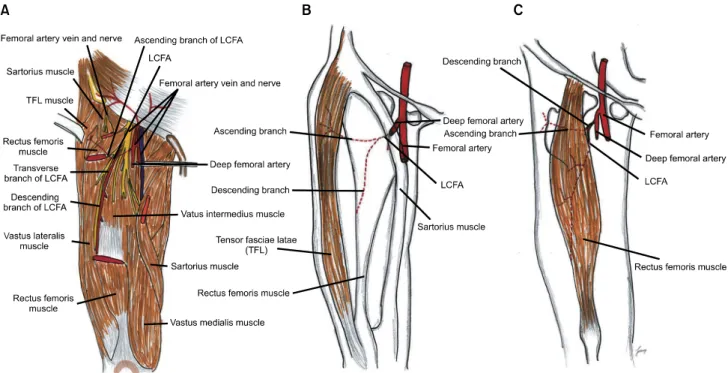
전외측대퇴피판의 근육 및 신경분포
대퇴부 표면의 대표적인 근육인 대퇴직근, 대퇴근막장근 (tensor fascia lata muscle) 및 봉공근(sartorius muscle)은 모 두 전상장골극(anterior superior iliac spine)에서 기원한다. 대 퇴근막장근과 봉공근은 대퇴직근의 표층에 위치하여 무릎 부위에 서 ‘inverted V’ 형태로 벌어져서 각각 무릎의 외측과 내측에 부착된다. 외측광근은 전외측 대퇴부에서 가장 큰 근육으로 대퇴 골의 전하방 경계(anterior and inferior border of trochanter), 대둔근(gluteal tuberosity), 대퇴골 조선(linea aspera) 및 측방 근간중격(lateral intermuscular septum)에서 기시하여 슬개골 의 상측방 경계부와 경골(tibia)의 외측과두부에 부착한다[4,5].
대퇴직근, 외측광근, 내측광근(vastus medialis muscle) 및 중간 광근(vastus intermedius muscle) 등은 모두 하나의 건막을 이 루어 슬개골에 부착함으로써 무릎의 신장(extension)에 관여한 다(Fig. 1).
대퇴부 근육에서 맨 처음 고려해야 할 근육은 대퇴근막장근으 로서 이 근육만으로도 대퇴근막장근피판(tensor fascie latae musculocutaneous flap)을 거상할 수 있다. 대퇴근막장근으로 공급되는 혈관은 외측대퇴회선동맥(lateral circumflex femoral artery)의 수평지(transverse branch)가 대부분이고 일부 상행지 (ascending branch)와 연관되므로 이들 분지들을 찾아서 외측대 퇴회선동맥 및 정맥의 혈관을 결찰함으로써 근육피판을 추가적으 로 거상하거나 또는 전외측대퇴피판의 거상이 어렵거나 피부 천공 지를 찾는 것을 실패할 경우 이에 대한 대안으로서 고려할 수 있다[6]. Coskunfirat와 Ozkan[7]은 실제로 전외측대퇴피판을 계획하였다가 피부로의 중격천공지 또는 근육중격천공지가 약하 거나 심지어 결손인 경우에, 이의 대안으로서 대퇴근막장근 천공 지피판을 거상하여 두경부 재건에 활용한 예를 보고하였는데 특 히, 같은 공여부에서 채취할 수 있으므로 추가적인 공여부 합병증 을 줄일 수 있으므로, 전외측대퇴피판의 훌륭한 대안으로서 고려 될 수 있다고 하였다[7,8].
또한, 임상적으로 보다 자신 있는 준비를 위해서는 대퇴직근을
피부에 같이 거상하는 대퇴직근피판(rectus femoris myocuta-
neous flap)도 같이 고려해야 한다. 대퇴직근은 전상장골극 및
관골절구(groove of acetabulum)에서 기원하여 슬개골 상부로
이어지는 근육으로 대략적인 길이는 36 cm, 두께는 1.5 cm,
그리고 폭경은 5.5 cm에 해당하는 근육으로 다리의 신장과 대퇴
부의 굴절을 가능하게 한다. 주된 혈관 공급은 외측대퇴회선동맥
의 상행지로서 혈관경은 대략 4 cm 정도의 길이로 박리가 가능하
다[6,9,10]. Fig. 2에서는 기본적인 전외측대퇴피판과 비교하여
대퇴근막장근피판 및 대퇴직근 피판의 근육 및 혈관 분포를 보여
준다.
Fig. 2. (A) Schematic drawings of lateral circumflex femoral artery (LCFA) and its branches, femoral nerves and its related muscular
anatomies, and (B) tensor fascie latae musculocutaneous flap, and (C) rectus femoris myocutaneous flap.Fig. 3. Schematic drawings of vastus
lateralis myocutaneous anterolateral thigh (ALT) flap, (A) approach to the vascular bundles after elevation of medial portion, (B) elevation of myocutaneous ALT flap including the subcutaneous layer and fascia lata down to the vastus lateralis muscle, and (C) elevation of myocutaneous ALT flap including the suprafascial plane and deep fascia.이처럼 전외측대퇴피판은 결손부의 크기 및 두께에 따라 근막 피부 피판 및 외측광근을 같이 거상하여 근육-근막피부판으로도 거상할 수 있으며(Fig. 3), 동시에 또는 따로 대퇴근막장근피판 및 대퇴직근피판도 같이 고려할 수 있는 여러 다양성을 지니는 피판이라 할 수 있다[11,12].
전외측대퇴피판의 주 감각신경은 요추신경총(lumbar plexus) L2 및 L3에서 분지하는 외측대퇴피신경(lateral femoral cuta- neous nerve)으로 이 신경은 서혜인대(inguinal ligament) 후외 측부에서 서혜관(inguinal tunnel)을 통과하여 심회선장골동맥
(deep circumflex iliac artery)의 여러 분지를 따라 대퇴근막
하방으로 주행한다. 이 중 하지(descending branch)는 전상장골
극 1 cm 내측으로 주행하여 외측대퇴회선동맥의 하지와 같이
주행하여 근육간 중격을 통과하고 봉공근 전방부로 주행하여 대퇴
부에 이르게 되며, 대퇴부에서 전방과 후방 가지로 나뉘어 분포한
다. 전방가지는 전상장골극의 10 cm 정도 하방에서 대퇴근막을
뚫고 대퇴부의 전외측 피부를 지배하고, 후방가지는 전방가지보
다 위쪽에서 대퇴근막을 뚫고 나와서 대퇴 측방부의 피부 감각을
지배하게 되는데, 따라서 전외측대퇴피판 자체의 주된 감각신경
Fig. 4. Schematic drawing of the lateral (lateral side) and anterior
(medial side) femoral cutaneous nerve of the anterolateral skin of the thigh.Fig. 5. Schematic drawing of the
femoral artery, descending branch of lateral circumflex femoral artery, and its related anatomies, anterior view. LCFA, lateral circumflex femoral artery; TFL, tensor fascia lata.가지는 외측대퇴피신경의 전방가지라고 할 수 있다(Fig. 4). 그밖 에 대퇴신경의 가지인 대퇴전피신경(anterior cutaneous nerve of the thigh)은 서혜인대 중앙부보다 8 cm 하방에서 대퇴근막을 뚫고 주행하며 대퇴 전방부의 감각을 지배하게 된다[4-6].
외측대퇴회선동맥 및 정맥
전외측대퇴피판의 혈액공급은 외측대퇴회선동맥에서 기원하 는 분지들에서 이루어지는데, 외측대퇴회선동맥은 심대퇴동맥 (profunda femoral artery)으로부터 나와 대퇴직근과 외측광근
의 심부를 지나 상행지, 수평지 및 하행지(descending branch) 로 나뉜다(Fig. 5). Wong과 Wei[13]는 하행지와 수평지 사이에 비스듬히 아래쪽으로 주행하는 사선지(oblique branch)가 89 증례에서 34%인 경우에 발견되었음을 보고하였으며, 또한 14%
에서는 전외측대퇴피판술에 포함하여 거상하였음을 최근 2010년 에 보고한 바 있다. 이처럼 전외측대퇴피판의 혈관경은 8∼12 cm 정도, 동맥의 직경은 2.1 mm 정도이며, 정맥은 대부분 2개의 동반정맥(vena comitans)으로 2.3 mm 정도의 직경을 지닌다.
하행지는 외측광근과 대퇴직근의 외측근 중격으로 주행하면서 외측광근에 혈류를 공급하면서 전상장골극(anterior superior iliac spine)과 슬개골(patella)의 상외측연(superior lateral border)을 연결하는 선의 midpoint 주변에서 전외측 대퇴부에 천공지를 내게 된다[14,15].
하행지가 전외측 피부에 분지시키는 천공지 중에서 약 80%는 외측광근을 통과하여 피부로 주행하는 근육피부 천공지이고, 20%
정도만이 외측광근과 대퇴직근 사이의 근중격 사이로 주행하는 중격피부 천공지임을 반드시 명심해야 하며, 이러한 근육피부 천공지는 Fig. 5와 같이 슬개골의 상외측연과 전상장골극을 연결 하는 선의 전외측 피부의 근위부에 주로 분포한다[16-18]. 전술한 바와 같이 대퇴근막장근피판 및 대퇴직근피판도 피부 피판의 표시 와 이에 따른 표면 해부학적 지식을 같이 지녀야 한다(Fig. 6).
이러한 천공지는 전술한 바와 같이 정중부를 중심으로 반경 3 cm인 원의 하방 4분의 1에 주로 많이 분포하는데, Park 등[19]
은 한국인 사체 19구의 양쪽 대퇴부를 전상장골극과 슬개골의
상외측연을 연결하는 선을 10등분하였을 때 천공지의 85%가
3/10에서 8/10 사이에 분포함을 확인하였으며, 외측대퇴회선동
맥의 변이를 관찰한 결과 약 68.4%에서 외측대퇴회선동맥의 상행
Fig. 7. (A) Myocutaneous perforator from the descending branch of the lateral circumflex femoral artery (LCFA), penetrating the vastus
lateralis with an intramuscular length of ∼5 cm in 57% of cases. (B) Myocutaneous perforator from the transverse branch of the LCFA, penetrating the vastus lateralis with an intramuscular length of ∼7 cm in 27% of cases. (C) Septocutaneous perforator from the descending branch of the LCFA, penetrating the intermuscular septum between the vastus lateralis and retus femoris with a length of ∼4 cm in the septum in 11% of cases. (D) Septocutaneous perforator from the transverse branch of the LCFA, penetrating the intermuscular septum formed by the rectus femoris, vastus lateralis, and tensor fascia lata (TFL) with a length of ∼8 cm in the septum in 5% of cases.Fig. 6. Flap skin designs of (A) ante-
rolateral thigh flap, (B) tensor fascia lata flap, and (C) rectus femoris flap.지, 수평지 및 하행지로 나뉘었는데, 일부분에서는 하행지가 외측 대퇴회선동맥에서 수평지와 상행지와는 다른 기원에서 분지되거 나 또는 드물게 하행지가 대퇴동맥(femoral artery)에서 직접 분지되는 경우도 확인하였다(Fig. 7).
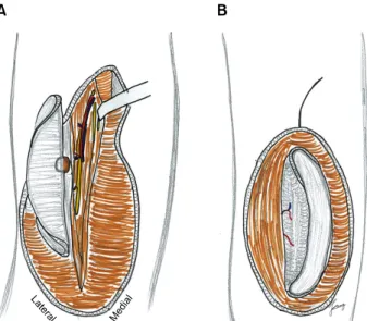
전외측대퇴피판을 채취하기 위한 기본 개념은 다른 근육기반
또는 근막기반의 피판과 달리 주된 혈행이 근육을 관통하고 주행
하는 천공지 피판임을 반드시 명심해야 하며, 근막 및 피부, 피하
층에 혈행을 공급하기 위한 천공지를 찾고, 이를 잘 유지하기
위한 과정으로서 이해할 수 있다. Fig. 6에서 소개된 바와 같이,
거상해야 할 피부에서 제일 잘 들리는 도플러를 확인하고, 이를
Fig. 9. Schematic drawings of septo-
fasciocutaneous anterolateral thigh (ALT) flap, (A) septocutaneous per- forator identification after elevation of medial portion, (B) elevation of lateral portion, and (C) vascular pedicle dissection exposing the de- scending branch of the lateral cir- cumflex femoral artery (LCFA).Fig. 8. Clinical view of two perforators (arrows) in the septofascio-
cutaneous anterolateral thigh (ALT) flap.기준으로 주변으로 영역을 넓혀가면서 거상하게 된다. 외측광근 과 대퇴직근 사이의 외측근간중격(lateral intermuscular sep- tum)을 가정하는 가상선으로 전상장골극과 슬개골의 상전외측연 을 연결하게 된다. 이 가상선의 중간 지점을 중앙점으로 하여 직경 3 cm의 원을 그리고 이 원을 4등분하였을 때 대부분 원의 하외측 부분에 전외측대퇴피판의 피부 천공지가 위치하므로, 도 플러 검사를 이 부위부터 확인하면서 혈류분포를 확인하게 된다.
피부 절개의 깊이는 피부와 피하지방층을 통과하여 근육 위 근막 이 나올 때까지 진행하며, 이 근막 하방의 근막하층(subfasical plane)을 포함하여 박리하는 것이 중요하며, 피하층의 두께를 보면서 아주 얇게 박리해야 할 경우에는 근막보다 표층으로 근막 상층(suprafasical plane)을 따라 박리하게 된다[6,17].
피부판의 크기는 한 개의 천공지로 35×25 cm까지 혈류 분포 가 가능하다고 보고되고 있으나, 피부판의 크기가 상대적으로
클 경우는 가능하면 2개 이상의 천공지를 포함시키는 것이 유리하 다(Fig. 8). 또한, 수술 중에 포함시킬 천공지를 선택할 때는 짧은 근육경로를 가지면서 직경이 굵은 천공지를 선택하는 것이 중요하며, 술자는 가능한 미세수술용 현미경이나 보안경을 사용 하여 작은 천공지를 찾은 후에 박리하는 것이 바람직하다. 디자인 한 피판의 내측연을 따라 대퇴근막의 깊이까지 절개하고 천공지가 피부판으로 들어가는 부분이 확인될 때까지 계속 박리하고, 상방 으로는 대퇴근막장근이 보일 때까지 박리하며 대퇴근막장근을 절개한 후에는 피판을 외측으로 당기면서 대퇴직근과 외측광근의 경계부까지 근막하로 박리하여 천공지를 찾게 된다(Fig. 9).
일단 천공지가 확인되면 수혜부의 결손 모양과 혈관의 위치에
맞게 피판을 디자인한 후, 천공지 주위의 연조직을 보존하면서
근막하 천공지에서 주 혈관까지 박리를 시작하게 된다. 피부판으
로 들어가는 천공지를 확인하면 대퇴직근과 외측광근 사이의 노란
지방층으로 확인될 수 있는 외측근간중격을 확인하고, 확인 후에
는 대퇴직근을 내측으로 견인하면서 근육외측경계부를 기준으로
외측광근의 내측을 따라 근심부로 올라가면서 외측대퇴회선동맥
의 하행지를 찾을 때까지 계속 박리하게 된다(Fig. 10). 하행지가
잘 보이면 좋으나, 대부분은 잘 보이지 않고 외측광근을 통과하는
근육피부천공지로 관찰될 수 있으므로 외측광근의 근육 내 박리를
천천히 조심하여 시행하여 피부로 이어지는 천공지가 혈관경과
만나는 지점까지 근육 내를 주행하는 혈관경을 박리하는 인내심이
필요하다. 이 때 박리하는 방법으로는 원심부에서 외측광근을
통과하여 중격으로 찾아 들어오는 방법과 또는 거꾸로 근간중격에
서 피부쪽 및 근심부로 외측광근을 통과하여 대퇴근막장근 내의
근심부로 찾아 나오는 방법이 가능한데, 어떤 방법이든 양측을
모두 살펴보면서 세심히 진행하고 가능하면 현미경으로 근육 내
혈관의 분포를 보면서 진행하는 것이 추천된다[6,13,17]. 또한,
Fig. 10. Schematic drawings of musculocutaneous anterolateral thigh (ALT) flap, (A) musculoutaneous perforator identification after
elevation of medial portion, (B) elevation of lateral portion by keeping the musculocutaneous perforator, (C) intramuscular pedicle dissection exposing the musculocutaneous perforator and muscular branch of femoral nerve, and (D) vascular pedicle dissection exposing the descending branch and the main trunk of lateral circumflex femoral artery (LCFA).Fig. 11. Schematic drawings of cutaneous anterolateral thigh
(ALT) flap by keeping the small patch of fascia, (A) cutaneous perforator identification from the septum or muscle substance, and (B) suprafascial elevation of lateral portion showing the fascia lata and musculocutaneous perforator.혈관경을 박리할 때는 혈관만을 유지하는 것이 아닌 혈관 주변의 근육조직과 일부 결합조직 등을 같이 포함하여 진행하는 것이 안전하며, 혈관경 주변을 박리할 경우 리도카인을 스프레이로 뿌려가면서 박리하면 혈관의 경축으로 인한 피판의 허혈 현상을 막을 수 있다. 피판의 두께는 전술한 바와 같이 근막상측 및 근막하층의 박리를 고려하여 진행함으로써 원하는 두께로 재단하 게 된다.
천공지의 박리 시 손상을 주지 않도록 주의하고 천공지가 손상 받아도 수술 중 발견이 되지 않을 수 있으므로 가능하면 2개 이상의 여러 개의 천공지를 찾아 박리하는 것이 좋으며, 혈관경이 쉽게 꼬일 수 있으므로 적절한 방향을 찾아가면서 박리하는 것이 필요하다. 따라서, 혈관경의 전방부를 표시하거나 대퇴근막을 일 부 보존하여 혈관경의 비틀림을 방지하게 되며, 경우에 따라 천공 지가 아주 작거나, 또는 박리가 어렵거나 심지어 천공지가 없는 경우는 전내측대퇴피판(anteromedial thigh flap)이나 좀 더 근 위부로 박리하여 대퇴근막장근피판이나 대퇴직근피판을 대신 이 용하려는 전환이 필요하며, 결손부위의 부피가 클 경우에는 근육 층을 일부 포함시킬 수 있고, 필요에 따라서는 피부와 피하 모세혈 관총을 제거하고 피하지방과 근막으로만 이루어진 피지방근막피 판(adipofascial flap)을 대신 고안할 수 있다[20,21](Fig. 11).
고 찰
전외측대퇴피판은 인체 내 다양한 재건상황에 적용될 수 있으
며, 구강 내, 외 재건을 위해서 저자 등이 기존에 보고한 광배근피
판(latissimus dorsi myocutaenous flap), 요골전완유리피판,
족배동맥피판(dorsalis pedis flap), 외측상완유리피판 및 복직근
유리피판(rectus abdominis myocutaneous free flap) 등과 같
이 구강악안면 영역의 연조직을 재건하기 위한 넓은 피부를 거상
할 수 있고, 해부학적인 변이가 상대적으로 적은 양상을 보이면서
공여부의 합병증도 최소화될 수 있는 장점을 지닌다. 특히, 경부청
소술을 포함하여 종물 제거에 있어 많은 시간이 소요되는 구강암
수술 및 재건수술에 있어서 두 팀이 동시에 수술을 할 수 있어서
수술 전체의 시간을 많이 줄일 수 있는 장점도 지닌다. 그러나, 여러 장점에도 불구하고 전외측대퇴피판은 피부천공지의 해부학 적인 변이가 많고, 주로 근육 내를 주행하는 근육피부천공지를 박리해야 하기 때문에 숙달되기 전에는 많은 인내심과 각고의 노력이 필요한 피판이라 할 수 있다[3,22,23].
최근 Park과 Miles[24]에 의하면 전외측대퇴피판은 보다 자유 로운 형태(free style)의 접근이 가능한 유용한 피판으로 설명되고 있다. 외측대퇴회선동맥의 하행지가 결손되거나 거상하기 여의치 않을 경우에는 내측의 하행지를 이용하는 전내측대퇴피판을 거상 할 수 있으며, 후인두 결손(laryngopharyngeal defect)부의 재 건 시 두 층의 피판층을 이용하여 충분한 부피로 거상하여 재건함 으로써 기능적으로도 좋은 결과를 보였음이 보고되었다. 또한, 구강 내의 여러 결손부의 재건 이외에도 하악골 등의 골이식 후에 연조직 결손부를 채우기 위해서도 활용되며, 두개저부 재건 및 이하선 및 경부 재건을 위해서도 유용하게 활용될 수 있음이 보고되었다[11,12,25].
전술한 바와 같이 전외측대퇴피판의 단점은 피부 천공지의 변형 및 결손이 있을 수 있다는 점으로 1998년 Kimata 등[26]은 74명의 환자에서 5.4%에 해당하는 4예에서 피부 천공지를 발견 하지 못하였다고 보고하였다. Celik 등[27]은 전외측대퇴피판을 시행한 439명의 환자에서 0.9%인 6명에서는 피부 천공지가 결손 이었으며, 이식 후에도 7.7%인 34명의 환자에서 재탐색이 필요하 였고, 다른 6명에서는 정맥에서의 순환 부전으로 실패하였음을 보고하였다. 이처럼 전외측대퇴피판의 실패 원인으로는 천공지를 박리할 때의 손상으로 혈관경이 다치거나, 천공지가 너무 가늘어 서 혈관문합 후 혈행유지가 어려운 경우, 천공지 혈관이 수축되거 나, 혈관경이 꼬여서 장력이 많이 걸려 있는 경우 등을 고려할 수 있다. 반면에 피판 거상이 익숙해지면 거상 방법을 다양하게 하면서 피판의 두께도 얇게 할 수 있는데, 경우에 따라 요골전완유 리피판 및 족배동맥피판 등과 유사하게 얇고 기능적인 피판으로도 재단할 수 있다. 실제 최근에는 이렇게 얇게 재단된 피판으로 활용함으로써 혀나 연구개부 재건을 위해 발음 및 연하운동 등의 기능적인 결과에서도 우수하다고 보고되고 있으며, 기존에는 주 로 요골전완유리피판을 사용하였던 구강 내 재건에 있어 점차 전외측대퇴피판이 더 추천되고 선택되는 경향을 보여왔다고 할 수 있다[28-30].
설절제술 및 구강저 절제술 등과 같이 구강 내의 여러 기능적인 재건이 필요한 경우 흔히 고려할 수 있는 유리피판이 요골전완유 리피판인데, 비심미적인 공여부 및 손목의 불편감을 고려하면 전외측대퇴피판이 대안으로서 고려되어 왔다. 공여부의 일차봉합 이 가능하고 심미적으로도 큰 반흔을 남기지 않는 우수함으로 선택될 수 있는 전외측대퇴피판은 대개 마른 동양인에서 웬만한 구강 내 결손부를 재건할 수 있으나, 비만이거나 여자 환자들에서 는 상대적으로 두꺼운 피부층이 단점이 될 수 있다. 또한, 인두부
및 혀의 뒷부분을 포함하는 광범위한 결손부에 대해서는 요골전완 유리피판만으로는 크기가 많이 부족할 수 있어서 피판의 길이가 대략 13 cm 이상 필요할 경우 및 넓은 피부면적이 필요할 경우 우선적으로 선택될 수 있다. 즉, 구강 내 점막 이장과 동시에 구강저의 사강(dead space) 부위도 지방층을 많이 포함시킬 수 있는 전외측대퇴피판을 사용함으로써 타액누공이 발생하거나 다 른 이차감염을 예방할 수 있는 장점도 지닐 수 있게 된다[28,31].
또한, 아주 광범위한 결손부에 활용할 수 있는 복직근유리피판 과 비교해서 혈관경을 상대적으로 길게 채취할 수 있고, 복직근유 리피판에서 생길 수 있는 탈장(hernia) 등의 합병증을 피하고, 복부의 지방층이 매우 두꺼워 피부판을 사용하기 어려운 경우에도 전외측대퇴피판은 훌륭한 대안으로 선택될 수 있는데, 특히 노인 환자에서 발생할 수 있는 탈장을 막을 수 있고, 부피를 회복하기 위해 외측장근을 동시에 거상할 수 있으며, 방사선치료 후에 일부 또는 과도하게 수축될 수 있는 복직근 하방의 지방층에 비해 대퇴부의 지방층은 상대적으로 수축률이 적은 장점을 지니게 된다 [32,33].
Song 등[1]은 피판 크기에 대해 대천 대퇴골 대전자부(greater trochanter)에서 슬개골의 3 cm 상방에 이르는 범위 내에서 전외 측 피부 영역에 최대 20×26 cm 크기의 피부판을 계획할 수 있다고 하였다. Koshima 등[2]은 최대 25×18 cm까지 피부판 거상이 가능하다고 하여 근육을 관통하는 피부 천공지의 혈행 분포에 따라 수평적으로는 20∼25 cm까지, 수직으로는 18∼26 cm까지의 다양한 크기의 피부조직판의 도안이 가능함을 알 수 있다. 피판 거상을 완료한 후, 전외측부인 공여부 결손은 대부분 일차 봉합을 시행하며, 거상 후 결손부의 좌우 폭경이 8∼10 cm 이상인 경우에는 피부의 두께 및 근육의 유연성에 따라 장력이 심하여 직접 봉합이 어려울 수 있으므로, 부분층 피부이식술을 고려하게 된다. 피부피판만 채취한 경우는 수술 후 바로 보행이 가능하고, 외측광근 및 다른 근육을 같이 채취한 경우는 1주일 정도 침상안정 이후 보행하도록 하며, 어떤 경우에서나 대개는 이차감염만 예방하면 잘 치유되는 것으로 알려져 있다[34,35].
전외측대퇴피판에서는 심막을 포함하여 피하조직 및 피부 피판
을 만드는 방법과 또는 심막은 제외하고 피하지방 피판만을 만드
는 방법도 있으며, 피부를 제외하고 피하지방과 근막으로만 이루
어진 근막 피판을 만드는 방법도 다양하게 있을 수 있다. 또한,
기존에 외측대퇴회선동맥의 분지와 함께 대퇴부 외측 피부와 근육
을 함께 거상하는 방식(Fig. 3)이 아니고, 반대로 피부 천공지를
근간으로 전외측대퇴부 천공지피판의 방식으로도 피판을 거상할
수 있게 되었다. 2002년 Wei 등[3]이 보고한 672명의 전외측대퇴
피판 환자에서 두경부 재건에 이용된 475명에 대해 95.68%의
성공률을 보였는데, 피판의 두께도 서양인에 비해 상대적으로
비만이 적은 동양인에서 피판의 두께를 적절히 재단할 수 있다고
보고되어 왔다.
천공지란 심부 혈관에서 기원하여 심층 근막(deep fascia)을 뚫고 피부에 혈액을 공급하는 혈관을 말하며, 천공지피판 (perforator flap)이란 이러한 천공지와 피부 및 피하지방으로만 이루어진 피판을 지칭하게 된다. 최근까지 전외측대퇴피판을 이 용한 다양한 부위의 재건이 보고되어 왔으며 천공지 분포 변이에 대한 해부학적 지식이 축적되고, 도플러를 이용하여 천공지의 위치를 찾을 수 있게 되면서 현재의 전외측대퇴피판은 전신의 어느 부위에서나 결손부 재건을 위해 유용하게 사용될 수 있으며, 두경부 영역의 재건에 있어서도 이제는 주요 재건 방법의 하나로 인정되고 있다.
Acknowledgements
This study was supported by a grant of the Korean Health Technology R&D Project, Ministry of Health & Welfare, Republic of Korea (A110405).
References
1. Song YG, Chen GZ, Song YL. The free thigh flap: a new free flap concept based on the septocutaneous artery. Br J Plast Surg 1984;37:149-59.
2. Koshima I, Fukuda H, Utunomiya R, Soeda S. The antero- lateral thigh flap; variations in its vascular pedicle. Br J Plast Surg 1989;42:260-2.
3. Wei FC, Jain V, Celik N, Chen HC, Chuang DC, Lin CH.
Have we found an ideal soft-tissue flap? An experience with 672 anterolateral thigh flaps. Plast Reconstr Surg 2002;
109:2219-26.
4. Putz R, Pabst R. Sobotta atlas of human anatomy. 14th ed.
Münich: Elsevier/Urban & Fischer; 2006.
5. Standring S. Gray’s anatomy: the anatomical basis of clinical practice. 39th ed. New York: Elsevier Churchill Livingstone;
2005.
6. Strauch B, Yu HL. Atlas of microvascular surgery: anatomy and operative approaches. 2nd ed. NewYork: Stuttgart; 2006.
p.372-87.
7. Coskunfirat OK, Ozkan O. Free tensor fascia lata perforator flap as a backup procedure for head and neck reconstruction.
Ann Plast Surg 2006;57:159-63.
8. Chalfoun CT, McConnell MP, Wirth GA, Brenner KA, Evans GR, Kobayashi M. Free tensor fasciae latae flap for abdomi- nal wall reconstruction: overview and new innovation. J Reconstr Microsurg 2012;28:211-9.
9. Choi SW, Park Jy, Hur MS,
et al. An anatomic assessment on perforators of the lateral circumflex femoral artery for anterolateral thigh flap. J Craniofac Surg 2007;18:866-71.
10. Cömert A, Altun S, Unlü RE, Orbay H, Tekdemir I.
Perforating arteries of the anteromedial aspect of the thigh:
an anatomical study regarding anteromedial thigh flap. Surg Radiol Anat 2011;33:241-7.
11. Camporro D, Fueyo A, Martín C, Carnero S, Llorente JL.
Use of lateral circumflex femoral artery system free flaps in skull base reconstruction. J Craniofac Surg 2011;22:888-93.
12. Iyer S, Chatni S, Kuriakose MA. Free tensor fascia lata-iliac crest osteomusculocutaneous flap for reconstruction of com- bined maxillectomy and orbital floor defect. Ann Plast Surg 2012;68:52-7.
13. Wong CH, Wei FC. Anterolateral thigh flap. Head Neck 2010;32:529-40.
14. Wong CH, Wei FC, Fu B, Chen YA, Lin JY. Alternative vas- cular pedicle of the anterolateral thigh flap: the oblique branch of the lateral circumflex femoral artery. Plast Reconstr Surg 2009;123:571-7.
15. Seth R, Manz RM, Dahan IJ,
et al. Comprehensive analysis of the anterolateral thigh flap vascular anatomy. Arch Facial Plast Surg 2011;13:347-54.
16. Lin DT, Coppit GL, Burkey BB. Use of the anterolateral thigh flap for reconstruction of the head and neck. Curr Opin Otolaryngol Head Neck Surg 2004;12:300-4.
17. Erba P, Raffoul W, Bauquis O. Safe dissection of the distally based anterolateral thigh flap. J Reconstr Microsurg 2012;28:
405-11.
18. Neligan PC, Lannon DA. Versatility of the pedicled antero- lateral thigh flap. Clin Plast Surg 2010;37:677-81.
19. Park JY, Park HD, Youn KH,
et al. Anterolateral thigh flap for head and neck reconstruction: anatomic study. J Korean Assoc Maxillofac Plast Reconstr Surg 2005;27:360-4.
20. Hanasono MM, Skoracki RJ, Silva AK, Yu P. Adipofascial per- forator flaps for "aesthetic" head and neck reconstruction.
Head Neck 2011;33:1513-9.
21. Agostini T, Agostini V. Adipofascial anterolateral thigh free flap for hemifacial atrophy. Acta Otorhinolaryngol Ital 2009;
29:103-7.
22. Lin PY, Chen CC, Kuo YR, Jeng SF. Simultaneous re- construction of head and neck defects following tumor re- section and trismus release with a single anterolateral thigh donor site utilizing a lateral approach to flap harvest.
Microsurgery 2012;32:289-95.
23. Kekatpure VD, Trivedi NP, Shetkar G, Manjula BV, Mathan Mohan A, Kuriakose MA. Single perforator based antero- lateral thigh flap for reconstruction of large composite de- fects of oral cavity. Oral Oncol 2011;47:517-21.
24. Park CW, Miles BA. The expanding role of the anterolateral thigh free flap in head and neck reconstruction. Curr Opin Otolaryngol Head Neck Surg 2011;19:263-8.
25. Posch NA, Mureau MA, Flood SJ, Hofer SO. The combined free partial vastus lateralis with anterolateral thigh perforator flap reconstruction of extensive composite defects. Br J Plast Surg 2005;58:1095-103.
26. Kimata Y, Uchiyama K, Ebihara S, Nakatsuka T, Harii K.
Anatomic variations and technical problems of the antero- lateral thigh flap: a report of 74 cases. Plast Reconstr Surg 1998;102:1517-23.
27. Celik N, Wei FC, Lin CH,
et al. Technique and strategy in anterolateral thigh perforator flap surgery, based on an anal- ysis of 15 complete and partial failures in 439 cases. Plast Reconstr Surg 2002;109:2211-6.
28. Camaioni A, Loreti A, Damiani V, Bellioni M, Passali FM,
Viti C. Anterolateral thigh cutaneous flap vs. radial forearm
free-flap in oral and oropharyngeal reconstruction: an analy-
sis of 48 flaps. Acta Otorhinolaryngol Ital 2008;28:7-12.
29. Kuo YR, Seng-Feng J, Kuo FM, Liu YT, Lai PW. Versatility of the free anterolateral thigh flap for reconstruction of soft-tissue defects: review of 140 cases. Ann Plast Surg 2002;
48:161-6.
30. Bianchi B, Ferri A, Ferrari S,
et al. The free anterolateral thigh musculocutaneous flap for head and neck reconstruction: one surgeon's experience in 92 cases. Microsurgery 2012;32:87-95.
31. Kesting MR, Hölzle F, Wales C,
et al. Microsurgical re- construction of the oral cavity with free flaps from the ante- rolateral thigh and the radial forearm: a comparison of peri- operative data from 161 cases. Ann Surg Oncol 2011;18:
1988-94.
32. Spyriounis PK. The extended approach to the vascular pedi-
cle of the anterolateral thigh perforator flap: anatomical and clinical study. Plast Reconstr Surg 2006;117:997-1001.
33. Liu WW, Yang AK, Ou YD. The harvesting and insetting of a chimeric anterolateral thigh flap to reconstruct through and through cheek defects. Int J Oral Maxillofac Surg 2011;
40:1421-3.
34. Agostini T, Lazzeri D, Spinelli G. Anterolateral thigh flap:
Systematic literature review of specific donor-site complica- tions and their management. J Craniomaxillofac Surg 2013;
41:15-21.
35. Townley WA, Royston EC, Karmiris N, Crick A, Dunn RL.
Critical assessment of the anterolateral thigh flap donor site.
J Plast Reconstr Aesthet Surg 2011;64:1621-6.
Appendix. Korean translational language of anatomical muscles, vessels and nerves in the anterolateral thigh region
Achilles tendonAdipofascial flap Anterolateral thigh flap
Anterior cutaneous nerve of thigh Anterior superior iliac spine (ASIS) Anteromedial thigh flap
Ascending branch of LCFA Deep femoral artery Descending branch of LCFA Femoral artery
Femoral nerve Gluteal tuberosity Groove of acetabulum Inferior gluteal artery Inguinal ligament
Lateral circumflex femoral artery (LCFA) Lateral cutaneous nerve of thigh Lateral femoral cutaneous nerve Lateral superior genicular artery Lateral intermuscular septum Linea aspera
Lumbar plexus
Medial circumflex femoral artery Muscular branch of the femoral nerve Myocutaneous perforator
Patella
Profunda femoral artery Quadriceps
Rectus femoris muscle Sartorius muscle Septocutaneou perforator Suprafascial vascular plexus Tensor fascia lata (TFL) Transverse branch of LCFA Vastus intermedius muscle Vastus lateralis muscle Vastus medialis muscle
아킬레스건 피지방근막피판 전외측대퇴피판 전대퇴피신경 전상장골극 전내측대퇴피판
외측대퇴회선동맥의 상행지 대퇴심동맥
외측대퇴회선동맥의 하행지 대퇴동맥
대퇴신경 대둔근 관골절구 하둔동맥 서혜인대 외측대퇴회선동맥 외측대퇴피신경 외측대퇴피신경 외측상슬동맥 외측근간중격 대퇴골 조선 요추신경총 내측대퇴회선동맥 대퇴신경의 근육가지 근육피부 천공지 슬개골 심대퇴동맥 대퇴사두근 대퇴직근 봉공근 중격피부 천공지 상근막혈관총 대퇴근막장근
외측대퇴회선동맥의 수평지 중간광근
외측광근 내측광근