14
Management of large soft tissue defects in the sacrum has advanced. Until recently, musculocutaneous flaps were widely used. The necessary loss of muscle and the main vessel has prompted the increasing use of perforator-based flaps.1
Several perforator-based flap techniques allow coverage of sacral defects. In 1993, Koshima et al.2 published the anatomy of the gluteal artery perforator flap for coverage of lumbosacral defects. They reported the superior and convenient features that included muscle sparing, versatility in design, and less donor site morbidity. The parasacral perforator flap can also be used to cover sacral defects. It originates from the lateral sacral artery or internal pudendal artery.3 The close location of sacral defects makes the pedicle length long enough to transfer to the defect site without dissecting any muscle. However, the parasacral perforator flap alone is unable to cover large
defects.4
So we designed super-flap using additional perforator considering the angiosomes territory concept. Angiosomes are synthetic blocks of tissue provided by the designated arteries, which expand between the skin and the bone.5 The angiosome territory concept posits the use of angiosomes served by different perforators to prevent postoperative complications, including flap failure.
We report two cases that used the superior gluteal artery perforator super-flap with additional parasacral perforator supported by different angiosomes to cover large sacral defects (Fig. 1).
Dual Perforator Flap for Reconstruction of Large Sacral Defects:
Superior Gluteal Artery Perforator Super-Flap with
Parasacral Perforator
Sang Pil Tae, Seong Yoon Lim*, Jin Kyung Song, Hong Sil Joo Department of Plastic and Reconstructive Surgery, Hanil General Hospital, Seoul, KoreaCC This is an open-access article distributed under the terms of the Creative Commons Attribution Non-Commercial License (http://creativecommons.org/licenses/by-nc/4.0) which permits
unrestricted noncommercial use, distribution, and reproduction in any medium, provided the original work is properly cited. Copyright © 2017 by the Korean Society for Microsurgery. All Rights Reserved.
Received March 7, 2017 Revised March 26, 2017 Accepted March 28, 2017
*Correspondence to: Seong Yoon Lim Department of Plastic and Reconstructive Surgery, Hanil General Hospital, 308 Uicheon-ro, Dobong-gu, Seoul 01450, Korea
Tel: +82-2-901-3109 Fax: +82-2-901-3104 E-mail: [email protected]
ORCID: http://orcid.org/0000-0002-8109-6213 Financial support: None.
Conflict of interest: None.
The superior gluteal artery perforator flap technique has increasingly been used for soft tissue defects in the sacral area following its introduction nearly 25 years ago. Advantages in covering sacral defects include muscle sparing, versatility in design, and low donor side morbidity. The bilateral superior gluteal artery perforator flap procedure is planned in cases of large sacral defects that cannot be covered with the unilateral superior gluteal artery perforator flap. Here, we report two cases of large sacral defects in which patient factors of poor general health, such as old age, pneumonia, and previous operation scar, led to use of a large unilateral superior gluteal artery perforator super-flap with parasacral perforator. The approach was utilized to reduce the operation time and prevent unpredictable flap failure due to the large flap size. Even though the parasacral perforator was included, the versatility of the large superior gluteal artery perforator flap was preserved because sufficient perforator length was acquired after adequate dissection.
Key Words: Dual perforator flap, Superior gluteal artery perforator, Sacral defect, Angiosome
ARMS
Archieves of Reconstructive MicrosurgeryCase Report
pISSN 2383-5257 eISSN 2288-6184 Arch Reconstr Microsurg 2017;26(1):14-17 https://doi.org/10.15596/ARMS.2017.26.1.14
Sang Pil Tae, et al. Superior Gluteal Artery Perforator Super-Flap with Parasacral Perforator
www.e-arms.org 15
CASE REPORT
Case 1
A 79-year-old female with a medical history of hypertension was referred for the treatment of 4th-degree burns on the sacral area, caused by an electric blanket. Serial debridement and vacuum assisted closure therapy was applied for 16 days, while the patient was consecutively treated for pneumonia. After debridement, the sacral wound size was 11×10 cm2
. We subsequently planned the superior gluteal artery perforator super-flap with additional parasacral perforator because of the large defect size. The perforators were identified and mapped with a hand-held Doppler instrument. The superior gluteal artery perforators were identified two-thirds along the line of the medial, which is between the posterior superior iliac spine
and great trochanter. The parasacral perforators were identified above the wound, between the bilateral posterior superior iliac spine. The flap was designed to include the superior gluteal perforator artery with the parasacral perforator artery (Fig. 2). Subfascial dissection was performed lateral to medial, with mapping guidance by the hand-held Doppler instrument. Dissection was carefully done to prevent any injuries to the perforator (Fig. 3). To obtain sufficient perforator length, the superior gluteal artery pedicle needed to be dissected within the muscle; parasacral perforator dissection was accomplished by meticulous subfascial dissection. After finishing the perforator dissection, the flap was transferred cautiously, taking care not to kink or stretch the dissected perforator. The flap size was 19×16 cm2
and the pedicle length of the superior gluteal artery perforator and parasacral artery perforator were 4.5 cm and 2.0 cm, respectively (Fig. 4). The flap was well accepted with Posterior superior iliac spine Dual perforator flap (orange line)
Sacral defect Coccyx
Superior gluteal artery perforator flap (blue line)
Fig. 1. This is the design of superior gluteal artery super-flap with parasacral perforator. This flap is designed to include parasacral perforator angiosome (dotted line) as well as superior gluteal artery perforator angiosome (full line) to cover large sacral defect.
Fig. 2. Preoperative photograph.
Fig. 3. Intraoperative photograph.
Fig. 4. Intraoperative photograph (pedicle of superior gluteal artery including perforator and parasacral perforator).
Arch Reconstr Microsurg Vol. 26. No. 1. May 2017
16
minimal postoperative complications.
Case 2
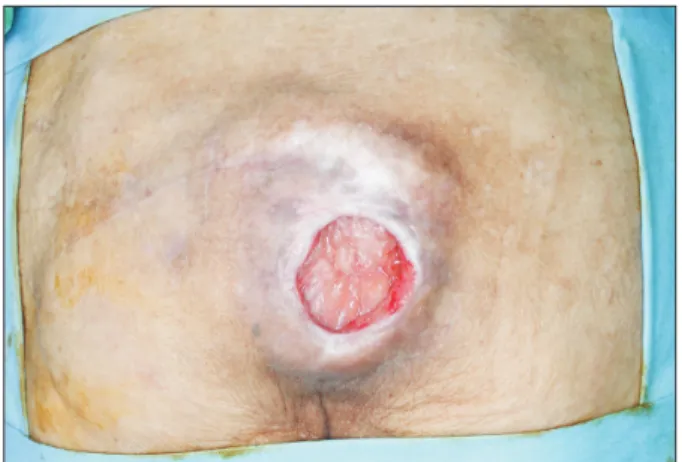
A 75-year-old female with a history of hypertension and Parkinson’s disease was referred for treatment of a grade 4 sacral pressure sore (9×10 cm2
) invading the muscle. Since a previous rotation flap scar was located on the left side, we planned a perforator flap procedure using the right superior gluteal artery flap including parasacral perforator (Fig. 5). The flap size was 18×15 cm2
(Fig. 6). The flap was well accepted with minimal postoperative complications (Fig. 7).
DISCUSSION
After introduction of the gluteal perforator-based flap in 1993, superior gluteal artery perforator flap has been used steadily.2
It
preserves the gluteus maximus muscle with consistent blood flow and the single perforator provides for large skin territories. Skin territories nourished by gluteal perforator-based flaps are almost same as gluteus maximus musculocutaneous flaps, which are estimated to be about 70 cm2
.6
Hence, a large defect can be covered by a superior gluteal artery perforator flap with low donor site morbidity.
Bilateral (double) superior gluteal artery perforator flap is usually used for large sacral defects.6
Alternately, as in our cases, the unilateral superior gluteal artery perforator super-flap with parasacral perforator can be substituted for certain conditions, such as poor general health or previous operation scars. Parasacral perforators originate from the lateral sacral artery or internal pudendal artery.3 We selected the parasacral perforator situated closest to the sacral defects. The parasacral perforator flap was 2.5 cm in one case and 2.0 cm in the other case. These lengths did not compromise flap versatility. Also, compared to bilateral superior gluteal artery perforator flap, this method can reduce the operation time and preserve contralateral side of the superior gluteal artery perforator and flap as well as ipsilateral side of inferior gluteal artery perforator and flap. The preserved area is available for future wound. Before using the perforator flap, we identified the location and variation of perforator arteries to visualize the angiosomes served by the main perforating branches.
Angiosomes are the synthetic blocks of tissue, named after the respective arteries which expand between the skin and the bone.5 According to the angiosome territory concept proposed in 1987, we designed the perforator flap using angiosomes to prevent from postoperative complications including flap failure.
Fig. 5. Preoperative photograph.
Fig. 6. Intraoperative photograph (pedicle of superior gluteal artery including perforator and parasacral perforator).
Sang Pil Tae, et al. Superior Gluteal Artery Perforator Super-Flap with Parasacral Perforator
www.e-arms.org 17
In both cases, the skin defect in sacrum was too large to include only the angiosome territory of the superior gluteal artery perforator flap. Hence, an additional perforator was needed to support the flap around the sacral defects.
A prior study reported the combined use of the superior gluteal artery perforator flap and lumbar artery perforator flap for coverage of a large sacral defect.7
Usually, the fourth lumbar artery perforators are used because they are large and reliable.8 Another paper also reported good results with a modified technique that used the nearest lumbar artery perforator from the defect site.9
This modified technique helps prevent injury to the perforator vessel, since it requires a shorter length of the pedicle, eliminating the need to skeletonize the perforator. In the same context, considering versatility of the flap, we designed a dual perforator close to the large defect site to cover the defect. Safe and large flaps with reliable perforators were obtained.
There are many treatment options for large soft tissue defects in the sacrum. Until recently, musculocutaneous flaps were principally used because of the short learning curve and reliability of the technique. Disadvantages include sacrifice of muscle and the main vessels.1
When nearby healthy tissue is not available because of repeated previous operation, free flap can be attempted. While the superior gluteal vessel can be used as the recipient for the free flap, the usage is restricted due to the lack of proper recipient vessel and the necessities of a long surgery time and advanced technical skills.10
In conclusion, the superior gluteal artery perforator super-flap with parasacral perforator with consideration of angiosome territories can be a good option in covering a large sacral defect unable to be covered by unilateral superior gluteal artery perforator artery flap. This procedure reduces the operation
time and prevents unpredictable flap failure. Even though the parasacral perforator is included, the versatility of the large superior gluteal artery perforator flap is preserved due to sufficient perforator length gained after adequate dissection.
REFERENCES
1. Kroll SS, Rosenfield L. Perforator-based flaps for low posterior midline defects. Plast Reconstr Surg 1988;81:561-6.
2. Koshima I, Moriguchi T, Soeda S, Kawata S, Ohta S, Ikeda A. The gluteal perforator-based flap for repair of sacral pressure sores. Plast Reconstr Surg 1993;91:678-83.
3. Lin CT, Chen SY, Chen SG, Tzeng YS, Chang SC. Parasacral perforator flaps for reconstruction of sacral pressure sores. Ann Plast Surg 2015;75:62-5.
4. Ahmadzadeh R, Bergeron L, Tang M, Morris SF. The superior and inferior gluteal artery perforator flaps. Plast Reconstr Surg 2007;120:1551-6.
5. Taylor GI, Palmer JH. The vascular territories (angiosomes) of the body: experimental study and clinical applications. Br J Plast Surg 1987;40:113-41.
6. Mahboub T. Superior gluteal artery perforator flap for closure of large sacral defects. Egypt J Plast Reconstr Surg 2004;28:175-9. 7. Lui KW, Hu S, Ahmad N, Tang M. Three-dimensional
angiography of the superior gluteal artery and lumbar artery perforator flap. Plast Reconstr Surg 2009;123:79-86.
8. Bissell MB, Greenspun DT, Levine J, Rahal W, Al-Dhamin A, AlKhawaji A, et al. The lumbar artery perforator flap: 3-dimensional anatomical study and clinical applications. Ann Plast Surg 2016;77:469-76.
9. Yoon CS, Yim JH, Kim MH, Ha W, Kim KN. Modified lumbar artery perforator flaps for gluteal pressure sore reconstruction. ANZ J Surg 2016. doi: 10.1111/ans.13494 [Epub].
10. Park S, Koh KS. Superior gluteal vessel as recipient for free flap reconstruction of lumbosacral defect. Plast Reconstr Surg 1998;101:1842-9.