Introduction
Soft tissue defects after total knee arthroplasty (TKR) are a daunting problem that have a high probability of revision. The medial gastrocnemius flap is often used to cover such soft tissue defects of the knee or upper third of the leg [1]. The gastrocne- mius flap was originally a muscle flap, but in 1978, McCraw et al. [2] demonstrated the possibility of a gastrocnemius myocutaneous flap (MCF). However, there are still few articles reporting knee coverage using a gastrocnemius MCF. Disadvantages of the medial gastrocnemius muscle flap are the reduced volume at the distal part of the flap and the short reach [3]. In order to overcome the limitations of the medial gastrocne- mius muscle flap, we combined a medial sural artery perforator flap with a medial gastrocnemius muscle flap, in four cases that required reconstruction of skin and soft tissue defects due to deep infection after TKR. The study was approved by the Institu- tional Review Board of Keimyung University School of Medicine (IRB No. 2021-02- 029) and performed in accordance with the principles of the Declaration of Helsinki.
Written informed consent was obtained from the patients for the use of their images.
Case
An 82-year-old woman visited our hospital and was admitted to the orthopedic sur- gery department because of a deep infection after TKR. Her underlying diseases were
Case Report
Received: November 12, 2020 Revised: December 11, 2020 Accepted: December 13, 2020 Corresponding author:
Jaehoon Choi, M.D., Ph.D.
Department of Plastic and Reconstructive Surgery, Keimyung University School of Medicine, 1035 Dalgubeol-daero, Dalseo-gu, Daegu 42601, Korea
Tel: +82-53-258-7815 Fax: +82-53-258-4590 E-mail: [email protected]
This is an Open Access article distributed under the terms of the Creative Commons Attribution Non-Commercial License (https://creativecommons.org/licenses/by-nc/4.0/) which permits unrestricted non-commercial use, distribution, and reproduction in any medium, provided the original work is properly cited.
© 2021 Korean Wound Management Society
Modified Medial Gastrocnemius Myocutaneous
Flap Technique for Knee Joint Coverage after Total Knee Arthroplasty
Dongseok Kim , Junhyung Kim , Woonhyeok Jeong , Taehee Jo , Jaehoon Choi
Department of Plastic and Reconstructive Surgery, Dongsan Medical Center, Keimyung University School of Medicine, Daegu, Korea
Abstract
While there are many reasons the medial gastrocnemius flap is often the favored treatment for soft tissue defects around the knee area, this flap has some disadvantages. Reduced volume at the distal part of the flap and a short reach complicate provision of sufficient coverage for soft tissue defects superior to the patella and the lateral knee. In order to overcome these shortcomings, we modified the typical surgical technique by combining a medial gastrocnemius muscle flap and a medial sural artery perforator flap. This approach was applied to four patients who had developed deep infections and skin and soft tissue defects around the knee joint after total knee arthroplasty. The surger- ies were successful. Dead space was well-filled and wounds healed without complications in all patients. This modified medial gastrocne- mius myocutaneous flap provides a new option for treating challenging skin and soft tissue defects caused by deep infection after total knee arthroplasty.
Keywords: Knee; Arthroplasty, replacement, knee; Reconstructive surgical procedures; Surgical flaps
hypertension and dementia. Incision and drainage were per- formed, followed by serial infection control surgery by the or- thopedic surgery department. Finally, the infection was con- trolled, but a skin and soft tissue defect resulted around the
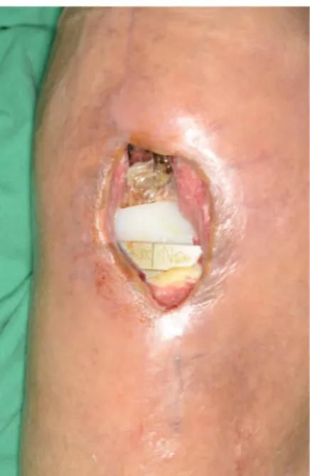
knee. Our department was consulted to cover the defect. The skin defect was located anterolateral to the knee joint (Fig. 1).
Since the defect was far from the pedicle of the medial gastroc- nemius muscle flap, modifications to the flap were required.
Our plan was to elevate the skin flap distal to the gastrocne- mius muscle by using a perforator from the most distal part of this muscle. Preoperatively, we used Doppler ultrasonography to find the medial sural artery perforators and marked the lo- cation with a skin stapler for visibility on computed tomogra- phy (CT). CT angiography of the lower extremities showed the presence of the appropriate perforator near our marking, and we designed the incision line based on the location of the ideal perforator which was located most distally (Figs. 2, 3).
Intraoperatively, a preliminary explorative incision was made on the skin and the subfascial plane was carefully dissected from anterosuperior to posteroinferior to locate the perforator.
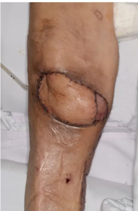
The viability of the perforator was assessed with Doppler ul- trasonography (Fig. 4). The flap was elevated and then tun- neled and inset to cover the defect (Fig. 5). Flap surgery was successful, and the patient was discharged on foot 2 weeks af-
Fig. 1. Preoperative clinical photograph. A 5 ×3-cm skin and soft tissue defect is seen on the anterolateral area of the knee.
The joint capsule is open and the prosthesis is visible.
Fig. 2. Preoperative computed tomography angiography of the lower extremities. Before scanning, we used Doppler ultrasonog- raphy to find the medial sural artery perforators and marked the location with a skin stapler for visibility on computed tomography.
(A) The medial sural artery perforator is observed just beneath the marking (red circle); left and right sides are reversed in the image. (B) Among three perforators (red arrows), we used the most distal perforator which is located about 165 mm below the popliteal crease; here the image is not reversed.
A B
Fig. 3. Design of the modified medial gastrocnemius myocuta-
neous flap. The marked skin flap (blue line) is elevated together
with the medial gastrocnemius muscle flap using the perforator
from the most distal part of the gastrocnemius muscle. The left
side of the picture is medial, and the right side is lateral.
ter surgery (Fig. 6).
Discussion
Defects on knees are difficult to cover. Among the reconstruc- tive options, local flaps are quite limited because of their small size; other muscular flaps, such as the reverse biceps femoris
flap, are not superior to the gastrocnemius flap, and have high- er donor morbidity. Free tissue transfers can be a good option to cover large and complex wounds, but require significant microsurgical skill and a longer operative time. The medial gastrocnemius muscle flap is, therefore, currently the work-
Fig. 5. Intraoperative photograph after the flap was elevated. (A) Upper side of the modified medial gastrocnemius myocutaneous flap.
(B) Lower side of the modified medial gastrocnemius myocutaneous flap. The skin flap is elevated more distally than the muscle flap.
Without perforator identification, this can lead to total necrosis of the skin paddle at worst. (C) View of the defect with the skin and muscle flap in place.
A
B C
Fig. 6. Postoperative photograph at 1 week after coverage.
Fig. 4. Intraoperative clinical photograph of case 2 (64-year-old
woman). The perforator came from the distal part of the medial
gastrocnemius muscle. The viability of the perforator was as-
sessed with Doppler ultrasonography. The left side of the picture
is proximal, and the right side is distal.
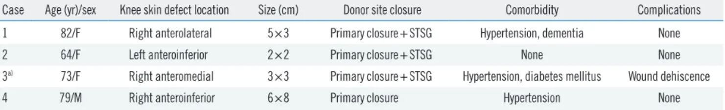
Table 1. Patient characteristics
Case Age (yr)/sex Knee skin defect location Size (cm) Donor site closure Comorbidity Complications
1 82/F Right anterolateral 5×3 Primary closure+STSG Hypertension, dementia None
2 64/F Left anteroinferior 2×2 Primary closure+STSG None None
3
a)73/F Right anteromedial 3×3 Primary closure+STSG Hypertension, diabetes mellitus Wound dehiscence
4 79/M Right anteroinferior 6×8 Primary closure Hypertension None
F, female; M, male; STSG, split-thickness skin graft.
a)