pISSN 1738-3544 eISSN 2288-1662
Rapid Detection of Mycobacterium tuberculosis Complex in Tissues by Using the Nested PCR
Jung-Yeon Park and Byoung-Seon Yang
Department of Medical Laboratory Science, Jinju Health College, Jinju 52655, Korea
Nested PCR을 이용하여 조직으로부터 Mycobacterium tuberculosis Complex 신속검출
박정연, 양병선
진주보건대학교 임상병리과
Due to the increase in incidence of infection of Mycobacterium tuberculosis complex (MTC), it is imperative that a rapid diagnosis accompanies the handling of MTC. This is due to the three to eight weeks it takes to culture Mycobacteria, and the lack of sensitivity of microscopic examination of AFB. Recently, nested PCR has been used to detect and diagnose mycobacteria. It is especially useful in complementing diagnosis by histological extra pulmonary. After culturing all the specimens and practicing the nested PCR, we did comparison analysis between nested PCR and culture. There were 76 specimens, 31 of which were positive. Of the 31 positive specimens in culturing, only 22 were positive in nested PCR. Of the 45 negative specimens, 36 were negative in nested PCR. As a result, Sensitivity was 71% and specificity was 80%. Furthermore, the positive predictive value was 71% and negative predictive value was 80%. These results indicate that nested PCR based techniques are sensitive, specific, and rapid methods for the detection of MTC.
Keywords: Mycobacterium tuberculosis , Positive predictive value, Nested PCR
Corresponding author: Jung-Yeon Park Department of Medical Laboratory Science, Jinju Health College, Jinju 52655, Korea Tel: 82-55-740-1851
E-mail: [email protected]
This is an Open Access article distributed under the terms of the Creative Commons Attribution Non-Commercial License (http://creativecommons.org/licenses/by-nc/4.0) which permits unrestricted non-commercial use, distribution, and reproduction in any medium, provided the original work is properly cited.
Copyright © 2015 The Korean Society for Clinical Laboratory Science. All rights reserved.
Received: October 6, 2015 Revised: October 29, 2015 Accepted: October 30, 2015
서 론
결핵은
Mycobacterium tuberculosis
,M.africanum
,M.bovis
,M.microti
,M.caprae
,M.mungi
,M.orygis
,M.pinnipedii
그리고M.cannetii
을 포함한M.tuberculosis
complex (MTC)에 의해 일 으키는 만성감염질환이다(Brosch 등, 2002; Cristina 등, 2014).과거 5년 동안, 개발도상국과 후진국에서 결핵균과 비결핵균의 감염이 증가되었다. 개발도상국가에서 결핵균 감염증의 주된 증가 원인은 Human immunodeficiency virus (HIV) 감염, 결핵균의 높은 유병률, 면역억제제 사용 그리고 다재내성 mycobacteria 출
현 때문이다. 그러므로 민감하며 신속하고 정확한 결핵균의 검출은 결핵의 진단과 치료에 필수적이다(WHO, 1995; WHO, 1996).
액체배지와 고체배지에 결핵균을 배양하는 방법이 표준화 방법 으로 사용되어 왔지만, 결핵균을 배양 하는 데는 3주에서 8주정도 의 오랜 배양기간이 걸린다. 그리고 항산성 염색에 의한 현미경적 진단 방법은 임상검체 1 mL당 10,000개의 세균을 필요로 한다 (Bannister 등, 1996). 특히 결핵이 의심되는 조직 검체의 경우 배 양하기가 어려워 조직의 형태학적 관찰이나 항산성 염색에 의한 현 미경적 방법으로 진단하기 때문에 mycobacteria의 검출을 위한 빠르고 정확한 방법이 필요하다(Eisenach 등, 1990; Osaki 등,
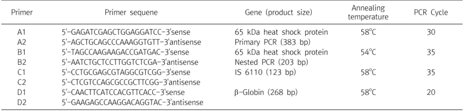
Table 1. Primer sequence for PCR
Primer Primer sequene Gene (product size) Annealing
temperature PCR Cycle
A1 5'-GAGATCGAGCTGGAGGATCC-3'sense 65 kDa heat shock protein 58
oC 30
A2 5'-AGCTGCAGCCCAAAGGTGTT-3'antisense Primary PCR (383 bp)
B1 5'-TAGCCAAGAAGACCGATGAC-3'sense 65 kDa heat shock protein 54
oC 35
B2 5'-AATCTGCTCCTTGGTCTCGA-3'antisense Nested PCR (203 bp)
C1 5'-CCTGCGAGCGTAGGCGTCGG-3'sense IS 6110 (123 bp) 58
oC 35
C2 5'-CTCGTCCAGCGCCGCTTCGG-3'antisense
D1 5'-CAACTTCATCCACGTTCACC-3'sense -Globin (268 bp) 58
oC 20
D2 5'-GAAGAGCCAAGGACAGGTAC-3'antisense
1997).
Hence 등(1995)은 65 kDa heat shock protein (HSP)의 383 bp mycobacterial DNA 유전자를 표적으로 한 polymerization chain reaction (PCR)방법을 사용하였다. 최근에는 DNA가 coding된 mycobacteria 항원, ribosomal RNA,
Mycobacterium tuberculosis
complex의 반복 염기서열을 대상으로 PCR을 실시 하였다(Boddinghaus 등, 1990; Jonas 등, 1993). 대부분의 병원에 서는 PCR방법을 호흡기 검체, 소변 등 배양 검체와 고정 되지 않은 조직검체에 사용하였다(Hermans 등, 1998; Thierry 등, 1990).Nested PCR방법은 두쌍의 primer을 사용하며 첫 번째 primer 로 유전자의 외측부위를 증폭시킨 후 두 번째 primer로 첫 번째 primer로 증폭시킨 유전자를 표적유전자로 사용하여 증폭시킨다.
Nested PCR의 장점은 특이유전자의 특이성 및 감수성을 증가시킨다.
본 연구에서는 76개의 조직검체를 nested PCR을 실시하였고 배양결과와 함께 비교 분석 하였다
재료 및 방법
1. 재료
2014년 3월부터 7월까지 부산소재의 B병원 으로부터 채취한 lung 37개, bronchus 2개, omentum 1개, lymph node 12개, colon 2개, soft tissue 7개, bone marrow 1개, liver 1개, breast 1개, skin 3개, jonint knee 2개, urinary blader 1개, esophagus 1 개, chest wall 1개, duodenum 1개, prostate 1개, pleura 1개, kidney 1개의 검체 총 76개의 조직 검체를 대상으로 배양과 nested PCR을 실시하였다.
2. 방법
1) 결핵균 도말 및 배양
조직검체를 직접 슬라이드에 도말하여 Ziel-Neelsen 염색을 실 시하였다. 배양용은 항산균 도말검사 후 나머지 검체를 3% Ogawa 배지에 접종하여 37
o
C에서 최대 8주간 관찰하였다.2) Nested PCR
포르말린에 고정된 파라핀 포매 조직을 5 m로 자른 후 슬라이 드 글라스에 부착시키고 자일렌을 이용해서 탈 파라핀 시켰다.
Alcohol (99-50%)을 이용해서 용매를 제거한 후 탈수 시켰다. 100
L 소화용액(200 g/mL proteinase K, 50 mM tris/HCl, pH 8.5, 1 mM ethylenediaminetetra acetic acid, 0.5% Tween 20)을 사 용하여 슬라이드 글라스에 있는 검체를 용해시킨 다음 이 혼합액을 56
o
C에서 24시간 방치 시켰다. 혼합액을 94o
C에서 10분 반응 시 켜 proteniase K에서 활성을 정지시켜 PCR반응 주형DNA로 사용 하였다. 1차 PCR master 혼합액(100 pmol A1 forward primer, A2 reverse primer, 100 ng DNA, 2 mM dNTP, 25 mM MgCl2
, 10X reaction buffer, 2.5 unitTaq
DNA polymerase) 최종부피 20 L가 되도록 하여 PCR (Perkin-Elmer Centus Corp., USA)을 실시하였고 2차 PCR master 혼합액(100 pmol B1 forward primer, B2 reverse primer, 100 ng DNA, 2 mM dNTP, 25 mM MgCl2
, 10X reaction buffer, 2.5 unitTaq
DNA polymerase) 최 종부피 20 L가 되도록 하여 1차 PCR 생성물을 1 L을 사용하여 표적으로 2차 PCR (Perkin-Elmer Centus Corp., USA)을 실시하 였다(Table 1).1차 PCR은 1시간 40분정도 소요되었고 94
o
C에서 15분간 1 cycle 실시, 94o
C에서 30초, 58o
C에서 30초, 72o
C에서 30초로 총 30 cycle 실시, 마지막으로 72o
C 60초 1 cycle 실시하였다. 2차 PCR은 94o
C에서 30초 , 54o
C에서 30초, 72o
C에서 30초에서 35 cycle 실시하였다. PCR 결과를 확인하기 위해서, PCR 산물은 1.5% agarose gel에서 40분간 전기영동 하였으며 전기영동 후 ethidium bromide (50 g/mL)에 10분간 염색하여 DW로 세정 후 BioDoc-14TM
Imaging system (UVP, UK)에 옮겨 PCR 증폭 산물 의 유무와 크기를 관찰하고 사진 촬영하였다.결 과
76개의 조직 검체를 사용하여 항산성염색과 배양 그리고
Table 3. Results of amplification of DNA fragment in mycobacteria of the tuberculosis complex using pulmonary
PCR Culture results
Total Positive Negative
Positive 11 5 16
Negative 5 16 21
Total 16 21 37
Sensitivity, 69% (11 of 16); specificity, 76% (16 of 21); positive predictive value, 69% (11 of 16); negative predictive value, 76%
(16 of 21).
Table 2. Results of amplification of DNA fragment in mycobacteria of the tuberculosis complex
PCR Culture results
Total Positive Negative
Positive 22 9 31
Negative 9 36 45
Total 31 45 76
Sensitivity, 71% (22 of 31); specificity, 80% (36 of 45); positive predictive value, 71% (22 of 31); negative predictive value, 80%
(36 of 45).
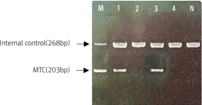
Internal control(268bp)
MTC(203bp)
Fig. 1. Electrophoresis of the amplified products of MTC by PCR in a 1.5% agarose gel. M, size marker; Lanes 1 and 3, positive;
lanes 2 and 4, negative; lane N, negative control. The amplified products and their sizes are indicated on the left.
Table 4. Results of amplification of DNA fragment in mycobacteria of the tuberculosis complex using nonpulmonary
PCR Culture results
total positive negative
positive 12 6 18
negative 3 18 21
total 15 24 39
Sensitivity, 80% (12 of 15); specificity, 75% (18 of 24); positive predictive value, 66% (12 of 18); negative predictive value, 85%
(18 of 21).
nested PCR을 실시하였다. 76개 검체 중 37개의 폐 검체(48.5%), 39개의 폐외 검체(51.5%)를 사용하여 검사를 실시하였다.
1. 배양과 nested PCR결과 비교 분석
76개의 검체 중 배양 결과 31개(41%)의 검체가 양성, 45개 (59%)의 검체가 음성으로 나타났다. 이중에 배양결과 양성인 31개 의 검체를 nested PCR을 실시한 결과 양성으로 나온 검체는 22개 (71%) 검체가 양성으로 나타났고, 배양 결과 음성인 45개의 검체 중 36개(80%) 검체가 음성으로 나타났다. 조직검체 MTC검출에 대 한 PCR민감도는 71%, 특이도는 80%로 나타났고, 양성 예측율은 배양 양성 31개 검체 중 PCR결과 양성으로 나타낸 22개의 검체로 71%로 나타났다. 음성예측률은 PCR결과 배양 음성 45개의 검체 중 PCR결과 음성으로 나타낸 32개의 검체로 80%로 나타났다(Fig.
1, Table 2).
2. 검체에 따른 배양과 Nested PCR결과 비교 분석
총 76개의 검체를 37개의 폐 검체와 39개 폐 외의 검체로 나누어 nested PCR과 비교 분석 하였다. 폐 검체의 경우 37개의 검체 중 배 양 결과 16개(43%)의 검체가 양성, 21개(57%)의 검체가 음성으로 나타났다. 이중에 배양결과 양성인 16개의 검체를 nested PCR을
실시한 결과 양성으로 나온 검체는 11개(69%) 검체가 양성으로 나 타났고, 배양 결과 음성인 21개의 검체 중 16개(76%) 검체가 음성 으로 나타났다. 폐검체 MTC검출에 대한 PCR민감도는 69%, 특이 도는 76%로 나타났고, 양성 예측율은 배양 양성 16개 검체 중 PCR 결과 양성으로 나타낸 11개의 검체로 69%로 나타났다. 음성예측 률은 PCR결과 배양 음성 21개의 검체 중 PCR결과 음성으로 나타 낸 16개의 검체로 76%로 나타났다(Table 3).
폐 외의 검체의 경우 39개의 검체 중 배양 결과 15개(38%)의 검 체가 양성, 24개(62%)의 검체가 음성으로 나타났다. 이중에 배양 결과 양성인 15개의 검체를 nested PCR을 실시한 결과 양성으로 나온 검체는 12개(80%) 검체가 양성으로 나타났고, 배양 결과 음성 인 24개의 검체 중 18개(75%) 검체가 음성으로 나타났다. 폐외 검 체 MTC검출에 대한 PCR민감도는 80%, 특이도는 75%로 나타났 고, 양성 예측율은 배양 양성 18개 검체 중 PCR결과 양성으로 나타 낸 12개의 검체로 66%로 나타났다. 음성예측률은 PCR결과 배양 음성 21개의 검체 중 PCR결과 음성으로 나타낸 18개의 검체로 85%로 나타났다(Table 4).
고 찰
결핵 진단 분야에 있어서 PCR방법을 이용한 분자 유전학적 연 구가 활발히 진행되어왔다. 특히 폐결핵 진단을 위한 객담과 기관 지 세척액 검체에서 PCR 방법을 실시하였다(Jang, 2015). 폐 이외
의 검체에서 결핵진단을 위해 흉수액이나 소변과 같은 체액검체, 조직검체, 세침흡입검체 등에서 PCR검사를 실시하였다(De 등, 1990). 폐 외의 검체에서 결핵의 진단은 주로 생검을 통한 조직괴사 를 동반한 만성육아종성 병변이 중요한 소견이다(Choi 등, 2006).
현재까지 국내에서는 건락성괴사를 동반한 만성육아종성 염증 동 반 시 결핵의 가능성이 가장 크다고 알려져 있지만, 만성육아종성 병변이 지속적인 항원자극에 대한 면역학적 반응이라 진균성 감염 이물질에 대한 반응 또는 매독등과 같은 여러 다른 요인들의 가능 성도 배제할 수 없다. 또한 현미경적 소견 상 육아종성 염증 소견과 균 자체의 확진을 위한 항산성균 도말 검사, 결핵균 배양 검사와의 상관관계는 그리 높지 않아 조직 검체의 결핵 진단을 보안해 줄 수 있는 또 다른 검사 방법으로 민감도가 높은 PCR의 방법에 대한 관 심이 증가되었다(Choi 등, 2006).
본 연구에서는 76개의 조직 검체를 이용해서 배양과 PCR을 실 시하였다. MTC 배양은 31개의 검체에서 양성을 보였지만 PCR방 법에선 9개의 검체에서 음성을 나타났다. Chan 등(1998)의 보고 에 의하면 formalin 고정액, 탈회 등이 DNA를 분해시켜 PCR반응 을 억제 한다고 하였다. 또한 뼈 같은 경우 DNA를 추출하기 힘든 검 체이기 때문에 배양에서는 양성으로 나타나지만 PCR방법에선 음 성을 나타낼 수 있다. 또 다른 예로 채취한 부위에 따라서 배양은 양 성이지만 PCR에서 음성을 보일 수 있다(Totsch 등, 1996). 반면에, 배양결과는 음성이지만 PCR방법에서 양성으로 나타나는 경우가 있다. 이 경우는 결핵약을 투여해서 이미 결핵균이 죽었지만 DNA 는 남아있어서 배양에서는 균이 자라지 않지만 PCR 방법 결과 양 성으로 나타날 수 있다(Brisson-Noel등, 1991; Savic 등, 1992).
또한 피부암과 같은 병을 앓고 있을 때 파괴된 조직으로부터 DNA가 방출될 수 있는데 이 경우에 PCR은 양성, 배양결과는 음성 으로 나타났다(Vago 등, 1998).
배양 결과 양성으로 나타난 검체 31개의 검체 중 PCR방법 양성 으로 나타난 검체는 22개로 PCR의 방법의 민감도가 71%, 그리고 배양 결과 음성으로 나타난 검체 45개 검체 중 PCR음성으로 나타 난 검체 36개 PCR의 방법의 특이도이 80%로 나타났다. 또한 양성 예측률과 음성예측률은 각각 71%, 80%를 보였다. Frevel 등 (1999)의 보고에 의하면 PCR방법의 의한 감수성이 90%, 특이성은 92%를 보여 본 연구 결과와 비교할 때 본 연구가 민감도와 특이도 가 낮았으나 특이도에서 높은 수치를 나타냈다. 앞으로 조직검체의 경우 한 부위 보다는 다양한 부위를 채취하여 DNA을 추출하고, 뼈 검체의 경우 많은 양의 검체를 사용하여 DNA을 추출하면 위 음성 문제를 해결할 수 있다고 생각 된다. Nested PCR방법은 조직검체 로부터 신속하고 정확하게 MTC을 검출 할 수 있고, 빠르게 결핵균 치료를 할 수 있는 정보를 제공할 수 있다.
요 약
결핵균의 감염이 증가함에 따라 결핵을 진단하고 치료하는 데 있어서 빠르고 민감한 진단방법이 필수적이다. Mycobacteria를 배양하는 것이 적어도 3주에서 8주정도 기간이 걸리고, 또한 AFB 의 현미경적 검경의 민감도가 낮다. 최근에는 PCR방법이 결핵균을 검출하고 진단하는데 사용되고 있다. 특히 병리조직학적 폐 외 감 염의 진단을 하기 위해 실시하고 있다.
병원에서 76개의 조직검체를 배양하고 nested PCR을 실시하여 배양결과와 비교 분석 하였다. 76개의 조직검체 중 배양 결과 양성 인 검체가 31개, 음성으로 나온 검체가 45개로 나타났다. 배양결과 양성인 31개 검체 중 nested PCR을 실시해서 양성으로 나온 검체 22개(71%)가 양성으로 나타났고, 배양결과 음성인 45개 검체 중 nested PCR을 실시해서 음성으로 나온 검체는 36개(80%)로 나타 났다. nested PCR의 민감도는 71%이고 특이도는 80%이다. 또한 양성 예측률은 71% 음성 예측률은 80%로 나타났다. Nested PCR 방법은 민감하고 신속하게 MTC을 검출 할 수 있다.
Acknowledgements: None Funding: None
Conflict of interest: None
References
1. Boddinghaus B, Rogall T, Flohr T,
et al. Detection and identi- fication of mycobacteria by amplification of rRNA.
J Clin Microbiol. 1990, 28:1751-1759.
2. Brisson-Noel A, Nguyen S, Bonete R,
et al. Diagnosis of tuber- culosis by DNA amplification in clinical practice evaluation.
Lancet