INTRODUCTION
Analysis of uncultured fetal cells by fluorescence in situ hybridization (FISH) has been used as a rapid alternative in screening for common chromosomal aneuploidies (1, 2).
Although FISH usually takes only 24 to 48 hr, it is expensive and intensively laborious. Recently, quantitative fluorescent PCR (QF-PCR) of single tandem repeats (STRs) has been developed for rapid prenatal diagnosis of aneuploidies (3, 4).
This approach has been used extensively by several groups on research basis (5-10), and the diagnosis of aneuploidies by QF-PCR of STRs has now been validated as a reliable method applicable in many laboratories.
In this study, we investigated the clinical feasibility of QF- PCR for rapid prenatal diagnosis of trisomy 21.
MATERIALS AND METHODS
Genomic DNA extraction was performed on the cell pel- let obtained from 1 to 2 mL of amniotic fluid from 200 nor- mal samples and 21 samples with trisomy 21. The karyotypes of all amniotic fluid samples were performed previously by conventional cytogenetic analysis. DNA was extracted by incubating cell pellets with InstaGene Matrix (Bio-Rad Labo- ratories, Hercules, CA, U.S.A.). A single tube multiplex PCR
was carried out by STR markers specific for chromosome 21.
D21S11, D21S1411, and D21S1270 (10) were used to test all samples for chromosome 21. D21S1412 (11) was added to test samples found to be homozygous for the first three STR (Table 1). The QF-PCR amplification of STR markers was performed in a total volume of 25 L containing 1.5 mM MgCl2, 200 m/L dNTP, 10-30 pmol of each primer, PCR buffer, 2U Taq polymerase, and 10 L genomic DNA. After the initial denaturation at 94℃for 5 min, hot start PCR was followed by 25 cycles of 94℃for 35 sec, 58℃for 35 sec, 72
℃for 40 sec and final extension was for 5 min at 72℃. For fragment analysis of PCR products, ABI 3100 and GeneScan 3.7 (Applied Biosystems, Foster City, CA, U.S.A.) were used.
The statistical analysis of the heterozygosity for each STR marker between the published population (10) and the Korean population were analyzed using the Z-test. A p-value >1.96 was taken as significant.
RESULTS
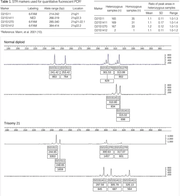
The majority of normal samples showed diallelic peaks with a ratio of 1:1 for each STR marker (Fig. 1). Among nor- mal samples, the ranges of diallelic peaks were 1.0-1.3 for D21S11, 1.0-1.4 for D21S1411 and 1.0-1.5 for D21S1270 (Table 2). Down syndrome samples that confirmed karyotypes
Moon-Hee Lee, Hyun-Mee Ryu*, Do-Jin Kim, Bom-Yi Lee, Eun-Hee Cho, Jae-Hyug Yang*, Moon-Young Kim*, Jung-Yeol Han*, So-Yeon Park
Laboratory of Medical Genetics, Department of Obstetrics and Gynecology*, Samsung Cheil Hospital
& Women’s Healthcare Center, Sungkyunkwan University, School of Medicine, Seoul, Korea
Address for correspondence So-Yeon Park, Ph.D.
Laboratory of Medical Genetics, Samsung Cheil Hospital & Women’s Healthcare Center, 1-19 Mookjung-dong, Chung-gu, Seoul 100-380, Korea Tel : +82.2-2278-4574, Fax : +82.2-2000-7679 E-mail : [email protected]
341 J Korean Med Sci 2004; 19: 341-4
ISSN 1011-8934
Copyright � The Korean Academy of Medical Sciences
Rapid Prenatal Diagnosis of Down Syndrome Using Quantitative Fluorescent PCR in Uncultured Amniocytes
Rapid prenatal diagnosis of common chromosome aneuploidies have been suc- cessful through quantitative fluoresent PCR (QF-PCR) assays and small tandem repeat (STR) markers. The purpose of our study was to investigate the clinical feasibility for rapid prenatal detection of Down syndrome using the quantitative flu- orescent PCR in uncultured amniocytes. DNA was extracted from uncultured amni- otic fluid of normal karyotype (n=200) and of Down syndrome (n=21). It was ampli- fied using QF-PCR with four STR markers located on chromosome 21. Among normal samples, the ranges of diallelic peaks for at least one STR marker were 1.0-1.3 for D21S11, 1.0-1.4 for D21S1411 and 1.0-1.5 for D21S1270. Down syn- drome samples showed trisomic triallelic patterns or trisomic diallelic patterns. The sensitivity, specificity, and efficiency of the assay for detecting Down syndrome were 95.4%, 100%, and 99.5%, respectively. Rapid prenatal diagnosis of Down syndrome using QF-PCR is a reliable technique that aids clinical management of pregnancy.
Key Words : Polymerase Chain Reaotion; Primed In Situ Labeling; Prenatal Diagnosis; Down Syndrome
Received : 12 December 2003 Accepted : 15 March 2004
342 M.-H. Lee, H.-M. Ryu, D.-J. Kim, et al.
with 47,+21 showed trisomic triallelic patterns with an allele peak ratio 1:1:1, or trisomic diallelic patterns with an allele peak ratio 2:1 (Fig. 1). However, one case that showed low- level mosaicism with a karyotype of 47,XX,+21[23]/46,XX [218] failed to be detected by QF-PCR. In complete trisom-
ic 21 samples, the ranges of diallelic peaks were 1.6-2.4 for D21S11, 2.2-2.7 for D21S1411, and 1.7-2.9 for D21S1270 (Table 3).
*Reference: Mann, et al. 2001 (10).
Marker Labeling Allele range (bp) Location
D21S11 6-FAM 214-242 21q21
D21S1411 NED 266-319 21q22.3
D21S1270 6-FAM 285-340 21q21-22.1
D21S1412 6-FAM 384-414 21q22.2
Table 1.STR markers used for quantitative fluorescent PCR*
Mean SD Range
Ratio of peak areas in heterozygous samples Homozygous
samples (n) Heterozygous
samples (n) Marker
D21S11 165 35 1.1 0.11 1.0-1.3
D21S1411 169 31 1.1 0.17 1.0-1.4
D21S1270 167 33 1.2 0.12 1.0-1.5
D21S1412 2 1 1.1 0.11 1.0-1.2
Table 2.Polymorphism and ratio of peak areas in 200 normal samples
Fig. 1.Electrophoregram of the QF-PCR products from normal and trisomy 21 samples. Fragment size in bp shown on horizontal axis, arbitrary fluorescence units shown on vertical axis. Markers labelled according to locus, size, and peak areas. Normal diploid sample: all markers are heterozygous and exhibit two peaks with a 1:1 ratio. Trisomy 21 sample: chromosome 21 markers exhibit three peaks with a 1:1:1 ratio or two peaks with a 2:1 ratio.
D21S11
Normal diploid
Trisomy 21
241.42 863
D21S11 244.65
3353
D21S1270 300.63
1457
D21S1411 297.59
845
D21S1411 305.79
687
D21S1411 326.13
564 D21S1270
317.07 601 D21S11
248.65 1659
D21S1270 301.53
829
D21S1270
800 600 400 200
800 600 400 200
800 600 400 200 3,000 2,000 1,000
190 200 210 220 230 240 250 260 270 280 290 300 310 320 330 340 350 360
190 200 210 220 230 240 250 260 270 280 290 300 310 320 330 340 350 360
313.99 802
D21S1411 310.80
834
D21S1411 315.02
698 D21S11
253.42 764
Rapid Prenatal Diagnosis 343
All but one normal samples were heterozygous and infor- mative for at least one STR marker (Table 4).
The sensitivity, specificity, and efficiency of QF-PCR for the detection of Down syndrome were 95.4%, 100%, and 99.5%, respectively. The false positive and the false nega- tive rates were 0% (0/200) and 4.7% (1/21), respectively.
All results were obtained within 24 to 48 hr.
DISCUSSION
QF-PCR for the detection of chromosome specific repeat- ed sequences has been improved to include several STR (3, 4). It is based on the incorporation of fluorochromes into the products of PCR amplification via oligonucleotide primers specific for each STR and on the assumption that, within the early exponential phase of PCR amplification, the amount of specific STR produced is proportional to the quantity of initial target sequence (12). In normal heterozygotes, the ratio of the fluorescent intensity of the two peaks should be close to 1:1. If the STR marker is highly polymorphic, few nor- mal subjects should be homozygotes and show one peak. In a trisomic patient, the three doses of an STR marker can be detected either as three peaks of fluorescent intensity with a ratio 1:1:1 (trisomic triallelic) or as a pattern of two peaks with a ratio 2:1 (trisomic diallelic). Using the high polymor- phism of the STR markers, very few trisomic patients should show a single peak of fluorescent intensity (4).
Our results present that the fluorescent intensity ratios showed a definite difference between normal disomic dial- lelic samples and diallelic trisomy 21 samples.
In the present study, we used the markers as published pre- viously. It was shown that heterozygosity of markers was low in our population compared to the published data (Table 5).
At the D21S11 and D21S1411 markers, a significant dif- ferences were found. We expected a difference of genetic diver- sity between an ethnic groups and it was supported by other previous reports (13-16).
Although 4 samples showed heterozygous for only one STR, even after using the extra marker, the use of combined STR markers could reduce the likelihood of homozygosity and consequently, the frequency of uninformative STR patterns.
Only one out of 220 samples was homozygous for all STR
tested (Table 4). As a results, we emphasize that the infor- mative rate of our experiment is prominent than other stud- ies so far (5, 8, 9, 16).
Yoon et al. reported three false positive in the multiplex QF-PCR using two of STR markers for Down syndrome (16).
As it is well known, the preferential amplification of multi- plex PCR is a potential problem that result in an incorrect genetic typing and is effected by low denaturation tempera- tures, low amount of DNA, and differential allelic priming.
In this study, there were no false positive results from mul- tiplex QF-PCR using three of STR markers. It is reported that a maximum primer set used in the multiplex QF-PCR is twelve up to now (10).
One case of low level mosaicism for trisomy 21 with a karyo- type of 47,XX,+21[23]/46,XX[218] resulted in a false nega- tive result. Several studies reported that mosaicism for trisomy was able to be detected by QF-PCR (10, 11), but based on our own and other published experiences, mosaicism, mater- nal contamination, structural abnormality, deletion and dupli- cation syndrome are not likely to be detected by the QF-PCR technique (8, 9). However, it is possible that future develop- ment, such as the use of extensive panel of markers to cover other key chromosomes, will result in significant improve- ments in the detection rate.
An advantage of using QF-PCR technique is that it is less time consuming and less laborious and this assay is useful on small volumes of amniotic fluid (1-2 mL). In this study, some results were obtained within 8 hr.
Our experiment demonstrates that QF-PCR can provide a rapid and accurate clinical method for prenatal identifica- tion of Down syndrome and also serve as an adjunctive test to help cytogenetics to reduce significant amounts of emo- tional stress experienced by patients and physicians. In fur-
Mean SD Range Ratio of peak areas in diallelic samples Diallelic
samples (n)
Monoallelic samples (n) Triallelic
samples (n) Marker
D21S11 9 9 2 2.0 0.34 1.6-2.4
D21S1411 5 9 6 2.3 0.14 2.2-2.7
D21S1270 5 15 0 2.2 0.55 1.7-2.9
*One case with mosaicism was excluded.
Table 3.Polymorphism and ratio of peak areas in trisomy 21 samples*
STR marker Homozygous % (n) Heterozygous % (n)
D21S11 16.8 (37/220) 83.2 (183/220)
D21S1411 16.8 (37/220) 83.2 (183/220)
D21S1270 15.0 (33/220) 85.0 (187/220)
D21S11+D21S1411+D21S1270 1.4 (3/220) D21S11+D21S1411+D21S1270
+D21S1412 0.45 (1/220)
*One of false negative was excluded.
Table 4.Evaluation for each STR marker and combined STR markers*
STR marker
Heterozygous frequency Reference
(Mann et al., 2001)
Reference (Mann et al.,
2001) Present study
(Korean group)
Present study (Korean
group) Allele range (bp)
D21S11 0.9 0.83 214-242 239-268
D21S1411 0.933 0.83 266-319 294-338
D21S1270 0.86 0.85 285-340 292-329
Table 5.Evaluation of markers in the Korean population
344 M.-H. Lee, H.-M. Ryu, D.-J. Kim, et al.
ther studies, we will test a larger scale population with expand- ed panel of markers for other numerical aberrations, such as trisomy 13 or 18 and sex chromosome aneuploidies.
ACKNOWLEDGEMENT
This study was supported by a grant of the Cheil Medical Encouragement Foundation.
REFERENCES
1. Klinger K, Landes G, Shook D, Harvey R, Lopez L, Locke P, Lern- er T, Osathanondh R, Leverone B, Houseal T. Rapid detection of chromosome aneuploidies in uncultured amniocytes by using fluores- cence in situ hybridization (FISH). Am J Hum Genet 1992; 51: 55-65.
2. Thilaganathan B, Sairam S, Ballard T, Peterson C, Meredith R. Effec- tiveness of prenatal chromosomal analysis using multicolor fluores- cent in situ hybridization. Bjog 2000; 107: 262-6.
3. Mansfield ES. Diagnosis of Down syndrome and other aneuploidies using quantitative polymerase chain reaction and small tandem repeat polymorphisms. Hum Mol Genet 1993; 2: 43-50.
4. Pertl B, Yau SC, Sherlock J, Davies AF, Mathew CG, Adinolfi M.
Rapid molecular method for prenatal detection of Down’s syndrome.
Lancet 1994; 343: 1197-8.
5. Pertl B, Weitgasser U, Kopp S, Kroisel PM, Sherlock J, Adinolfi M.
Rapid detection of trisomies 21 and 18 and sexing by quantitative fluorescent multiplex PCR. Hum Genet 1996; 98: 55-9.
6. Toth T, Findlay I, Papp C, Toth-Pal E, Marton T, Nagy B, Quirke P, Papp Z. Prenatal detection of trisomy 21 and 18 from amniotic fluid by quantitative fluorescent polymerase chain reaction. J Med Genet 1998; 35: 126-9.
7. Verma L, Macdonald F, Leedham P, McConachie M, Dhanjal S,
Hulten M. Rapid and simple prenatal DNA diagnosis of Down’s syndrome. Lancet 1998; 352: 9-12.
8. Schmidt W, Jenderny J, Hecher K, Hackeloer BJ, Kerber S, Kochhan L, Held KR. Detection of aneuploidy in chromosomes X, Y, 13, 18 and 21 by QF-PCR in 662 selected pregnancies at risk. Mol Hum Reprod 2000; 6: 855-60.
9. Levett LJ, Liddle S, Meredith R. A large-scale evaluation of amnio- PCR for the rapid prenatal diagnosis of fetal trisomy. Ultrasound Obstet Gynecol 2001; 17: 115-8.
10. Mann K, Fox SP, Abbs SJ, Yau SC, Scriven PN, Docherty Z, Ogilvie CM. Development and implementation of a new rapid aneuploidy diagnostic service within the UK National Health Service and impli- cations for the future of prenatal diagnosis. Lancet 2001; 358: 1057- 61.
11. Pertl B, Kopp S, Kroisel PM, Hausler M, Sherlock J, Winter R, Adi- nolfi M. Quantitative fluorescence polymerase chain reaction for the rapid prenatal detection of common aneuploidies and fetal sex. Am J Obstet Gynecol 1997; 177: 899-906.
12. Ferre F. Quantitative or semi-quantitative PCR: reality versus myth.
PCR Methods Appl 1992; 2: 1-9.
13. Han GR, Lee YW, Lee HL, Kim SM, Ku TW, Kang IH, Lee HS, Hwang JJ. A Korean population study of the nine STR loci FGA, VWA, D3S1358, D18S51, D21S11, D8S1179, D7S820, D13S317 and D5S818. Int J Legal Med 2000; 114: 41-4.
14. Lee JW, Lee HS, Hwang JJ. Statistical analysis for estimating het- erogeneity of the Korean population in DNA typing using STR loci.
Int J Legal Med 2002; 116: 153-60.
15. Kim YL, Hwang JY, Kim YJ, Lee S, Chung NG, Goh HG, Kim CC, Kim DW. Allele frequencies of 15 STR loci using AmpF/STR Iden- tifiler kit in a Korean population. Forensic Sci Int 2003; 136: 92-5.
16. Yoon HR, Park YS, Kim YK. Rapid prenatal detection of Down and Edwards syndromes by fluorescent polymerase chain reaction with short tandem repeat markers. Yonsei Med J 2002: 43: 557-66.