† These authors contributed equally to this work.
* For correspondence. E-mail: [email protected]; Tel: +82-2- 450-4121; Fax: +82-2-450-3037
Copyright ⓒ 2011, The Microbiological Society of Korea
Filtration과 Integrated Cell Culture/Real-Time Reverse Transcription PCR 기법을 이용한 채소류에서
Human Rotavirus 신속 검출
현지연1†·천정환1†·송광영1·황인균2·곽효선2·이정수2·김무상3·이중복4·서건호1*
1건국대학교 수의과대학 공중보건학, 2식품의약품안전청 식품의약품안전평가원 미생물과,
3서울시 보건환경연구원, 4건국대학교 수의과대학 전염병학
Rapid Detection Method for Human Rotavirus from Vegetables by a Combination of Filtration and Integrated Cell
Culture/Real-Time Reverse Transcription PCR
Ji-Yeon Hyeon1†, Jung-Whan Chon1†, Kwang-Young Song1, In-Gyun Hwang2, Hyo-Sun Kwak2, Jung-Soo Lee2, Moo-Sang Kim3, Jung-Bok Lee4, and Kun-Ho Seo1*
1Department of Public Health, 4Department of Infectious Disease, College of Veterinary Medicine, Konkuk University, Seoul 143-701, Republic of Korea
2Korea Food and Drug Administration, Osong Health Technology Administration Complex, Osong 363-951, Republic of Korea
3Seoul Metropolitan Government Research Institute of Public Health and Environment, Gwacheon 427-070, Republic of Korea
(Received March 14, 2011 / Accepted June 15, 2011)
The purpose of this study was to evaluate and compare different elution and concentration methods for optimization of human rotavirus (HRV) detection method using real-time RT-PCR and cell culture techniques. The leafy vegetable samples (lettuce, Chinese cabbage) were artificially inoculated with HRV. Viruses were extracted from the vegetables by two different elution buffers, buffer A (100 mM Tris-HCl, 50 mM glycine, 3% beef extract, pH 9.5) and buffer B (250 mM Threonine, 300 mM NaCl, pH 9.5), and the extracted viruses were concentrated by filtration and PEG precipitation sequentially. To determine infectivity of the viruses, the viruses recovered from the samples were infected to the MA-104 cells, and integrated cell culture real-time RT-PCR was performed at 1, 48, 72, 96, 120, 144, 168 h post-infection (p.i.). The elution buffer A was more efficient in extracting the virus from the produce samples tested than the buffer B, 29.54% and 18.32% of recoveries, respectively. The sensitivity of real-time RT-PCR method was markedly improved when the virus was concentrated by the filtration method. When the viruses were eluted and concentrated by buffer A and filtration, respectively, the average recovery rate was approximately 51.89%. When the viruses recovered from samples were infected to MA-104 cell, infectious HRV was detected within 48 h p.i. by ICC/real-time RT-PCR, whereas cytopathic effects were not observed until 72 h p.i. The optimized detection method evaluated in this study could be useful for rapid and reliable detection of HRV in fresh produce products and applied for detection of other food-borne viruses.
Keywords: cell culture, filtration, integrated cell culture/real-time reverse transcription PCR, rotavirus, vegetables
Human rotavirus (HRV)는 개발도상국뿐 아니라 선진국에 서도 영유아 급성 설사증의 가장 중요한 원인체로 인식되는 바이러스로서 전 세계적으로 영유아 급성 장관염의 30-50%
가량이 HRV에 의한 것이라고 보고되고 있다(21). 잠복기는 1-4일이며 주요 증상은 설사, 구토, 고열을 나타내고 특히 구 토의 경우는 평균 2.6일간 지속되어 다른 원인체에 비해 장시 간 유지된다(13). 미국에서는 HRV로 인해 연간 7만명까지 입 원하고 있으며 해마다 전세계적으로 5세 미만의 아이 중 약 44만명의 아이가 사망한다고 알려져 있다(16). 바이러스의 전 파경로로는 주로 식품을 통한 전파이고 그 외 수인성 또는 사 람간의 직접적인 접촉을 통해 전파된다(9). 최근 과일 및 신선 야채류 섭취와 관련된 집단 바이러스성 식중독 사고가 미국의 경우 급증하는 추세인데 사람 및 동물의 분변에 오염된 지하 수와 농업용수를 사용할 경우 신선야채류와 과일 등에 병원성 세균 및 바이러스 오염 가능성이 있다고 알려져 있다(6, 7, 14, 20).
바이러스성 식중독 사고를 예방하기 위해서는 식품에서의 바이러스 검출 및 모니터링이 중요하지만 다음과 같은 이유로 검출에 어려움을 겪고 있다. 첫 번째로 환자의 분변과는 달리 식품 내 존재하는 바이러스의 수가 매우 적어서 많은 양의 음 식을 샘플로 채취해야 한다(3). 두 번째로 식품의 다양성과 복 잡성 때문에 직접 검출 및 동정하기 어려우며 식품 내에 바이 러스가 균질하지 않게 분포되어 있다. 세 번째로 식품의 구성 성분이 바이러스 검출법을 방해하는 물질로 작용 가능하다.
이러한 문제점 외에도 바이러스는 저농도로도 질병을 유발 할 수 있기 때문에 민감하고 정확한 신속 검출법 개발이 필요한 상황이다(13).
식품에서 바이러스를 검출하기 위해서는 식품에서 바이러 스를 탈리하는 과정, 탈리액에서 바이러스를 농축하는 과정, 바이러스를 검출하는 과정을 거치게 된다. 탈리하는 과정에 사용되는 탈리액은 연구팀에 따라 매우 다양하게 사용되고 있 으며 농축 방법은 immunomagnetic capture, organic floccula- tion, polyethylene glycol (PEG) 침전법이 사용되었으나 이러 한 방법들은 시간이 오래 걸리고, 과정이 복잡하여 바이러스 손실이 많아 민감도가 떨어진다는 단점이 있다(2, 3, 12, 14, 15). 최근에는 전하를 띄는 필터를 이용하는 농축 방법인 filtration이 많은 용량의 물에서 바이러스를 농축하는데 사용 되고 있으며 이 방법을 이용하여 바이러스의 검출을 저해하는 물질도 제거 가능하다는 연구가 보고 되고 있다(3, 18). 하지 만 대부분의 연구결과가 물에서 바이러스를 검출하는데 초점 이 맞춰 있으며 식품에 적용한 연구나 filtration을 PEG 침전법 과 비교한 연구도 부족한 실정이다(2, 4).
바이러스를 검출하는 방법으로는 현재 reverse transcription- polymerase chain reaction (RT-PCR)이 가장 많이 사용되고 있으며 이 방법은 빠르고 민감한 검출법으로 알려져 있다(2, 7, 15, 21). 최근에는 real-time RT-PCR이 정성뿐 아니라 정 량까지 가능하기 때문에 HRV 외에 다른 바이러스의 검출법 으로 널리 사용되고 있다(5, 8, 10, 22). 하지만 이러한 분자생 물학적 방법은 감염성이 없는 바이러스까지 검출 가능하다는
단점이 있어 이를 극복하기 위해 세포배양을 이용한 감염성 평가도 필요하다(1, 2, 19). 세포배양은 주로 plaque, cytopa- thicity assays를 통하여 감염성을 평가하는데 이 방법은 시간 이 오래 걸리고 세포병변이 나타나지 않는 바이러스에서는 사 용이 불가능하다는 단점이 있다(11).
따라서 본 연구의 목적은 채소류에서 HRV를 검출하기 위 해 두 가지 탈리액을 선정하여 회수 능력을 비교하였고, 농축 방법으로 양전하를 띄는 필터(Nanoceram® matrix)를 이용한 filtration을 사용하여 농축 능력을 PEG 침전법과 비교하였다.
또한 세포배양을 통해 filtration으로 농축된 바이러스의 감염 력을 평가하였는데 이때 신속한 감염성 평가를 위하여 integrated cell culture/real-time RT-PCR (ICC/real-time RT-PCR)를 통 해서 시간대 별로 세포 내의 바이러스 증가를 정량하였다.
재료 및 방법
바이러스와 세포
Human rotavirus Wa (TC adapted) strain을 American Type Culture Collection (ATCC)에서 구입하였으며 건국대학 교 수의과대학 전염병학 실험실에서 분양 받은 MA-104 cell 를 이용하여 실험을 수행 하였다. MA-104 cell은 바이러스 감 염성 평가를 위해 배양하였으며 Dulbecco’s Modified Eagle Medium (DMEM, Gibco, USA)에 10% fetal bovine serum, 0.06% L-glutamine, 0.1% NaHCO3를 첨가하여 사용하였으며 유지배지로는 5% fetal bovine serum, 0.06% L-glutamine, and 0.25% NaHCO3를 첨가한 DMEM를 사용하였다. 세포는 37°C의 5% CO2 환경에서 배양되었다.
채소 샘플에 HRV 접종
채소류 중 엽채류인 배추, 상추, 깻잎을 선정하여 25 g씩 지퍼백에 넣은 후 4 log RNA copies로 희석된 1 ml의 바이 러스액을 샘플의 10개의 spot에 골고루 접종하였다. 바이러스 를 접종하지 않은 샘플은 음성 대조군으로 사용하며, 야채를 넣지 않은 탈리용액에 바이러스만을 첨가하여 양성 대조군으 로 사용하였다. 양성 대조군에서 회수된 바이러스 농도는 각 방법의 회수율을 계산 하는데 사용되었다. 접종을 마친 샘플 은 후드에서 60분 동안 건조 후에 4°C에서 하루 동안 안정화 를 시켰다.
탈리액 비교
탈리액은 기존의 연구에서 탈리효과가 뛰어나다고 평가된 두 가지를 선정하여 비교하였다(7, 17). 100 mM Tris-HCl, 50 mM glycine, 3% beef extract (pH 9.5)와 250 mM Threonine-300 mM NaCl (pH 9.5)(Sigma-Aldrich, Korea)를 비교하였으며 농축방법은 PEG 침전법을 사용하였다. 바이러 스가 접종된 샘플과 음성 대조군이 들어있는 지퍼백에 각각의 탈리용액을 각 샘플에 100 ml을 넣고 균질화를 30초간 실시 한 후 실온에서 60분간 교반하였다. 교반을 마친 탈리용액은 아래에 기술된 바와 같이 PEG 침전법을 사용하여 농축되었으
며 농축된 바이러스는 real-time RT-PCR로 정량하고 양성 대 조군과 비교하여 회수율을 구하였다. 이 실험을 두 번 반복 실 험하였고 각 실험에서 샘플은 4개씩 시행하였다.
농축방법 비교
바이러스가 접종된 샘플과 음성 대조군이 들어있는 지퍼백 에 탈리용액 100 mM Tris-HCl, 50 mM glycine, 3% beef extract pH 9.5을 샘플 당 100 ml을 넣고 균질화를 30초간 실 시한 후 실온에서 60분간 교반하였다. 교반을 마친 탈리 용액 을 4°C에서 10,000×g 15분간 원심분리 한 후에 상층액을 수 거하였다. 그 후 PEG 침전법 또는 filtration을 이용하여 농축 된 바이러스를 real-time RT-PCR로 정량하고 회수율을 비교 하였다. 이 실험을 두 번 반복 실험하였고 각 실험에서 샘플은 4개씩 시행하였다.
PEG 침전법의 과정은 기존 발표된 연구를 참고하였다(7).
탈리 후 원심 분리된 상층액을 5 N HCl을 이용하여 pH 7.0±0.2로 적정한 후 10,000×g 15분간 원심 분리하였다. 원심 분리 후 상층액은 버리고 pellet을 50 ml의 30% PEG 6000와 10 ml의 300 mM NaCl에 풀고 2시간 동안 4°C에서 반응하 였다. 4°C에서 10,000×g 15분간 원심분리 한 후 상층액을 버 리고 5 ml phosphate buffered saline (pH 7.5)로 pellet을 풀 어주었다. 그 후 동량의 chloroform: buthanol (1:1)을 넣고 5 분 반응 후 4°C에서 10,000×g 15분간 원심분리하고 상층액만 을 -20°C에 보관하였다. 농축된 바이러스는 real-time RT-PCR 로 정량하고 양성 대조군과 비교하여 회수율을 구하였다.
Filtration은 Nanoceram filter® (Argonide corporation)의 양전하 성질을 이용하여 시행하였다. 필터를 장치에 장착한 후 pH 6 멸균 증류수로 통과시켰다. 그 후 샘플의 탈리 후 원 심 분리된 상층액을 2번 반복 통과시켰다. 그 후, 5 ml의 1%
beef extract (pH 9.5)로 필터를 5분간 반응 시킨 후 back-flush 하였다. 필터를 통과한 바이러스가 포함된 용액을 다시 한 번 필터에서 5분간 반응 시킨 후 통과시켰다. 이 과정을 한번 더 반복한 후 마지막으로 통과된 용액을 -20°C에 보관하였다. 농 축된 바이러스는 real-time RT-PCR로 정량하고 양성 대조군 과 비교하여 회수율을 구하였다.
Real-time RT-PCR
RNA를 추출하는 방법은 식품공전방법에 있는 노로 바이러 스 검출법을 참고로 하였다(http://www.kfda.go.kr/index.kfda?mid
=92&division=식품). Magnetic silica bead RNA Extractor를 이용하여 MagExtractor Viral RNA kit (TOYOBO, Japan)나, GM Autoprep kit (Green Mate, Korea) 등을 사용하거나 이 와 동등 이상의 결과(재현성)를 얻을 수 있는 상업적 kit를 선 택하여 사용할 수 있는데 이에 본 연구에서는 식품공전법을 참조하여 GM Autoprep kit를 사용하여 RNA를 추출하였다.
세포에 감염된 HRV의 RNA를 추출하는 방법은 Viral Gene- spin™ Viral DNA/RNA Extraction kit (iNtRON Biotechnology, Korea)을 사용하였다. 바이러스의 정량은 HRV의 RNA를 10 배 연속 희석하여 real-time RT-PCR을 시행하여 standard
curve를 완성한 후 마지막으로 검출된 희석농도를 1 RT-PCR unit (PCRU)으로 결정하고 그 후 본래의 농도를 계산했다. 그 리고 GeneAmp® RNA PCR Control kit (Applied Bio- systems, USA)를 이용하였는데 이 시약에는 일정한 양의 internal RNA copies (6 log RNA copies/ml)가 들어있어 HRV RNA 의 비교 정량이 가능하였다.
Real-time RT-PCR은 QuantiTect SYBR Green Onestep RT-PCR kit (QIAGEN, USA)를 이용하였으며 0.5 μl의 Quanti- Tect RT mix, 25 μl의 QuantiTect SYBR Green RT-PCR Master mix, 0.5 μM의 forward와 reverse primer, 5 μl의HRV RNA, RNase free water 를 섞어 총 50 μl으로 반응을 진행하 였다. Primer는 HRV의 VP6 gene (NCBI Accession no.
X94617)을 증폭하는 것으로 서열은 다음과 같다. Forward primer (5′-ggakgtyctgtactcmttgtcaaaaa-3′), reverse primer (5′- tccagtttgaaastcatttccatt-3′)(16). Real-time RT-PCR은 ABI 7500® real-time PCR System (Applied Biosystems)을 사용하 였으며 반응 조건은 50°C에서 30분, 95°C에서 10분간 반응시 킨 후 94°C에서 15초, 50°C에서 30초, 72°C에서 1분을 1회 로 하여 40 cycle을 반응시켰다. Ct value (threshold cycle)는 형광커브와 역치선이 만나는 cycle 값으로 ABI 7500® software (Applied Biosystems)에서 분석되었다.
Integrated real-time RT-PCR을 이용한 감염성 평가 MA-104 cell을 6 well에 70% 정도 monolayer 되도록 배 양 한다. 샘플에서 회수된 HRV는 세포의 오염을 예방하기 위 하여 0.22 μm인 cellulose-ester membrane (Millex-GS, Milli- pore)에 필터하고 trypsin 10 μg/ml과 37°C에서 30분 반응 시 킨 후 세포에 접종했다. 접종 후 한 시간 동안 20분마다 흔들 어 주어 바이러스가 세포에 골고루 감염될 수 있게 하고 유지 배지를 넣고 배양했다. 접종 후 1, 48, 72, 96, 120, 144, 168 시간 후에 cell scraper (Gibco)를 이용하여 세포를 수거한 후 RNA를 추출하여 real-time RT-PCR을 시행하였다. 그리고 7 일간 현미경으로 세포병변을 관찰 하였고 세포병변을 나타낸 세포는 감염을 확인하기 위해 real-time RT-PCR을 시행하였다.
통계학적 분석
바이러스의 탈리 방법과 농축방법에 대한 회수율은 InStat Software (Version 3.05; GraphPad, USA)을 이용하여 분석되 었다. 각 trial별 회수율의 평균값을 구하여 두 가지 방법 간의 통계학적 유의차(p value)를 분석하였다. 이 때 p value가 0.05 이하일 때 통계학적 유의차가 있는 것으로 판단하였다. HRV 의 감염성 평가를 위하여 1시간 후의 Ct value와 각 시간별 Ct value의 통계학적 유의차를 비교하였다.
결과 및 고찰
본 실험에서는 탈리용액 100 mM Tris-HCl, 50 mM glycine, 3% beef extract pH 9.5과 250 mM Threonine, 300 mM NaCl pH 9.5의 바이러스의 탈리효과를 비교하였고 alumina
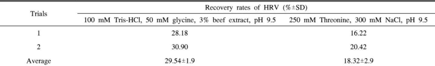
Table 1. Comparison of different types of buffers for elution of human rotavirus (HRV) from vegetables quantifying by real-time RT-PCR. Vegetables were inoculated with HRV (4 log RNA copies) and recovered by PEG precipitation (n=4).
Trials Recovery rates of HRV (%±SD)
100 mM Tris-HCl, 50 mM glycine, 3% beef extract, pH 9.5 250 mM Threonine, 300 mM NaCl, pH 9.5
1 28.18 16.22
2 30.90 20.42
Average 29.54±1.9 18.32±2.9
* P value is less than 0.0419; the difference in recovery rates between the two different types of buffers is considered statistically significant.
Table 2. Comparison of PEG precipitation and filtration for human rotavirus (HRV) concentration from vegetables quantifying by real‐time RT‐PCR. Vegetables were inoculated with HRV (4 log RNA copies) and recovered by PEG precipitation (n=4).
Trials Recovery rates of HRV (%±SD)
PEG precipitation Filtration
1 18.2 51.3
2 19.9 52.5
Average 19.05±1.2 51.89±0.8
* P value is less than 0.0049; the difference in recovery rates between the two concentration methods is considered statistically significant.
strand로 이루어진 electropositive filter를 이용한 filtration과 PEG 침전법의 농축효과를 비교하였다. 100 mM Tris-HCl, 50 mM glycine, 3% beef extract, pH 9.5과 250 mM Threonine, 300 mM NaCl pH 9.5을 이용하여 탈리 하였을 때 각각의 회 수율은 29.54%와 18.32%로 100 mM Tris-HCl, 50 mM glycine, 3% beef extract, pH 9.5이 더 뛰어난 탈리효과를 보 였다(Table 1). 각 실험의 회수율을 통계학적으로 비교해본 결 과 p value는 0.0419으로 큰 유의차를 보였다. 100 mM Tris- HCl, 50 mM glycine, 3% beef extract pH 9.5는 Dubios 등 의 연구(7)에서 pH 완충능이 뛰어나고 탈리효과가 우수한 것 으로 보고된 용액으로서 채소류 외에 pH가 낮은 샘플인 과일 에도 적용 가능할 것으로 보인다. 또한 Butot 등의 연구(4)에 서 본 연구와 비슷한 연구 결과를 나타냈는데 라즈베리와 딸 기에서 glycine-tris buffer 1% beef extract와 tris buffer의 탈 리효과를 비교하였을 때 glycine-tris buffer 1% beef extract을 사용한 탈리 방법이 라즈베리에서 7.5%, 딸기에서 2.2%로 tris buffer 보다 우수했다.
농축방법을 비교했을 때 PEG 침전법과 filtration의 각각의 회수율은 양성 컨트롤과 비교했을 때 19.05%와 51.89%로 filtration이 농축에 더 효과적인 것으로 나타났다(Table 2). 통 계학적으로 비교해 본 결과 p value는 0.0049로 큰 유의차를 보였다. 또한 filtration 시행 시 필터를 1회 시행했을 때 보다 2회 시행했을 때 바이러스의 손실이 현저히 줄어들었으며(자 료 미제시) filtration이 기존의 PEG 침전법에 비해서 시간이 적게 소요되었고 절차가 간단하여 효율적이었다(4). 본 연구와 유사한 연구로서 딸기와 양배추에서 hepatitis A virus (HAV) 를 검출하기 위해 electropositive virosorb filters (Cuno Inc., USA)와 immunomagnetic beads로 농축한 연구에서는 62%의
filter-captured HAV 중 34.8%가 탈리된다는 결과가 나타났 다. 그리고 filer와 magnetic bead를 모두 사용했을 때에는 10 PFU 이하의 HAV가 검출되는 효과를 보였다(2). Butot 등의 연구(4)에서는 농축방법으로 ultrafiltration과 PEG 침전법을 비교하였는데 ultrafiltration은 라즈베리에서 3%, 딸기에서 15%의 회수율을 보이면서 PEG 침전법 보다 높은 회수율을 나타냈다. 이는 본 연구의 filtration의 회수율과 비교하였을 때 낮은 회수율이었지만 각 농축법에서 사용한 샘플, 탈리용액, 농축법의 final volume, RNA 추출방법 및 real-time PCR법이 다르기 때문에 추가적인 비교연구가 필요할 것으로 보인다.
하지만 농축에 필요한 시간을 비교하였을 경우, filtration은 탈 리 시간을 제외하고 한 샘플당 약 20분 정도 소요되었으나 ultrafiltration은 30-60분 정도 소요되어 시간적인 면으로 효율 적일 것으로 보이며 각 농축법에 필요한 기구의 경우 본 연구 에서 사용한 filter는 syringe filter 형식으로 비용이 저렴하여 ultrafiltration에 사용된 Centricon Plus-70 centrifugal filter device (100K NMWL; Millipore, France) 보다 경제적일 것 으로 여겨진다.
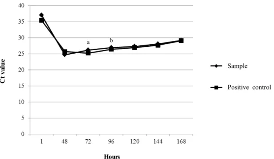
본 연구에서는 세포배양법과 real-time RT-PCR을 복합적 으로 사용하는 방법으로 ICC/real-time RT-PCR을 개발하여 바이러스의 감염성 평가에 사용하였는데, 이 방법은 세포 내 에서 증식하는 바이러스를 real-time RT-PCR로 정량함으로써 현미경으로 세포병변이 확인되기 이전에 바이러스의 감염성을 평가할 수 있는 방법이다. 채소에서 filtration을 이용하여 회수 한 HRV를 감염성 평가를 하기 위해 MA-104 cell에 접종하 고 접종한 직후 1시간째에 세포를 수거하여 real-time RT-PCR 을 시행하였다. 이 때의 Ct value는 37.14이었으며 48시간 후 에는 통계학적 유의차를 보이며 Ct value가 24.7로 감소하기
(A) (B)
(C)
Fig. 2. Microscopic view of MA-104 cells. (A) Monolayer of MA-104 cells before infection. (B) Cytopathic effects were observed in MA-104 cells infected with the HRV recovered from inoculated vegetables by filtration at day 3 post-infection.
The cytopathic effects consisted of cell rounding, shrinking, and detachment from the surface. (C) No cytopathic effects appeared on MA-104 cells inoculated with negative controls, solution recovered from uninoculated vegetables by filtration to 7 days post-infection.
Fig. 1. Detection of infectious human rotavirus (HRV) recovered from vegetables by ICC/real-time RT-PCR. Recovered HRV (3 log RNA copies) from vegetables by filtration and cultured HRV (3 log RNA copies) as positive control were used to infect MA-104 cells, and cells were collected after 1, 48, 72, 96, 120, 144, and 168 h post-infection (n=2).
a, Statistically significant reduction of Ct value was evident. b, Cytopathic effects began to appear.
a b
Hours
Ct value
Positive control Sample
시작하여 바이러스의 증가가 확인 가능하였다(Fig 1). 72시간 이후에는 세포병변이 나타나기 시작하면서 Ct value가 26.17 로 약간 증가하였다(Figs. 1 and 2). 양성 컨트롤로써 HRV를 동일한 농도로 세포에 접종하였을 때 샘플과 유사한 Ct value 를 보이며 바이러스의 증식이 확인되었다(Fig. 1). 음성컨트롤 샘플을 접종한 세포는 세포변성 효과 없이 7일간 유지되었다 (Fig. 2). 따라서 ICC/real-time RT-PCR을 이용하였을 경우 빠른 시간 내에 바이러스의 감염성을 평가 할 수 있을 것으로 보이며 이 방법은 세포병변이 잘 나타나지 않고 천천히 증식 하는 HRV외 여러 바이러스의 감염성 평가에도 응용 가능 할 것으로 보인다.
본 연구 결과 100 mM Tris-HCl, 50 mM glycine, 3% beef extract (pH 9.5)이 채소류에서 HRV를 탈리하는데 적합하였 으며 또한 filtration을 이용한 농축법이 기존의 PEG 침전법에 비해 검출 시간을 줄일 수 있고 효율 또한 우수한 방법인 것 을 확인하였다. 또한 ICC/real-time RT-PCR을 이용하여 세포 배양법 보다 빠른 시간 내에 HRV의 감염성 평가가 가능하였다.
적요
본 연구는 human rotavirus (HRV)의 검출법을 최적화하기 위해 real-time RT-PCR과 세포 배양법을 이용하여 여러 가지 탈리·농축법을 비교 및 평가하는 것을 목적으로 하였다. 채소 류 중 배추, 상추, 깻잎을 선정하여 바이러스 희석액을 접종하 고 탈리액 비교를 위하여 buffer A (100 mM Tris-HCl, 50 mM glycine, 3% beef extract, pH 9.5)와 buffer B (250 mM Threonine, 300 mM NaCl, pH 9.5)를 이용하여 탈리하였고, 농축방법을 비교하기 위하여 PEG (polyethylene glycol) 침전 법 또는 filtration [Nanoceram filter® (Argonide corporation)]
을 이용하여 농축하였다. 또한 바이러스의 감염성 평가를 위 하여 MA-104 cell을 배양하여 탈리, 농축 방법을 거쳐 회수된 HRV를 접종하고 1, 48, 72, 96, 120, 144, 168시간 후 세포 를 수거하여 real-time RT-PCR을 시행하고 세포병변을 관찰 하였다. 탈리 용액은 buffer A가 회수율 29.54%로 buffer B의 18.32%보다 더 뛰어난 탈리효과를 보였으며 농축방법을 비교 했을 때 filtration 방법이 회수율 51.89%를 나타내며 PEG 침 전법에 비해서 바이러스의 농축에 효과적이었으며 검출 소요 시간이나 간단한 과정 면에서 효율적이었다. ICC/real-time RT-PCR을 시행하였을 때 세포병변 72시간 후부터 나타나기 시작했지만 Ct value는 48시간부터 감소하기 시작하여 더 빠 른 시간 내에 감염성을 평가할 수 있었다. 따라서, filtration과 integrated/cell culture real-time RT-PCR을 이용하면 기존의 검출방법보다 빠른 시간 내에 바이러스 검출이 가능할 것으로 여겨진다.
감사의 말
본 논문은 식품의약품안전청 용역과제(08082식품안030)에 의해 이루어졌으며, 현지연과 천정환은 교육과학기술부 BK21
프로그램의 장학금을 수혜 받았습니다. 또한 실험에 도움을 주신 김윤경, 박준호, 김동현, 김홍석, 오아사, 임진혁에게 감 사를 표합니다.
참고문헌
1. Bhattacharya, S.S., M. Kulka, K.A. Lampel, T.A. Cebula, and B.B. Goswami. 2004. Use of reverse transcription and PCR to discriminate between infectious and non-infectious hepatitis A virus. J. Virol. Methods 116, 181-187.
2. Bidawid, S., J.M. Farber, and S.A. Sattar. 2000. Rapid concen- tration and detection of hepatitis A virus from lettuce and strawberries. J. Virol. Methods 88, 175-185.
3. Brassard, J., K. Seyer, A. Houde, C. Simard, and Y.L. Trottier.
2005. Concentration and detection of hepatitis A virus and rotavirus in spring water samples by reverse transcription-PCR.
J. Virol. Methods 123, 163-169.
4. Butot, S., T. Putallaz, and G. Sanchez. 2007. Procedure for rapid concentration and detection of enteric viruses from berries and vegetables. Appl. Environ. Microbiol. 73, 186-192.
5. Casas, N., F. Amarita, and I.M. de Maranon. 2007. Evaluation of an extracting method for the detection of Hepatitis A virus in shellfish by SYBR-Green real-time RT-PCR. Int. J. Food Microbiol. 120, 179-185.
6. Dentinger, C.M., W.A. Bower, O.V. Nainan, S.M. Cotter, G.
Myers, L.M. Dubusky, S. Fowler, E.D. Salehi, and B.P. Bell.
2001. An outbreak of hepatitis A associated with green onions.
J. Infect. Dis. 183, 1273-12176.
7. Dubois, E., C. Agier, O. Traore, C. Hennechart, G. Merle, C.
Cruciere, and H. Laveran. 2002. Modified concentration method for the detection of enteric viruses on fruits and vegetables by reverse transcriptase-polymerase chain reaction or cell culture. J.
Food Prot. 65, 1962-1969.
8. Dubois, E., C. Hennechart, G. Merle, C. Burger, N. Hmila, S.
Ruelle, S. Perelle, and V. Ferre. 2007. Detection and quantifi- cation by real-time RT-PCR of hepatitis A virus from inoculated tap waters, salad vegetables, and soft fruits: characterization of the method performances. Int. J. Food Microbiol. 117, 141-149.
9. Hernandez, F., R. Monge, C. Jimenez, and L. Taylor. 1997.
Rotavirus and hepatitis A virus in market lettuce (Latuca sativa) in Costa Rica. Int. J. Food Microbiol. 37, 221-223.
10. Houde, A., E. Guevremont, E. Poitras, D. Leblanc, P. Ward, C.
Simard, and Y.L. Trottier. 2007. Comparative evaluation of new TaqMan real-time assays for the detection of hepatitis A virus.
J. Virol. Methods 140, 80-89.
11. Jiang, Y.J., G.Y. Liao, W. Zhao, M.B. Sun, Y. Qian, C.X. Bian, and S.D. Jiang. 2004. Detection of infectious hepatitis A virus by integrated cell culture/strand-specific reverse transcriptase- polymerase chain reaction. J. Appl. Microbiol. 97, 1105-1112.
12. Jothikumar, N., D.O. Cliver, and T.W. Mariam. 1998. Immuno- magnetic capture PCR for rapid concentration and detection of hepatitis A virus from environmental samples. Appl. Environ.
Microbiol. 64, 504-508.
13. Koopmans, M. and E. Duizer. 2004. Foodborne viruses: an emerging problem. Int. J. Food Microbiol. 90, 23-41.
14. Le Guyader, F.S., A.C. Schultz, L. Haugarreau, L. Croci, L.
Maunula, E. Duizer, F. Lodder-Verschoor, C.H. von Bonsdorff, E. Suffredini, W.M. van der Poel, and et al. 2004. Round-robin comparison of methods for the detection of human enteric viruses in lettuce. J. Food Prot. 67, 2315-2319.
15. Leggitt, P.R. and L.A. Jaykus. 2000. Detection methods for
human enteric viruses in representative foods. J. Food Prot. 63, 1738-1744.
16. Logan, C., J.J. O'Leary, and N. O'Sullivan. 2006. Real-time reverse transcription-PCR for detection of rotavirus and adenovirus as causative agents of acute viral gastroenteritis in children. J. Clin. Microbiol. 44, 3189-3195.
17. Mullendore, J.L., M.D. Sobsey, and Y.C. Shieh. 2001. Improved method for the recovery of hepatitis A virus from oysters. J.
Virol. Methods 94, 25-35.
18. Queiroz, A.P., F.M. Santos, A. Sassaroli, C.M. Harsi, T.A.
Monezi, and D.U. Mehnert. 2001. Electropositive filter membrane as an alternative for the elimination of PCR inhibitors from sewage and water samples. Appl. Environ. Microbiol. 67, 4614- 4618.
19. Reynolds, K.A., C.P. Gerba, and I.L. Pepper. 1996. Detection of
infectious enteroviruses by an integrated cell culture-PCR procedure. Appl. Environ. Microbiol. 62, 1424-1427.
20. Rosenblum, L.S., I.R. Mirkin, D.T. Allen, S. Safford, and S.C.
Hadler. 1990. A multifocal outbreak of hepatitis A traced to commercially distributed lettuce. Am. J. Public Health 80, 1075-1079.
21. Schwarz, B.A., R. Bange, T.W. Vahlenkamp, R. Johne, and H.
Muller. 2002. Detection and quantitation of group A rotaviruses by competitive and real-time reverse transcription-polymerase chain reaction. J. Virol. Methods 105, 277-285.
22. Shan, X.C., P. Wolffs, and M.W. Griffiths. 2005. Rapid and quantitative detection of hepatitis A virus from green onion and strawberry rinses by use of real-time reverse transcription-PCR.
Appl. Environ. Microbiol. 71, 5624-5626.