Asia Pacific
allergy
pISSN 2233-8276 · eISSN 2233-8268
Educational & Teaching Material Case Report
http://dx.doi.org/10.5415/apallergy.2016.6.1.67 Asia Pac Allergy 2016;6:67-69
Childhood allergic bronchopulmonary aspergillosis presenting as a middle lobe syndrome
Ashok Shah*, Kamal Gera, and Chandramani Panjabi†
Department of Pulmonary Medicine, Vallabhbhai Patel Chest Institute, University of Delhi, Delhi 110 007, India
Allergic bronchopulmonary aspergillosis (ABPA) is infrequently documented in children with asthma. Although collapse is not uncommon, middle lobe syndrome (MLS) as a presentation of ABPA is rather a rarity. A 9-year-old female child with asthma presented with increase in intensity of symptoms along with a right midzone patchy consolidation on a chest radiograph. In addition, an ill-defined opacity abutting the right cardiac border with loss of cardiac silhouette was noted. A right lateral view confirmed a MLS, which was further corroborated by high resolution computed tomography. Central bronchiectasis was also observed, which prompted a work-up for ABPA. The child met 7/8 major diagnostic criteria for ABPA. She was then initiated on oral prednisolone that resulted in a marked clinical improvement within a fortnight. Radiological clearance occurred at 3 months with inflation of the middle lobe. ABPA presenting with MLS in a child is yet to be reported. A high index of suspicion is required to establish the diagnosis of ABPA in a child presenting with MLS. This would obviate the invasive investigations usually done to ascertain the cause of MLS.
Key words: Allergic Bronchopulmonary Aspergillosis; Asthma; Central Bronchiectasis; Middle Lobe Syndrome; Paediatrics
INTRODUCTION
Allergic bronchopulmonary aspergillosis (ABPA), a hypersensitivity respiratory disorder, is predominantly seen in asthmatics in the third and fourth decades of life. It is now well- recognised in India and is reported to occur in 7.6% of Indian adults with asthma [1]. Although ABPA is infrequently seen in children with asthma [2], it has been documented in infants as
young as 6 months [3]. However, in children with cystic fibrosis, this entity is not as uncommon as in children with asthma [2].
The term “middle lobe syndrome” (MLS) was coined by Graham et al. [4] in 1948 to describe a clinical entity characterised by chronic or recurrent collapse of the right middle lobe while describing 12 patients with middle lobe atelectasis due to enlarged lymph nodes of nontuberculous origin. Brock et al. [5]
originally described eight patients with recurrent atelectasis
Correspondence: Ashok Shah
Department of Pulmonary Medicine, Vallabhbhai Patel Chest Institute, University of Delhi, P.O. Box 2101, Delhi 110 007, India
Tel: +91-11-2543-3783 Fax: +91-11-2766-6549
E-mail: [email protected]
†Current affiliation: Department of Respiratory Medicine, Mata Chanan Devi Hospital, New Delhi 110058, India
Received: October 9, 2015 Accepted: January 20, 2016
This is an Open Access article distributed under the terms of the Creative Commons Attribution. Non-Commercial License (http://creativecommons.
org/licenses/by-nc/4.0/) which permits unrestricted non-commercial use, distribution, and reproduction in any medium, provided the original work is properly cited.
Shah A, et al.
Asia Pacific
allergy
68 http://dx.doi.org/10.5415/apallergy.2016.6.1.67 apallergy.org
of the right middle lobe due to extrinsic compression by enlarged tuberculous lymph nodes and MLS caused by enlarged tuberculous lymph nodes is frequently known as “Brock’s syndrome”.
In ABPA, collapse, both lobar and segmental due to mucoid impaction is not infrequent but a MLS due to this disease is rather rare [6-8]. To our knowledge, ABPA as a cause of MLS has been documented in 3 adult patients [6-8] but never in children. We describe a 9-year-old female child with ABPA who presented with a MLS.
CASE REPORT
A 9-year-old female child was referred to our Institute for evaluation of right midzone patchy consolidation. She had episodic wheezing dyspnoea and nonproductive cough for 4 years, which had increased over the last fortnight with no acute distress, pallor, clubbing or cyanosis. Bilateral expiratory rhonchi were audible in all areas of the chest. The total leucocyte count was 7,190 cells/cumm, with neutrophils 68%, lymphocytes 25%, monocytes 2.6%, and eosinophils 4.4%. The absolute eosinophil count was 350 cells/cumm. In addition to the midzone patchy consolidation, an ill-defined opacity abutting the right cardiac border with loss of cardiac silhouette was detected on the chest radiograph (Fig. 1A). A right lateral view showed a wedge shaped density extending from the hilum anteriorly and inferiorly along with loss of volume confirming a MLS (Fig. 1B). High resolution computed tomography (HRCT) of the thorax confirmed MLS
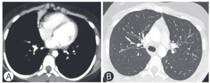
and also revealed central bronchiectasis (Fig. 2) which prompted investigations for ABPA. Skin prick test with antigens of Aspergillus fumigatus and Aspergillus flavus elicited a type I reaction. Strong bands of serum precipitins were detected against the same antigens. Specific IgE and IgG were positive for A. fumigatus and total serum IgE levels were elevated (1,488 IU/mL). Spirometry was suggestive of moderate obstruction with significant reversibility.
A diagnosis of ABPA presenting as MLS was made as she met 7/8 major diagnostic criteria (Table 1; Rosenberg-Patterson criteria [9, 10]) and was initiated on oral prednisolone in the dosage of 0.5 mg/kg daily, which was tapered to alternate day dosages after a fortnight and tapered off after 6 months as the patient improved. She also received inhaled steroids for asthma.
Marked symptomatic relief was noted within a fortnight with radiographic resolution of the MLS and reduction in total IgE levels to 903 IU/mL after 3 months. Informed written consent was obtained from mother.
DISCUSSION
MLS refers to chronic or recurrent collapse of the right middle lobe and has a distinct radiological presentation which continues to confound clinicians. Although, MLS is well documented in adults [11], this clinical entity is often overlooked in children with resultant diagnostic delay [12]. The occurrence of MLS in children with asthma is not infrequent and it is thought that this is caused by mucus hypersecretion which plug the bronchi [12].
It has been postulated that the propensity of the middle lobe to collapse may be due to the narrow diameter and long length of the middle lobe bronchus as well as the angular take-off of this bronchus. Furthermore, inadequate clearance of the impacted mucous along with the anatomically poor collateral ventilation
Fig. 1. (A) Chest radiograph posteroanterior view showing a right midzone patchy consolidation and an ill-defined opacity abutting the right cardiac border with loss of cardiac silhouette. (B) Chest radiograph right lateral view showing a wedge shaped density extending from the hilum anteriorly and inferiorly along with loss of volume confirming a middle lobe syndrome.
Fig. 2. (A) High resolution computed tomography (HRCT) (mediastinal window) of the thorax showing middle lobe syndrome. (B) HRCT (lung window) of the thorax showing central bronchiectasis.
A B
A B
Childhood ABPA presenting as MLS
apallergy.org http://dx.doi.org/10.5415/apallergy.2016.6.1.67 69 contributes to the susceptibility of the middle lobe to collapse in isolation [11, 12].
The initial diagnosis of MLS is based on imaging and is distinctly recognised on a lateral radiograph as a wedge-shaped density extending from the hilum, anteriorly and inferiorly. However, it must be suspected on a posteroanterior radiograph, where the right cardiac border is obscured (Silhouette sign). A lordotic view would show the MLS as a wedge shaped density in the basal central zone of the right lower lung field, due to parenchymal involvement of the middle lobe. This view is often of help when there is diagnostic confusion. However, the HRCT can confirm the diagnosis as a trapezoidal or broad triangular opacity is visible with its base towards the hilum and is contiguous with the right cardiac border. In addition, it can detect other abnormalities like central bronchiectasis, as was seen in our patient [11, 12].
Although, ABPA is not an uncommon cause of lobar or segmental collapse, MLS has rarely been associated with this clinical entity [1]. A search of the literature revealed only three adult patients with ABPA who presented with MLS [6-8] but MLS as a presentation of childhood ABPA is yet to be documented.
Our patient, a female child met 7/8 major diagnostic criteria for ABPA including central bronchiectasis, a pathognomic feature central to the diagnosis [1]. All 3 documented patients had
marked symptomatic improvement after therapy with oral corticosteroids [6-8]. In 2 patients [6, 7], there was radiological clearance but in the patient [8] documented by us in 2014, MLS persisted till the time of publication. However, a review at 9 months revealed an inflated middle lobe and radiological clearance. Our patient too had a marked symptomatic and radiological clearance after therapy with oral corticosteroids.
In an appropriate setting, all children with Aspergillus sensitive asthma should be evaluated for ABPA.
REFERENCES
1. Shah A, Panjabi C. Allergic aspergillosis of the respiratory tract. Eur Respir Rev 2014;23:8-29.
2. Shah A, Kala J, Sahay S. Allergic bronchopulmonary aspergillosis with hilar adenopathy in a 42-month-old boy. Pediatr Pulmonol 2007;42:747-8.
3. Imbeau SA, Cohen M, Reed CE. Allergic bronchopulmonary aspergillosis in infants. Am J Dis Child 1977;131:1127-30.
4. Graham EA, Burford TH, Mayer JH. Middle lobe syndrome. Postgrad Med 1948;4:29-34.
5. Brock RC, Cann RJ, Dickinson JR. Tuberculous mediastinal lymphadenitis in childhood: secondary effects on the lungs. Guy Hosp Rep 1937;87:295-317.
6. Eisenberg RS, Valdesuso C. Middle lobe syndrome secondary to allergic bronchopulmonary aspergillosis. Ann Allergy 1980;44:217-9.
7. Shah A, Bhagat R, Panchal N, Jaggi OP, Khan ZU. Allergic bronchopulmonary aspergillosis with middle lobe syndrome and allergic Aspergillus sinusitis. Eur Respir J 1993;6:917-8.
8. Shah A, Behera S, Panjabi C. Middle lobe syndrome: a rare presentation of allergic bronchopulmonary aspergillosis. Eur Ann Allergy Clin Immunol 2014;46:147-51.
9. Rosenberg M, Patterson R, Mintzer R, Cooper BJ, Roberts M, Harris KE. Clinical and immunologic criteria for the diagnosis of allergic bronchopulmonary aspergillosis. Ann Intern Med 1977;86:405-14.
10. Wang JL, Patterson R, Rosenberg M, Roberts M, Cooper BJ. Serum IgE and IgG antibody activity against Aspergillus fumigatus as a diagnostic aid in allergic bronchopulmonary aspergillosis. Am Rev Respir Dis 1978;117:917-27.
11. Gudbjartsson T, Gudmundsson G. Middle lobe syndrome: a review of clinicopathological features, diagnosis and treatment. Respiration 2012;84:80-6.
12. Romagnoli V, Priftis KN, de Benedictis FM. Middle lobe syndrome in children today. Paediatr Respir Rev 2014;15:188-93.
Table 1. Diagnostic criteria for allergic bronchopulmonary aspergillosis fulfilled by our patient (Rosenberg-Patterson criteria [9, 10])
Major criteria
1. Asthma +
2. Presence of transient pulmonary infiltrates (fleeting shadows)
+ 3. Immediate cutaneous reactivity to Aspergillus fumigatus,
Aspergillus flavus +
4. Elevated total serum IgE +
5. Precipitating antibodies against Aspergillus niger
and Aspergillus fumigatus +
6. Peripheral blood eosinophilia –
7. Elevated serum IgG to Aspergillus fumigatus + 8. Central bronchiectasis with normal tapering of distal
bronchi +
Minor criteria
1. Expectoration of golden brownish sputum plugs – 2. Positive sputum culture for Aspergillus niger – 3. Late (Arthus - type ) skin reactivity to Aspergillus
fumigatus –
![Table 1. Diagnostic criteria for allergic bronchopulmonary aspergillosis fulfilled by our patient (Rosenberg-Patterson criteria [9, 10])](https://thumb-ap.123doks.com/thumbv2/123dokinfo/5206757.119332/3.892.84.448.185.553/diagnostic-criteria-allergic-bronchopulmonary-aspergillosis-fulfilled-rosenberg-patterson.webp)
