ORIGINAL ARTICLE
급성 담석성 췌장염에서 조기 내시경 역행성 담췌관조영술 후 일시적 췌관삽입술의 유용성
이준규, 장동기, 강현우, 이상협1
동국대학교 일산병원 내과, 서울대학교병원 내과 및 간연구소1
Feasibility of Temporary Pancreatic Stenting after Early Endoscopic Retrograde Cholangiopancreatography in Patients with Acute Biliary Pancreatitis
Jun Kyu Lee, Dong Kee Jang, Hyun Woo Kang and Sang Hyub Lee1
Department of Internal Medicine, Dongguk University Ilsan Hospital, Goyang, Department of Internal Medicine and Liver Research Institute, Seoul National University Hospital1, Seoul, Korea
Background/Aims: To assess the safety and effectiveness of temporary pancreatic stenting after early endoscopic retrograde chol- angiopancreatography (ERCP) in patients with acute biliary pancreatitis regardless of the severity or concomitant cholangitis.
Methods: Temporary pancreatic stenting was performed in 79 patients with visualized pancreatic duct during ERCP. The outcomes of 64 patients with adequate pancreatic stenting (PS) and 15 patients with inadequate pancreatic stenting (no PS) were compared in this prospective, observational trial.
Results: The baseline characteristics were similar. Development of systemic inflammatory response syndrome (7.8% for PS vs. 13.3%
for no PS; p=0.50) and mortality (none for both groups; p=0.99) did not differ. However, fewer local complications occurred in PS than in no PS (4.7% for PS vs. 20.0% for no PS; p=0.04) and the difference was most outstanding in necrosis (1.6% for PS vs. 13.3% for no PS; p=0.03).
Conclusions: Temporary pancreatic stenting after early ERCP should be considered safe, as complications did not increase even in cases of inadequate stenting. However, if successful, there appears to be a reduction in local complications. (Korean J Gastroenterol 2017;70:247-252)
Key Words: Acute biliary pancreatitis; Endoscopic retrograde cholangiopancreatography; Pancreatic stenting
Received July 9, 2017. Revised August 11, 2017. Accepted August 31, 2017.
CC This is an open access article distributed under the terms of the Creative Commons Attribution Non-Commercial License (http://creativecommons.org/licenses/
by-nc/4.0) which permits unrestricted non-commercial use, distribution, and reproduction in any medium, provided the original work is properly cited.
Copyright © 2017. Korean Society of Gastroenterology.
교신저자: 이상협, 03080, 서울시 종로구 대학로 101, 서울대학교병원 내과 및 간연구소
Correspondence to: Sang Hyub Lee, Department of Internal Medicine and Liver Research Institute, Seoul National University Hospital, 101 Daehak-ro, Jongro-gu, Seoul 03080, Korea. Tel: +82-2-2072-4892, Fax: +82-2-2072-4892, E-mail: [email protected]
Financial support: None. Conflict of interest: None.
INTRODUCTION
Early endoscopic retrograde cholangiopancreatography (ERCP) has proven to be helpful in patients with acute biliary pancreatitis (ABP), especially when severe disease is pre- dicted or combined with cholangitis.1-7 However, early ERCP remains to be controversial for other patients since improve- ment in prognosis has not always been observed when com-
pared with conservative treatment. The rationale behind conservative treatment is that the stones that are small enough to obstruct the pancreatic duct tend to pass sponta- neously and manipulations of ERCP itself might aggravate the papillary edema and do more harm to the vulnerable pancreas. However, the development of complications from conservative treatment is not negligible. Neoptolemos et al.1 and Fan et al.2 reported that 12% and 7% of their patients, re-
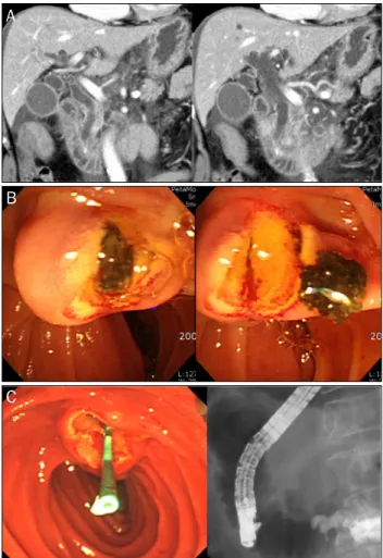
Fig. 1. Representative images. (A) A bile duct stone was impacted in the ampulla of vater and swollen of pancreas with multiple fluid collections was noticed on computed tomography. (B) The stone was removed by endoscopic retrograde cholangiopancreatography. (C) A 3 Fr stent was inserted into the pancreatic duct after successful removal of the stone.
spectively, experienced systemic and local complications, even when mild disease was predicted. Furthermore, be- cause all of the patients with ABP presents with upper ab- dominal pain, and leukocytosis and jaundice are seen in most of them regardless of severity, concomitant cholangitis, which is diagnosed on the basis of Charcot’s triad, cannot easily be excluded. In practice, rescue ERCP is frequently per- formed during conservative treatment for patients with pre- dicted mild disease. In addition, more complications develop if the duration of ampullary obstruction by a sustained stone exceeds 48 hours.4 Therefore, if feasible, it is crucial to per- form ERCP as early as possible, irrespective of severity, for pa- tients with ABP.
Meanwhile, 1% to 7% of patients who receive ERCP in the absence of pancreatitis experience post-ERCP pancreatitis (PEP),8-12 and the risk may exceed 25% amongst the high risk populations.13 There are several potential procedure-related risk factors; among them, pancreatic injection, which fre- quently occurs during biliary cannulation and removal of stones, is considered to be a definitive one.9,10,14-17 To over- come this risk factor, temporary pancreatic stenting after ERCP has been attempted, with success, to reduce the devel- opment of PEP; this method has been supported with con- crete evidence.18-24 Moreover, its use for patients who are at high risk is recommended by the guidelines put forth by the European Society of Gastrointestinal Endoscopy.25 However, to the best of our knowledge, there has only a limited number studies evaluating the safety of leaving a pancreatic stent temporarily after ERCP in a patient who was already affected by acute pancreatitis. Therefore, this current study was de- signed to explore this safety and to determine its influences on clinical outcomes.
SUBJECTS AND METHODS
1. Patients
Patients who underwent early ERCP under the diagnosis of ABP with cholangitis were prospectively enrolled. The diag- nosis of acute pancreatitis was made if there were two or more of the following features: (1) abdominal pain consistent with acute pancreatitis; (2) at least a 3-fold elevation of se- rum amylase lipase and/or amylase activity; and (3) charac- teristic imaging findings. Pancreatitis was causally attributed to the biliary origin if common bile duct (CBD) stones were dis-
covered on an ultrasonography or computed tomography in the absence of other causes of acute pancreatitis, such as ex- cessive alcohol intake or hypertriglycemia. Patients with the following conditions were excluded from the study: under the age of 18 years, pregnancy, hepatic cirrhosis, history of pan- creaticobiliary surgery other than cholecystectomy, and pre- vious ERCP. The study population was categorized into two groups in accordance with the success of insertion (the PS group and no PS group). The local institutional review boards approved this study (DUIHIRB 2013-105).
2. Procedures
Unified procedural strategy was maintained throughout the study period for all patients as follows. Once the diagnosis was made, ERCP was performed within 24 hours from hospi-
A
B
C
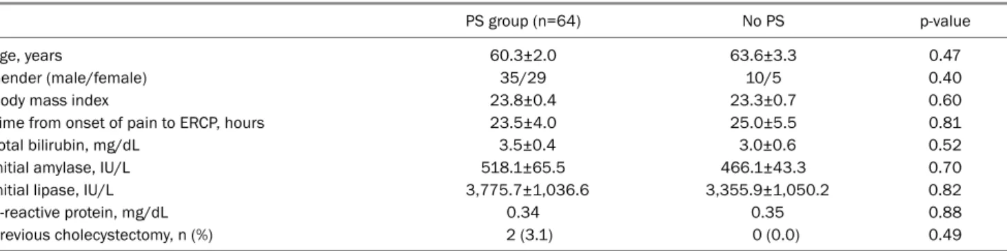
Table 1. Baseline Characteristics
PS group (n=64) No PS p-value
Age, years 60.3±2.0 63.6±3.3 0.47
Gender (male/female) 35/29 10/5 0.40
Body mass index 23.8±0.4 23.3±0.7 0.60
Time from onset of pain to ERCP, hours 23.5±4.0 25.0±5.5 0.81
Total bilirubin, mg/dL 3.5±0.4 3.0±0.6 0.52
Initial amylase, IU/L 518.1±65.5 466.1±43.3 0.70
Initial lipase, IU/L 3,775.7±1,036.6 3,355.9±1,050.2 0.82
C-reactive protein, mg/dL 0.34 0.35 0.88
Previous cholecystectomy, n (%) 2 (3.1) 0 (0.0) 0.49
Values were expressed as means±standard errors (SE).
PS, pancreatic stenting; ERCP, endoscopic retrograde cholangiopancreatography.
talization with a side-view of duodenoscope (JF-240, TJF-260, Olympus Corporation, Tokyo, Japan). Patients were sedated with midazolam (2.5-5.0 mg) and meperidine (25-50 mg), which were administered by registered nurses. Duodenal re- laxation was obtained with scopolamine butylbromide.
Continuous cardiopulmonary monitoring was used for all patients. The operator chose freely the device and technique for cannulation and removal of stones. When stones were visible after selective probing of CBD, endoscopic sphinctert- omy was commonly performed with a pull-type sphincter- otome, and stones were removed with baskets and/or re- trieval balloons. If the pancreatic duct was unexpectedly vi- sualized by a contrast injection during the procedure, a pan- creatic stent was inserted after removing the stones. To mini- mize possible insults, recannulation of the pancreatic duct by a guide-wire was limited to 5 attempts. If successful, a 3 cm-long, 3 Fr polyethylene stent with 2 flanges on both the pan- creatic ductal and duodenal sides was inserted (GPSO-3-3, Cook Endoscopy, Inc., Winston- Salem, NC, USA). Placement of the pancreatic stent was regarded adequate if the stent was adequately positioned in the pancreatic duct with its distal end in the duodenal lumen. Fig. 1 shows the typical computed to- mography image and describes the procedure. Spontaneous dislodgement of the pancreatic stent was assessed with plain abdominal radiographs taken on the following day, 7th day, and 14th day after ERCP. If the pancreatic stent was not passed out spontaneously by the 14 days, it was removed endoscopically.
3. Outcome measure
The primary outcomes of interests were mortality and over- all complications within 90 days after enrollment. Complications
of pancreatitis were classified, and organ failure was defined ac- cording to the revised Atlanta classification.26
The clinical courses were assessed as secondary out- comes; the mean changes in serum amylase and lipase lev- els between the initial and the next day of ERCP, time elapsed for patients to become eligible for cholecystectomy or discharge. Moreover, adverse events from the procedure, such as bowel-wall and sphinctertomy-related perforations or bleeding, were closely monitored. Significant bleeding was defined as the need for blood transfusion; a decrease in the hemoglobin level of greater than 2 g/dL; or hematochezia, me- lena or hematemesis within 24 hours after the procedure.27
4. Statistical analysis
The differences in categorical variables were analyzed us- ing chi-square test with Yates’ correction or Fisher’s exact test, as applicable. The mean values were expressed as the means±standard errors (SE) and compared using the Student’s t-test. All data were analyzed using SPSS version 12.0 for Windows (SPSS Inc., Chicago, IL, USA). Differences were consid- ered statistically significant when p-values were less than 0.05.
RESULTS
During the study period, ERCP was successfully performed in 587 patients with ABP and insertion of a pancreatic stent was attempted in 79 patients, in whom the pancreas was injected. Pancreatic stenting was adequate in 64 (81.0%) pa- tients (the PS group); they were compared with 15 patients with unsuccessful―or inadequate―stenting (the no PS group).
The two groups showed no meaningful differences in the baseline demographic and clinical characteristics (Table 1).
Table 2. Primary Outcome of Interest
PS group (n=64) No PS group (n=15) p-value
Overall complications 6 (9.4) 3 (20.0) 0.24
Local complications 3 (4.7) 3 (20.0) 0.04
Acute peripancreatic fluid collection or pseudocyst 2 (3.1) 2 (13.3) 0.10
Acute necrotic collection or walled-off necrosis 1 (1.6) 2 (13.3) 0.03
Infected necrosis 1 (1.6) 1 (6.7) 0.26
Presence of SIRSa 5 (7.8) 2 (13.3) 0.50
Persistent organ failure >48 hours 4 (6.7) 2 (13.3) 0.35
Mortality 0 (0.0) 0 (0.0) 0.99
Values are presented as n (%).
PS, pancreatic stenting; SIRS, systemic inflammatory response syndrome.
aSIRS was defined by presence of two or more criteria: heart rate >90 beats/min; core temperature <36℃ or >38℃; white blood count <4,000 or
>12,000/mm; respirations >20/min or PCO2 <32 mmHg.
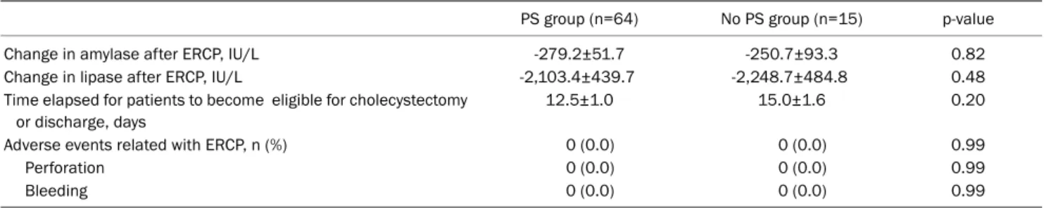
Table 3. Secondary Outcome of Interest
PS group (n=64) No PS group (n=15) p-value
Change in amylase after ERCP, IU/L -279.2±51.7 -250.7±93.3 0.82
Change in lipase after ERCP, IU/L -2,103.4±439.7 -2,248.7±484.8 0.48
Time elapsed for patients to become eligible for cholecystectomy or discharge, days
12.5±1.0 15.0±1.6 0.20
Adverse events related with ERCP, n (%) 0 (0.0) 0 (0.0) 0.99
Perforation 0 (0.0) 0 (0.0) 0.99
Bleeding 0 (0.0) 0 (0.0) 0.99
Values were expressed as means±standard errors (SE).
PS, pancreatic stenting; ERCP, endoscopic retrograde cholangiopancreatography.
1. Primary outcome measure
There were no significant differences in the overall compli- cations and systemic inflammatory response syndrome (SIRS) between the two groups (Table 2). However, fewer local complications occurred in the PS group than in the no PS group (4.7% for PS vs. 20.0% for no PS; p=0.04) and the dif- ference was the most remarkable in necrosis (1.6% for PS vs.
13.3% for no PS; p=0.03). There was no case of mortality in both groups (p=0.99).
2. Secondary outcome measure
Serum amylase and lipase levels measured on the day af- ter ERCP tended to decrease in both groups; no patient showed an increase in the levels after ERCP. There were no significant differences in time elapsed for patients to become eligible for cholecystectomy or discharge between the two groups. There was no case of perforation or significant bleed- ing (Table 3).
DISCUSSION
Although the benefit of early ERCP for patients with ABP other than predicted severe disease or concomitant chol- angitis is debatable, the development of complications is not infrequent. Moreover, concurrent acute cholangitis is not ex- cluded readily, and despite its absence at the time of diag- nosis, rescue ERCP during conservative treatment is fre- quently performed due to the emergence of cholangitis not only in daily practices, but also in clinical trials. Neoptolemos et al.1 and Fölsch et al.3 reported 23% and 20% of patients from the conservative group, respectively, received rescue ERCP within 4 weeks from randomization, although the num- ber of patients with predicted mild disease is not exactly specified. Remarkably, Fan et al. reported that 9 (15.6%) out of 58 patients with predicted mild disease from the con- servative group received emergency ERCP with a median of 48 hours after admission.2 Among the 40 patients with pre- dicted severe disease from the conservative group, 18 (45.0%) received emergency ERCP, with a median of 60 hours after admission. Because early ductal decompression
is a mandatory part of the treatment of acute cholangitis, it is critical to perform ERCP as early as possible, if it can be per- formed relatively safely. Procedure-related complications, such as bleeding or perforation, were not reported to be in- creased by almost all of the relevant studies.1-4,28-31 However, further injury by manipulations of ERCP itself is still a matter of concern. The exact mechanism by which ERCP insults the pancreas is not fully understood. However, a decrease in the flow of pancreatic juice due to papillary edema caused during manipulations is believed to be one of the most important causes.32 The papilla of a patient with ABP in whom ERCP is going to be performed is obstructed by a gallstone, in which point in time, the flow of pancreatic juice has already been disturbed. In such an unfavorable circumstance, even the very successful extraction of the stone with the finest manip- ulations always leaves a chance that might bring about fur- ther swelling of the papilla, which would surely impose an un- beneficial influence on ABP even after stone removal. We as- sumed that this can be overcome by temporary pancreatic stenting, which has been proven for preventing PEP.
There is still much to be elucidated about the use of a pan- creatic stent in patients with acute pancreatitis, unlike pa- tients with chronic pancreatitis in whom pancreatic stenting is most commonly used. Madácsy et al., in a case series, de- scribed the use of rescue pancreatic stenting to prevent the evolution of severe PEP.33 Fejes et al. reported that temporary pancreatic stenting can serve as a good bridging procedure in 27 patients with severe ABP in whom biliary EST was unsuccessful.34 However, the number patients in the study was small, and the lack of a control group limited the full un- derstanding of the consequences of failed stenting.
In our study, it is admitted that patients in the no PS group might have a smaller and more tortuous pancreatic duct, making the stent insertion more difficult. Moreover, we can- not deny the possibility that a worse outcome in the no PS group resulted due to the failed attempts, as Freeman et al.
suggested.35 To minimize possible insults, however, we lim- ited the number of attempts for recannulation of the pancre- atic duct to less than 5 times. More importantly, the develop- ment of overall complication of ‘unfavorable’ no PS group was not increased compared with the previous series. The complication rates of the early ERCP group of the prospective randomized trials were 10/59 (17%) by Neoptolemos et al.,1 17/97 (18%) by Fan et al.,2 and 58/126 (46%) by Fölsch et
al.3 This implies that our new strategy, in which early ERCP is performed for all patients with ABP having radiologically-proven CBD stones regardless of severity, can be helpful if pancre- atic stent can be inserted successfully after ERCP, as ex- pected for most of them, with minimal harm even in failed cases. This is supported by our data, as no patient experi- enced paradoxical elevation of serum amylase and lipase af- ter ERCP. By our strategy, unnecessary prolongation of hospi- tal stays and increased medical costs, which are unavoidable in ‘the wait-and-see principle’, can be evaded.
Ideally, a study in which patients were randomized into ei- ther the PS group or the no PS group would have been a better comparison. However, such an approach can be achieved on- ly after knowing the feasibility of pancreatic stenting in pa- tients with ABP. We firmly believe that our study is a good cor- nerstone for future studies.
In conclusion, temporary pancreatic stenting after early ERCP for patients with ABP appears to be safe since there was no increase in the occurrence of complications even in the failed cases. However, if successful, local complications are significantly reduced.
ACKNOWLEDGEMENTS
The authors thank Mr. Ho Jung Lee for providing English proofreading of the manuscript.
REFERENCES
1. Neoptolemos JP, Carr-Locke DL, London NJ, Bailey IA, James D, Fossard DP. Controlled trial of urgent endoscopic retrograde cholangiopancreatography and endoscopic sphincterotomy ver- sus conservative treatment for acute pancreatitis due to gallstones.
Lancet 1988;2:979-983.
2. Fan ST, Lai EC, Mok FP, Lo CM, Zheng SS, Wong J. Early treatment of acute biliary pancreatitis by endoscopic papillotomy. N Engl J Med 1993;328:228-232.
3. Fölsch UR, Nitsche R Lüdtke R, Hilgers RA, Creutzfeldt W. Early ERCP and papillotomy compared with conservative treatment for acute biliary pancreatitis. The German study group on acute bili- ary pancreatitis. N Engl J Med 1997;336:237-242.
4. Acosta JM, Katkhouda N, Debian KA, Groshen SG, Tsao-Wei DD, Berne TV. Early ductal decompression versus conservative man- agement for gallstone pancreatitis with ampullary obstruction:
a prospective randomized clinical trial. Ann Surg 2006;243:33-40.
5. Sharma VK, Howden CW. Metaanalysis of randomized controlled trials of endoscopic retrograde cholangiography and endoscopic sphincterotomy for the treatment of acute biliary pancreatitis.
Am J Gastroenterol 1999;94:3211-3214.
6. Heinrich S, Schäfer M, Rousson V, Clavien PA. Evidence-based treatment of acute pancreatitis: a look at established paradigms.
Ann Surg 2006;243:154-168.
7. Petrov MS, van Santvoort HC, Besselink MG, van der Heijden GJ, van Erpecum KJ, Gooszen HG. Early endoscopic retrograde chol- angiopancreatography versus conservative management in acute biliary pancreatitis without cholangitis: a meta-analysis of random- ized trials. Ann Surg 2008;247:250-257.
8. Mehta SN, Pavone E, Barkun JS, Bouchard S, Barkun AN.
Predictors of post-ERCP complications in patients with suspected choledocholithiasis. Endoscopy 1998;30:457-463.
9. Loperfido S, Angelini G, Benedetti G, et al. Major early complica- tions from diagnostic and therapeutic ERCP: a prospective multi- center study. Gastrointest Endosc 1998;48:1-10.
10. Masci E, Toti G, Mariani A, et al. Complications of diagnostic and ther- apeutic ERCP: a prospective multicenter study. Am J Gastroenterol 2001;96:417-423.
11. Vandervoort J, Soetikno RM, Tham TC, et al. Risk factors for com- plications after performance of ERCP. Gastrointest Endosc 2002;
56:652-656.
12. Cotton PB, Garrow DA, Gallagher J, Romagnuolo J. Risk factors for complications after ERCP: a multivariate analysis of 11,497 procedures over 12 years. Gastrointest Endosc 2009;70:80-88.
13. Fogel EL, Eversman D, Jamidar P, Sherman S, Lehman GA. Sphincter of oddi dysfunction: pancreaticobiliary sphincterotomy with pan- creatic stent placement has a lower rate of pancreatitis than bili- ary sphincterotomy alone. Endoscopy 2002;34:280-285.
14. Freeman ML, Nelson DB, Sherman S, et al. Complications of en- doscopic biliary sphincterotomy. N Engl J Med 1996;335:909-918.
15. Freeman ML, DiSario JA, Nelson DB, et al. Risk factors for post- ERCP pancreatitis: a prospective, multicenter study. Gastrointest Endosc 2001;54:425-434.
16. Williams EJ, Taylor S, Fairclough P, et al. Risk factors for complica- tion following ERCP; results of a large-scale, prospective multi- center study. Endoscopy 2007;39:793-801.
17. Masci E, Mariani A, Curioni S, Testoni PA. Risk factors for pan- creatitis following endoscopic retrograde cholangiopancreatog- raphy: a meta-analysis. Endoscopy 2003;35:830-834.
18. Tarnasky PR, Palesch YY, Cunningham JT, Mauldin PD, Cotton PB, Hawes RH. Pancreatic stenting prevents pancreatitis after biliary sphincterotomy in patients with sphincter of oddi dysfunction.
Gastroenterology 1998;115:1518-1524.
19. Fazel A, Quadri A, Catalano MF, Meyerson SM, Geenen JE. Does a pancreatic duct stent prevent post-ercp pancreatitis? A pro- spective randomized study. Gastrointest Endosc 2003;57:291-294.
20. Sofuni A, Maguchi H, Itoi T, et al. Prophylaxis of post-endoscopic retrograde cholangiopancreatography pancreatitis by an endo- scopic pancreatic spontaneous dislodgement stent. Clin Gas- troenterol Hepatol 2007;5:1339-1346.
21. Tsuchiya T, Itoi T, Sofuni A, et al. Temporary pancreatic stent to
prevent post endoscopic retrograde cholangiopancreatography pancreatitis: a preliminary, single-center, randomized controlled trial. J Hepatobiliary Pancreat Surg 2007;14:302-307.
22. Singh P, Das A, Isenberg G, et al. Does prophylactic pancreatic stent placement reduce the risk of post-ERCP acute pancreatitis?
A meta-analysis of controlled trials. Gastrointest Endosc 2004;
60:544-550.
23. Andriulli A, Forlano R, Napolitano G, et al. Pancreatic duct stents in the prophylaxis of pancreatic damage after endoscopic retro- grade cholangiopancreatography: a systematic analysis of bene- fits and associated risks. Digestion 2007;75:156-163.
24. Mazaki T, Masuda H, Takayama T. Prophylactic pancreatic stent placement and post-ERCP pancreatitis: a systematic review and meta-analysis. Endoscopy 2010;42:842-853.
25. Dumonceau JM, Andriulli A, Deviere J, et al. European Society of Gastrointestinal Endoscopy (ESGE) guideline: prophylaxis of post-ERCP pancreatitis. Endoscopy 2010;42:503-515.
26. Banks PA, Bollen TL, Dervenis C, et al. Classification of acute pan- creatitis--2012: revision of the Atlanta classification and defi- nitions by international consensus. Gut 2013;62:102-211.
27. Macintosh DG, Love J, Abraham NS. Endoscopic sphincterotomy by using pure-cut electrosurgical current and the risk of post- ERCP pancreatitis: a prospective randomized trial. Gastrointest Endosc 2004;60:551-556.
28. Oria A, Cimmino D, Ocampo C, et al. Early endoscopic inter- vention versus early conservative management in patients with acute gallstone pancreatitis and biliopancreatic obstruction: a randomized clinical trial. Ann Surg 2007;245:10-17.
29. Rosseland AR, Solhaug JH. Early or delayed endoscopic papil- lotomy (EPT) in gallstone pancreatitis. Ann Surg 1984;199:165-167.
30. Aiyer MK, Burdick JS, Sonnenberg A. Outcome of surgical and en- doscopic management of biliary pancreatitis. Dig Dis Sci 1999;
44:1684-1690.
31. Chen WX, Li YM, Gao DJ, et al. Application of endoscopic sphinc- terotomy in acute pancreatitis with fluid collection: a prospective study. World J Gastroenterol 2005;11:3636-3639.
32. Saari A, Kivisaari L, Standertskjöld-Nordenstam CG, Brackett K, Schröder T. Experimental pancreatography: a comparison of three contrast media. Scand J Gastroenterol 1988;23:53-58.
33. Madácsy L, Kurucsai G, Joó I, Gódi S, Fejes R, Székely A. Rescue ERCP and insertion of a small-caliber pancreatic stent to prevent the evolution of severe post-ERCP pancreatitis: a case-controlled series. Surg Endosc 2009;23:1887-1893.
34. Fejes R, Kurucsai G, Székely A, Székely I, Altorjay A, Madácsy L.
Feasibility and safety of emergency ERCP and small-caliber pan- creatic stenting as a bridging procedure in patients with acute biliary pancreatitis but difficult sphincterotomy. Surg Endosc 2010;24:1878-1885.
35. Freeman ML, Overby C, Qi D. Pancreatic stent insertion: con- sequences of failure and results of a modified technique to max- imize success. Gastrointest Endosc 2004;59:8-14.