491
서 론
췌관 내 유두상 점액성 종양은 1982년 일본의 Ohashi 등 이 처음으로 점액 생산 췌종양으로 보고한 이래 이 종양의 개념, 분류, 명칭, 조직발생, 진전 양식 등에 관해 많은 토의 가 이루어지다가 1996년 WHO가 정의와 분류를 한 종양이 다.(1) 특징으로는 다량의 점액 생산, 점액의 저류에 의한 주 췌관의 확장, 유두 개구부의 확장, 유두부로부터 점액의 배설 등 내시경 소견을 중심으로 보고되었다. 또한 이 종양 은 췌관 내에서 발육 진행하고 침윤 경향이 적어 절제율이 높고 예후가 좋은 췌종양으로 알려져 있다. 전체 췌암의 5%
이내를 점하며 60∼70세의 고령 남성에 많이 발생하고 췌 두부에 호발하는 것으로 알려져 있다. 과형성 이하의 병변 에서는 경과 관찰을 보고하는 경우도 있지만 수술 시에는 췌 절제가 원칙이다. 췌 두부에 병변이 있는 경우에는 췌십 이지장 절제술을 일반적으로 실시하게 되는데 이 종양은 악성도가 낮고 고령자가 많아서 침습이 적은 축소 수술 방 법을 주장하는 보고들도 있다. 이러한 다양성 이외에도 외 과의사로서는 수술 시 절제의 범위를 결정하는 데 다음과 같은 이유로 어려움이 있다. 췌관의 점액 때문에 췌관이 막 혀서 췌장염이 동반된 경우가 많고 췌관 내에 연속 진행되 는 특징과 남아 있는 췌관의 atypical hyperplasia가 발생모지 (母地)가 돼서 다중심성 발생, 이시성(異時性) 발생의 근거
췌관 내 유두상 점액성 종양의 외과적 치료
성균관대학교 의과대학 외과학교실, 삼성서울병원 외과 장원영․허진석․노재형․손태성․최성호․김용일
책임저자:김용일, 서울시 강남구 일원동 50번지 ꂕ 135-710, 삼성서울병원 외과 Tel: 02-3410-3479, Fax: 02-3410-0040 E-mail: [email protected]
접수일:2002년 4월 13일, 게재승인일:2002년 5월 29일
Surgical Treatment for Intraductal Papillary Mu- cinous Tumor of the Pancreas
Weon Young Chang, M.D., Jin Seok Heo, M.D., Jae Hyung Noh, M.D., Tae Sung Sohn, M.D., Seong Ho Choi, M.D. and Yong Il Kim, M.D.
Purpose: The surgical strategy for patients with a pancreatic intraductal papillary mucinous tumor (IPMT) is still contro- versial. In this study the clinicopathologic findings in a series of patients were used to rationalize surgical choice and reassess the need for a total pancreatectomy.
Methods: Between Oct. 1994 and Nov. 2001, 25 patients with IPMT underwent surgery. We retrospectively examined the clinicopathologic features and surgical treatment. The factors evaluated included: symptoms, tumor site, operation type, histological findings, resection margin, follow-up and survival.
Results: Pancreaticoduodenectomy was the most frequent surgical treatment (10 patients: 40%) followed by distal pan- createctomy (6), pylorus-preserving-pancreatico-duodenec- tomy (5) and total pancreatectomy (4). Histological assess- ment revealed the tumors to be an adenoma in 11 patients (44%), a borderline tumor in 8 patients (32%) and a car- cinoma in 6 patients (24%). There were no operative or hospital deaths. All of the cases with hyperplasia, adenoma and noninvasive carcinoma survived. Only two of the pa- tients with invasive carcinoma died. Mild to moderate dys- plasia was present at the resection margin in two patients (8%), and carcinoma in one. A total pancreatectomy was performed in four patients. Invasive carcinoma patient sur- vival was significantly associated with the presence of peri- pancreatic lymph node involvement.
Conclusion: Our study and review of the literature indicates that preoperative indicators of malignancy in IPMT are still lacking. These results suggest that resection should be the
treatment for IPMT. Sometimes IPMT is best treated by a total pancreatectomy, although lesser subtotal resections should definitely be considered. When selecting a surgical procedure for treating these tumors, it is useful to confirm the tumors’ extent by intra-operative imaging modalities. In the cases with invasion, a radical resection is required. (J Korean Surg Soc 2002;62:491-495)
Key Words: Pancreas, Intraductal papillary mucinous tumor (IPMT), Total pancreatectomy
중심 단어: 췌장, 췌관 내 유두상 점액성 종양, 췌 전 절제술
ꠏꠏꠏꠏꠏꠏꠏꠏꠏꠏꠏꠏꠏꠏꠏꠏꠏꠏꠏꠏꠏꠏꠏꠏꠏꠏꠏꠏꠏꠏꠏꠏꠏꠏꠏꠏꠏꠏꠏꠏꠏꠏꠏꠏꠏꠏꠏꠏꠏꠏ Department of Surgery, Sungkyunkwan University, School of Medicine, Samsung Medical Center, Seoul, Korea
가 되고있다.(2-4) 재발 양식에서도 간 전이, 림프절 전이 외 에 남아 있는 절단면 종양 양성 때문에 재발의 예가 비교적 많다.(5) 이러한 문제점 때문에 췌 전절제술이 필요한 경우 도 있다. 본 연구에서는 수술 예를 후향적으로 조사하여 적 절한 절제 범위를 결정하는 방법을 알아보고자 하였다.
방 법 1) 대상
1994년 10월부터 2001년 11월까지 삼성서울병원 외과에 서 영상의학적으로 IPMT 진단 후 개복술로써 확진된 25명 (남:여=20:5)의 환자를 대상으로 하였다.
2) 방법
대상 환자들에 대해 성별, 연령, 주소, 내원 시 동반증상, 동반된 전신질환, 전신 신체검사 소견 등의 임상소견과 종 양 표시자인 CA19-9의 측정치를 조사하였다. 주췌관의 직 경은 내시경적 역행성 췌관조영술 소견과 복부전산화 단층 촬영 소견에서 측정하였다. 대상 환자들에 대해 수술명을 포함한 수술 소견 및 병리학적 소견을 조사하였고, 병변의
위치는 내시경적 역행성 췌관 조영술, 복부전산화 단층촬 영, 복부초음파 등의 소견과 함께 최종적으로 수술 및 병리 학적 소견을 통해 확인하였다. 아형은 Kuroda의 분류에 따 라 주췌관형(main duct type), 분지췌관형(branch duct type) 및 혼합형(combined type)으로 분류하였다. 통계분석은 SPSS/
PC (10.0) program을 이용하여 각 군 간의 빈도 차이는 Chi- square 또는 Fisher's exact test로 검정하였고, P값이 0.05 미 만일 때 통계적으로 유의한 차이가 있다고 판단하였다.
결 과
대상 환자 25명의 평균연령은 60.7±10.8세이었으며, 성비 는 20:5로써 남자가 80%를 차지하였다. 수술을 통한 췌장 절제 후 병리조직검사에 따르면 종양의 위치는 두부와 구 상돌기에 위치하는 경우가 14예, 체부와 미부에 위치하는 경우가 8예, 전체 췌장에 분포하는 경우가 3예 있었다 (Table 1). 수술 전 종양의 악성여부 및 절제가능성 여부를 예측하기 위해서 이용되는 임상적 소견과 방사선학적 검사 의 결과 중 통계학적으로 의미를 갖는 지표는 Kuroda에 의 한 아형분류에서 주췌관형의 경우와(P=0.012) 췌장관의 확 장이 있는 경우에(P<0.00003) 양성의 병변보다는 경계성내 지는 악성의 병변으로 가정할 수 있는 P값을 보였다. 그러 나 이 수치는 경계성 병변을 악성 병변과 함께 묶었을 때
Table 2. Clinicopathologic features of IPMT of pancreas ꠚꠚꠚꠚꠚꠚꠚꠚꠚꠚꠚꠚꠚꠚꠚꠚꠚꠚꠚꠚꠚꠚꠚꠚꠚꠚꠚꠚꠚꠚꠚꠚꠚꠚꠚꠚꠚꠚꠚꠚꠚꠚꠚꠚꠚꠚꠚꠚꠚꠚꠚꠚꠚꠚꠚ
P- IPBB†/ IPMA*
value IPMC‡ ꠏꠏꠏꠏꠏꠏꠏꠏꠏꠏꠏꠏꠏꠏꠏꠏꠏꠏꠏꠏꠏꠏꠏꠏꠏꠏꠏꠏꠏꠏꠏꠏꠏꠏꠏꠏꠏꠏꠏꠏꠏꠏꠏꠏꠏꠏꠏꠏꠏꠏꠏꠏꠏꠏꠏ
Sex (M:F) 9:2 0.840 11:3
Age (years) 61.8 0.462 60.4
Symptom (presence:abscence) 6:5 0.056 13:1 Body weight change
(presence:absence) 1:10 0.341 4:10
DM (presence:absence) 1:10 0.180 5:9
CA19-9 (normal range:increase) 9:2 0.661 10:4 Type (main:branch:combined) 2:8:1 0.012 7:2:5 Duct dilatation (presence:absence) 2:9 0.00003 13:1 ꠏꠏꠏꠏꠏꠏꠏꠏꠏꠏꠏꠏꠏꠏꠏꠏꠏꠏꠏꠏꠏꠏꠏꠏꠏꠏꠏꠏꠏꠏꠏꠏꠏꠏꠏꠏꠏꠏꠏꠏꠏꠏꠏꠏꠏꠏꠏꠏꠏꠏꠏꠏꠏꠏꠏ
* = intraductal papillary mucinous adenoma; † = intraductal papil- lary mucinous borderline tumor; ‡ = intraductal papillary mucinous carcinoma.
Table 1. Age, sex location and classification of IPMT in pancreas ꠚꠚꠚꠚꠚꠚꠚꠚꠚꠚꠚꠚꠚꠚꠚꠚꠚꠚꠚꠚꠚꠚꠚꠚꠚꠚꠚꠚꠚꠚꠚꠚꠚꠚꠚꠚꠚꠚꠚꠚꠚꠚꠚꠚꠚꠚꠚꠚꠚꠚꠚꠚꠚꠚꠚ
Sex (M:F) 20 (80%):5 (20%)
Age range 38∼78 years
Mean age 60.7±10.8 years
Location of tumor
Head & uncinate process 14
Body & tail 8
Total pancreas 3
ꠏꠏꠏꠏꠏꠏꠏꠏꠏꠏꠏꠏꠏꠏꠏꠏꠏꠏꠏꠏꠏꠏꠏꠏꠏꠏꠏꠏꠏꠏꠏꠏꠏꠏꠏꠏꠏꠏꠏꠏꠏꠏꠏꠏꠏꠏꠏꠏꠏꠏꠏꠏꠏꠏꠏ
Table 4. Types of operation for IPMT of pancreas ꠚꠚꠚꠚꠚꠚꠚꠚꠚꠚꠚꠚꠚꠚꠚꠚꠚꠚꠚꠚꠚꠚꠚꠚꠚꠚꠚꠚꠚꠚꠚꠚꠚꠚꠚꠚꠚꠚꠚꠚꠚꠚꠚꠚꠚꠚꠚꠚꠚꠚꠚꠚꠚꠚꠚ
IPMC/IPMB IPMA
ꠏꠏꠏꠏꠏꠏꠏꠏꠏꠏꠏꠏꠏꠏꠏꠏꠏꠏꠏꠏꠏꠏꠏꠏꠏꠏꠏꠏꠏꠏꠏꠏꠏꠏꠏꠏꠏꠏꠏꠏꠏꠏꠏꠏꠏꠏꠏꠏꠏꠏꠏꠏꠏꠏꠏ Types of operation
Total pancreatectomy 4 0
Whipple's procedure 3 7
PPPD 3 2
Distal pancreatectomy 4 2
ꠏꠏꠏꠏꠏꠏꠏꠏꠏꠏꠏꠏꠏꠏꠏꠏꠏꠏꠏꠏꠏꠏꠏꠏꠏꠏꠏꠏꠏꠏꠏꠏꠏꠏꠏꠏꠏꠏꠏꠏꠏꠏꠏꠏꠏꠏꠏꠏꠏꠏꠏꠏꠏꠏꠏ Table 3. Histological finding of resected specimens of IPMT of
pancreas
ꠚꠚꠚꠚꠚꠚꠚꠚꠚꠚꠚꠚꠚꠚꠚꠚꠚꠚꠚꠚꠚꠚꠚꠚꠚꠚꠚꠚꠚꠚꠚꠚꠚꠚꠚꠚꠚꠚꠚꠚꠚꠚꠚꠚꠚꠚꠚꠚꠚꠚꠚꠚꠚꠚꠚ Main duct Branch Combined Total
type duct type duct type ꠏꠏꠏꠏꠏꠏꠏꠏꠏꠏꠏꠏꠏꠏꠏꠏꠏꠏꠏꠏꠏꠏꠏꠏꠏꠏꠏꠏꠏꠏꠏꠏꠏꠏꠏꠏꠏꠏꠏꠏꠏꠏꠏꠏꠏꠏꠏꠏꠏꠏꠏꠏꠏꠏꠏ
Benign 11 2 8 1
Borderline 8 3 1 4
Malignant 6 4 1 1
Invasion depth
In situ 2 1 1
Pancreas parenchyme 2 2
Peripancreatic tissue 2 1 1 Lymph node metastasis 2 1 1
ꠏꠏꠏꠏꠏꠏꠏꠏꠏꠏꠏꠏꠏꠏꠏꠏꠏꠏꠏꠏꠏꠏꠏꠏꠏꠏꠏꠏꠏꠏꠏꠏꠏꠏꠏꠏꠏꠏꠏꠏꠏꠏꠏꠏꠏꠏꠏꠏꠏꠏꠏꠏꠏꠏꠏ
얻을 수 있는 수치로써 8예의 경계성 병변을 별도로 놓았을 때는 아형분류와 주췌관 확장 모두 의미없는 P값을 나타냈 다. 이외에 성별, 나이, 주관적 증상 유무, 체중변화, 당뇨, CA19-9 수치의 상승 등은 통계학적인 의미가 없었다(Table 2). 절제 후 조직검사 결과를 토대로 한 분류에서는 악성종 양이 6예로써 24%였으며, 이 중 췌장의 관을 넘어서 침범한 4예 중 2예에서는 임파절 전이가 있었다(Table 3). 수술방법 은 악성과 경계성 병변의 4예에서 췌 전절제술을 시행하였 으며, Whipple씨 수술이 가장 많은 10예, 췌장 원위부 절제 술 6예, 유문보존 췌십이지장 절제술 5예를 시행하였다 (Table 4). 수술 후 병리검사에서 절제면에 종양세포가 남아 있는 경우는 총 3예로써 악성, 경계성, 양성종양 각각 1예이 며 수술방법은 췌장 원위부 절제술 2예, Whipple씨 수술 1 예이었으며 추적 관찰 당시에는 재발의 증거 없이 모두 생 존해 있었다(Table 5). 췌장 전절제술은 4예에서 시행하였으 며 수술 후 병리결과를 보면 1예는 주변조직 침윤이 있는 악성 병변이고 나머지 3예는 경계성 병변이었다. Kuroda 분 류로는 주췌관형이 2예, 복합형이 2예이었으며, 3예는 만성 췌장염이 동반된 소견이었으나, 1예는 췌장염이 없이 10×
2 cm의 종양이 있었다(Table 6). 췌장절제 후 추적 관찰 기 간이 짧아서 생존율을 통계화할 수는 없지만 악성화한 경 우만이 재발이 있고 특히 림프절 침범이 있는 경우에만 종 양 재발에 의한 사망이 발생하였다(Table 7).
고 찰
췌관 내 유두상 점액성 종양은 1982년 일본의 Ohhashi 등
(6)이 처음으로 점액생산 췌종양으로 보고한 이래 이 종양 의 개념, 명칭, 분류, 조직발생, 진전 양식 등에 대하여 많은 토의가 이루어지다가 1996년 WHO분류가 정해졌다. 이전 에는 여러 가지 다른 이름으로 불리었는데(mucinous ductal ectasia, intraductal papillary neoplasm, mucin-hypersecreting intraductal neoplasm, and intraductal papillary adenocarci- noma) 최근 병리학적 이해가 축적되면서 예전의 생각보다 발병률이 높은 것으로 보고되고 있다.(7) 본원에서는 1994 년 10월부터 2001년 11월까지 췌장 종양으로 췌장 절제술 을 시행한 305예 중에서 25예의 IPMT가 있었다. McDonald 등(8)은 한 해에 인구 281,000명당 1건씩 발생하며 췌장종양 으로 췌장절제술을 시행한 환자 중 약 19%가 IPMT로 보고 하고 있다. 유 등(9)은 IPMT의 치료방법에 대해서는 증상이 없으면서 방사선학적으로 분지췌관형이면서 주췌관의 직 경이 정상이거나 7 mm 이하의 주췌관 확장 소견을 보이는 예에서는 병리학적으로는 과형성 병변으로 예측할 수 있다 는 연구들에 근거해서 수술을 하지 않고 반복적으로 방사 선학적 검사를 시행하면서 경과를 관찰하자는 주장을 했으 며, Obara 등(10)은 9예의 IPMT 환자를 진단 후 ERP (endo- scopic retrograde pancreatography)를 반복적으로 시행하면서 수술을 하지 않고 6∼50개월(평균 30개월)을 추적 관찰 후 분지 췌관형 6예에서는 변화가 없었다고 보고를 하였다. 한 편 IPMT의 발생장소에 따른 악성도를 예측하려는 연구가 있었는데 Terris 등(11)은 분지형의 IPMT 경우 13예 중 단지 2예만 carcinoma in situ 상태이며 나머지는 양성 병변이었다 고 발표하였다. Kobori 등(12)도 분지형은 16예 중 1예만 췌 장실질 침범이 있고 4예는 악성병변이지만 췌장 실질침범 이 없는 상태이므로 분지형에서 축소 수술의 정당성에 대 해서 주장하였다. 그러나, 본원의 자료에서는 분지형 10예 Table 5. Resection margin positive patient
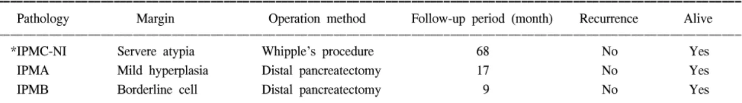
ꠚꠚꠚꠚꠚꠚꠚꠚꠚꠚꠚꠚꠚꠚꠚꠚꠚꠚꠚꠚꠚꠚꠚꠚꠚꠚꠚꠚꠚꠚꠚꠚꠚꠚꠚꠚꠚꠚꠚꠚꠚꠚꠚꠚꠚꠚꠚꠚꠚꠚꠚꠚꠚꠚꠚꠚꠚꠚꠚꠚꠚꠚꠚꠚꠚꠚꠚꠚꠚꠚꠚꠚꠚꠚꠚꠚꠚꠚꠚꠚꠚꠚꠚꠚꠚꠚꠚꠚꠚꠚꠚꠚꠚꠚꠚꠚꠚꠚꠚꠚꠚꠚꠚꠚꠚꠚꠚꠚꠚꠚꠚꠚꠚꠚꠚ
Pathology Margin Operation method Follow-up period (month) Recurrence Alive
ꠏꠏꠏꠏꠏꠏꠏꠏꠏꠏꠏꠏꠏꠏꠏꠏꠏꠏꠏꠏꠏꠏꠏꠏꠏꠏꠏꠏꠏꠏꠏꠏꠏꠏꠏꠏꠏꠏꠏꠏꠏꠏꠏꠏꠏꠏꠏꠏꠏꠏꠏꠏꠏꠏꠏꠏꠏꠏꠏꠏꠏꠏꠏꠏꠏꠏꠏꠏꠏꠏꠏꠏꠏꠏꠏꠏꠏꠏꠏꠏꠏꠏꠏꠏꠏꠏꠏꠏꠏꠏꠏꠏꠏꠏꠏꠏꠏꠏꠏꠏꠏꠏꠏꠏꠏꠏꠏꠏꠏꠏꠏꠏꠏꠏꠏ
*IPMC-NI Servere atypia Whipple's procedure 68 No Yes
IPMA Mild hyperplasia Distal pancreatectomy 17 No Yes
IPMB Borderline cell Distal pancreatectomy 9 No Yes
ꠏꠏꠏꠏꠏꠏꠏꠏꠏꠏꠏꠏꠏꠏꠏꠏꠏꠏꠏꠏꠏꠏꠏꠏꠏꠏꠏꠏꠏꠏꠏꠏꠏꠏꠏꠏꠏꠏꠏꠏꠏꠏꠏꠏꠏꠏꠏꠏꠏꠏꠏꠏꠏꠏꠏꠏꠏꠏꠏꠏꠏꠏꠏꠏꠏꠏꠏꠏꠏꠏꠏꠏꠏꠏꠏꠏꠏꠏꠏꠏꠏꠏꠏꠏꠏꠏꠏꠏꠏꠏꠏꠏꠏꠏꠏꠏꠏꠏꠏꠏꠏꠏꠏꠏꠏꠏꠏꠏꠏꠏꠏꠏꠏꠏꠏ
* = intraductal papillary mucinous carcinoma-non-invasive.
Table 6. Total pancreatectomy
ꠚꠚꠚꠚꠚꠚꠚꠚꠚꠚꠚꠚꠚꠚꠚꠚꠚꠚꠚꠚꠚꠚꠚꠚꠚꠚꠚꠚꠚꠚꠚꠚꠚꠚꠚꠚꠚꠚꠚꠚꠚꠚꠚꠚꠚꠚꠚꠚꠚꠚꠚꠚꠚꠚꠚ Age/Sex Pathology Pancreatitis Type Tumor
size (cm)
ꠏꠏꠏꠏꠏꠏꠏꠏꠏꠏꠏꠏꠏꠏꠏꠏꠏꠏꠏꠏꠏꠏꠏꠏꠏꠏꠏꠏꠏꠏꠏꠏꠏꠏꠏꠏꠏꠏꠏꠏꠏꠏꠏꠏꠏꠏꠏꠏꠏꠏꠏꠏꠏꠏꠏ
39/F *IPMC-CI Yes Main 8×1
64/F IPMB Yes Main 2×1.2
62/M IPMB Yes Combined 6×4.3
68/M IPMB No Combined 10×2
ꠏꠏꠏꠏꠏꠏꠏꠏꠏꠏꠏꠏꠏꠏꠏꠏꠏꠏꠏꠏꠏꠏꠏꠏꠏꠏꠏꠏꠏꠏꠏꠏꠏꠏꠏꠏꠏꠏꠏꠏꠏꠏꠏꠏꠏꠏꠏꠏꠏꠏꠏꠏꠏꠏꠏ
* = intraductal papillary mucinous carcinoma-carcinoma invasive.
Table 7. Outcome after surgical resection of IPMT ꠚꠚꠚꠚꠚꠚꠚꠚꠚꠚꠚꠚꠚꠚꠚꠚꠚꠚꠚꠚꠚꠚꠚꠚꠚꠚꠚꠚꠚꠚꠚꠚꠚꠚꠚꠚꠚꠚꠚꠚꠚꠚꠚꠚꠚꠚꠚꠚꠚꠚꠚꠚꠚꠚꠚ
IPMA IPMB/IPMC
(N=11) (N=14)
ꠏꠏꠏꠏꠏꠏꠏꠏꠏꠏꠏꠏꠏꠏꠏꠏꠏꠏꠏꠏꠏꠏꠏꠏꠏꠏꠏꠏꠏꠏꠏꠏꠏꠏꠏꠏꠏꠏꠏꠏꠏꠏꠏꠏꠏꠏꠏꠏꠏꠏꠏꠏꠏꠏꠏ Mean follow-up (month) 28.0 17.5
Recurrence (case number) 0 3
Death (caes number) 0 2
ꠏꠏꠏꠏꠏꠏꠏꠏꠏꠏꠏꠏꠏꠏꠏꠏꠏꠏꠏꠏꠏꠏꠏꠏꠏꠏꠏꠏꠏꠏꠏꠏꠏꠏꠏꠏꠏꠏꠏꠏꠏꠏꠏꠏꠏꠏꠏꠏꠏꠏꠏꠏꠏꠏꠏ
중 2예가 carcinoma 상태이며 1예는 주변조직 침윤이 있으 면서 주변림프절 전이가 있었다. Zamora 등(13)은 26예의 임상적 자료, 종양표지검사, 영상의학적 검사 어느 것도 수 술 전 악성종양이 있는 상태를 말 할 수 있는 지표는 없었 다고 발표했다. Yamaguchi 등(14)은 악성종양과 양성종양을 구별하기는 힘들지만 종양의 크기가 크고 췌장관이 심하게 늘어난 주췌관형이면 악성종양을 시사한다고 주장하였다.
본원의 결과도 주췌관형과 췌관확장은 악성을 예측할 수 있는 것으로 나왔으나 8예의 경계성 병변을 악성병변에 포 함시켰을 때의 결과이다. 종양의 크기는 주췌관형의 경우 10 mm 이상 분지췌관의 경우에는 50 mm 이상의 경우와, 췌관확장이 10 mm 이상인 경우, 주췌관과 분지췌관이 함께 확장된 경우, 황달이 동반된 경우는 강력하게 악성을 시사 하므로 악성종양에 준하는 수술방법을 고려하자는 주장도 있다. 그러나 지금까지의 어떤 검사도 80% 이상의 정확도 를 가지고 종양의 침습성 여부를 예측할 수 있는 방법은 없는 것으로 되어 있다.(15,16) 또 다른 문제점은 IPMT는 60∼70대 환자에서 췌장의 두부에 주로 발생하는데, 본 연 구에서도 평균 연령이 약 61세이며, 췌장 두부를 전체 25예 중 17예에서 침범하였다. IPMT의 이러한 특징은 수술적 치 료 시 Whipple씨 수술처럼 합병증 발생률이 높은 수술을 필 요로 하게 된다. 종합하면 IPMT는 양성 악성의 구분도 분 명하지 않고, 고령에서 그리고 축소 수술이 곤란한 부위에 발생하는 종양이다.
Kojima 등(2)은 IPMT가 다발성으로 발생한 경우를 보고 했으며, Obara 등(4)은 IPMT가 다발성으로 발생하면서 췌 장관의 diffuse atypical papillary hyperplasia에 대해서 보고를 했으며 따라서 절제면에 종양 세포를 남기지 않기 위해서 는 경우에 따라서 췌장 전절제술을 고려해야 하는 경우가 있다는 보고들이 있다.(17) 본 연구에서도 3예에서 절제면 에 종양세포가 잔존하였으며 3예 중 2예는 췌장 원위부 절 제술을 시행한 경우이었다. 반면 4예에서 췌장 전 절제술을 시행했으며 3예는 거의 전체 췌장에 걸쳐서 종양이 존재 했으나, 1예는 췌장의 체부에 국한된 2.0×1.5 cm의 종양이 었으나 췌장염이 동반되어 전체 췌장이 딱딱한 상태이었 다. 이처럼 췌장 원위부 절제술 시 절제면에 종양 세포가 남을 수 있는 가능성이 높다는 문제점과, 췌장염이 동반된 경우 종양의 위치와 범위를 정하기 힘들다는 문제를 수술 전, 수술 도중 어떻게 해결할 것인지가 IPMT 수술 시 힘든 점 이다.
IPMT의 수술 후 재발의 양상에 대해 Sho 등(5)은 8예의 재발 중 절제 후 남은 췌장에 발생한 예가 6예 이며, 나머지 2예는 간, 복강 등에 재발된 경우였다. Cuillerier 등(18)은 췌 장 부분절제술을 시행한 35명의 환자에서 비침습성 종양 20명 중 절제면에 종양이 남지 않은 경우는 재발이 없었으 나 절제면에 종양이 발견된 환자의 7명 중 2명은 재발이 있 었고, 침습성 종양 15예 중 임파절 전이가 있는 7예는 전
예에서 재발하였으나 임파절 전이가 없는 절제면에 종양이 남지 않은 경우 3예 중 2예에서, 절제면에 종양이 남아 있는 5예 중 2예가 재발을 하였다. 본 연구의 결과에서는 절제면 에는 종양세포가 남지 않았지만 임파절 전이가 있는 2예는 모두 재발하여 사망하였으나, 수술 후 병리 조직 검사 절제 면에 종양 세포가 남아있는 3예의 환자는 모두 재발 없이 생존해 있으나 추적관찰 기간이 짧아서 속단할 수 없는 상 태이다.
Yamao 등(19)은 수술 전 초음파 검사와 복부 단층촬영 만으로는 IPMT의 악성화 여부, 범위, 주변조직 침범 유무 등을 파악하는 데 한계가 있으므로 Endoscopic Ultrasonogra- phy나 췌관 내 초음파를 이용하자고 주장했으며, Kaneko 등 (20)은 췌장관을 길이 방향으로 따라 가면서 검사해서 종양 의 범위를 결정할 수 있는 수술 중 시행하는 Intraoperative Annular Array Ultrasonography가 효과적이라고 발표하였다.
이 방법을 이용해서 수술 중 조사한 바에 따르면 주병변 이외에 mural nodule들이 3 mm 이하에서는 75%에서 과형 성만 있는 병변이지만 3 mm가 넘어가면 100%에서 선종 또 는 악성종양이 존재하므로 다중심성 병변으로 간주해서 치 료해야 한다고 주장하였다. 수술 중 동결절편 조직검사를 이용해서 췌 전절제술의 빈도를 줄이면서 절제면에 종양 세포를 남기지 않으려는 노력 중 Paye 등(21)은 동결절편 조직검사를 통해서 92%의 정확도가 있었다고 발표하면서 도 췌관 폐쇄에 의한 염증성 과형성과 low-grade IPMT의 구 분이 힘들다는 점과, 췌 전절제술의 위험과 기능적 후유증 을 악성 종양이 발생했을 때의 심각성과 비교했을 때 동결 절편 조직 검사의 유용성에 대해서 말하고 있다.
결 론
본 연구의 결과와 문헌 고찰을 토대로 할 때 수술 전 IPMT의 악성화 여부를 속단하는 것은 위험하다. 좋은 예 후를 위해서는 악성 병변이 되기 전, 특히 림프절 전이가 되기 전 수술적 제거를 해야만 한다고 생각된다. 또한, 절 제면에 종양 세포를 남기지 않기 위해서는 종양의 경계가 불분명할 때는 췌장 원위부 절제술은 좋은 수술 방법이 아니며 췌 전절제술만이 종양 세포를 남기지 않는 방법 일 때가 있다. 그러나 가능한 한 췌장의 기능을 충분하게 보존하기 위해서는 수술 도중 영상기기, 동결절편 조직검 사 등을 이용해서 종양의 범위를 평가하는 것이 필요하다 고 생각된다.
REFERENCES
1) Klhöppel G, Solcia E, Longnecker DS, Capella C, Sobin LH.
Histological typing of tumours of the exocrine pancreas. In World Health Organization Histological Classification of
Tumours. Berlin: Springer, 1996.
2) Kojima Y, Akiyama T, Saito H, Kosaka T, Kita I, Takashima S. Multifocal intraductal adenocarcinoma of pancreas: Report of a case. Jpn J Surg 1993;23:471-5.
3) Conley CR, Scheithauer BW, Weiland LH, van Heerden JA.
Diffuse intraductal papillary adenocarcinoma of the pancreas.
Ann Surg 1987;205:246-9.
4) Obara T, Saito Y, Maguchi H, Ura H. Multicentric develop- ment of pancreatic intraductal carcinoma through atypical papillary hyperplasia. Hum Pathol 1992;23:82-5.
5) Sho M, Nakajima Y, Kanehiro H, Hisanaga M. Pattern of recurrence after resection for intraductal papillary mucinous tumors of the pancreas. World J Surg 1998;22:874-8.
6) Ohhashi K, Murakami Y, Takekoshi M. Four cases of
“mucin-producing” cancer of the pancres on specific findings of the papillar Vater. Prog Dig Endosc 1982;20:348-51(in Japanese; abstract in English).
7) Yamaguchi K, Tanaka Masao. Intraductal papillary-mucinous tumor of pancreas: a historical review of the nomenclature and recent controversy. Pancreas 2001;23:12-9.
8) McDonald JM, Williard W, Mais D, Beitler A. The incidence of intraductal papillary mucinous tumors of pancres. Current Surgery 2000;57:610-3.
9) Yoo KS, Park ET, Lim BC. An intrductal papillary mucinous tumors (IPMT) of the pancreas: clinical, radiologic, and patho- logic findings according to its subtypes. Korean J Gastrointest Endosc 2000;20:443-8.
10) Obara T, Maguchi H, Saito Y, Itoh A, Arisato S. Mucin- producing tumor of the pancreas: Natural history and serial pancreatogram changes. Am J Gastroenterol 1993;88:564-9.
11) Terris B, Ponsot P, Paye F, Hammel P, Sauvanet A, Molas G. Intraductal papillary mucinous tumor of the pancreas con- fined to secondary duct show less aggressive pathologic fea- tures as compared with those involving the main pancreatic duct. Am J Surg Pathol 2000;24:1372-7.
12) Kobori M, Egawa SI, Shibuya K, Shimamura H. Intraducatal papillary mucinous tumor of the pancreas comprise 2 clinical
subtypes. Arch Surg 1999;134:1131-6.
13) Zamora C, Sahel J, Cantu DG, Heyries L, Bernard JP. Intra- ductal papillary or mucinous tumors of the pancreas: Report of a case series and review of the literature. Am J Gastro- enterol 2001;96:1441-7.
14) Yamaguchi K, Ogawa Y, Chijiiwa K, Tanaka M. Mucin- hypersecreting tumors of the pancreas: Assessing the grade of malignancy preoperatively. Am J Surg 1996;171:427-31.
15) Cellier C, Cuillerier E, Palazzo L, Rickaert F. Intraductal papillary and mucinous tumors of the pancreas: accuracy of preoperative computed tomography, endoscopic retrograde pancreatography and endoscopic ultrasonography, and long- term outcome in a large surgical series. Gastrointest Endosc 1998;47:42-9.
16) Paye F, Sauvanet A, Terris B, Ponsot P. Intraductal papillary mucinous tumors of the pancreas: pancreatic resections guided by preoperative morphological assessment and intraoperative frozen section examination.
17) Falconi M, Salvia R, Bassi C. Clinicopathological features and treatment of intraductal papillary mucinous tumor of the pancreas. Br J Surg 2001;88:376-81.
18) Cuillerier E, Cellier C, Palazzo L. Outcome after surgical re- section of intraductal papillary and mucinous tumors of the pancreas. Am J Gastroenterol 2000;95:441-5.
19) Yamao K, Ohashi K, Nakamura T, Suzuki T, Watanabe Y.
Evaluation of various imaging methods in the differential diagnosis of intraductal papillary mucinous tumor (IPMT) of the pancreas. Hepatogastro-enterology 2001;48:962-6.
20) Kaneko T, Nakao A, Inoue S, Sugimoto H, Hatsuno T. Intrao- perative ultrasonography by high-resolution annular arraytrans- ducer for intraductal papillary mucinous tumors of the pan- creas. Surgery 2001;129:55-65.
21) Paye F, Sauvanet A, Terris B, Ponsot P, Vilgrain V, Hammel P. Intraductal papillary mucino us tumors of the pancreas: pan- creatic resections guided by preoperative morphological asse ssment and intraoperative frozen section examination. Surgery 2000;127:536-44.