Branch duct intraductal papillary mucinous neoplasm of the pancreas: single-center experience with 324 patients
who underwent surgical resection
Young Il Kim*, Sang Hyun Shin*, Ki Byung Song, Dae Wook Hwang, Jae Hoon Lee, Kwang-Min Park, Young-Joo Lee, and Song Cheol Kim
Division of Hepato-Biliary and Pancreatic Surgery, Department of Surgery, Asan Medical Center, University of Ulsan College of Medicine, Seoul, Korea
Backgrounds/Aims: International treatment guidelines for branch duct intraductal papillary mucinous neoplasm (BD-IPMN) of the pancreas have been proposed, for features associated with malignancy and invasiveness. We inves- tigated the clinicopathological characteristics that are predictive of malignancy or invasiveness and disease recurrence.
Methods: A review of 324 patients with resected and pathologically confirmed BD-IPMN, between March 1997 and December 2013, was conducted. Results: There were 144 (44.4%) low grade dysplasia (LGD), 138 (42.6%) inter- mediate grade dysplasia (IMGD), 17 (5.3%) high grade dysplasia (HGD), and 25 (7.7%) invasive carcinoma (invIPMC) cases. The 5-year survival rates were 98.1% for LGD, 95.3% for IMGD, 100% for HGD, and 71.8% for invIPMC.
Through a univariate analysis, the male sex was associated with malignancy, and CA19-9 was related to both malignant and invasive IPMN. The high risk or worrisome features of the international guidelines were associated with both malig- nant and invasive IPMN: the total bilirubin of the head/uncinate lesion, tumor size, mural nodule, and the size of the main pancreatic duct (MPD). Through a multivariate analysis, the male sex, elevated CA19-9, mural nodule, and dilated MPD diameter were independently correlated with the malignant IPMN. The elevated CA19-9 and dilated MPD diameter were also correlated with invasive carcinoma. The patient age and the initial pathological diagnosis were strongly asso- ciated with disease recurrence following surgical resection. Conclusions: The high risk or worrisome features in the current treatment guidelines for BD-IPMN are confined to the morphological characteristics of the disease. Patient fac- tors and biological features should also be considered in order to develop optimal therapeutic or surveillance strategies.
(Korean J Hepatobiliary Pancreat Surg 2015;19:113-120)
Key Words: Intraductal papillary mucinous neoplasm; Branch duct; Pancreas; Malignancy
Received: July 31, 2015; Revised: August 7, 2015; Accepted: August 15, 2015 Corresponding author: Song Cheol Kim
Division of Hepato-Biliary and Pancreatic Surgery, Department of Surgery, Asan Medical Center, University of Ulsan College of Medicine, 388-1 Pungnap-dong, Songpa-gu, Seoul 05505, Korea
Tel: +82-2-3010-3936, Fax: +82-2-474-9027, E-mail: [email protected]
*These two authors equally contributed to this study.
Copyright Ⓒ 2015 by The Korean Association of Hepato-Biliary-Pancreatic Surgery
This is an Open Access article distributed under the terms of the Creative Commons Attribution Non-Commercial License (http://creativecommons.org/
licenses/by-nc/4.0) which permits unrestricted non-commercial use, distribution, and reproduction in any medium, provided the original work is properly cited.
Korean Journal of Hepato-Biliary-Pancreatic Surgery ∙ pISSN: 1738-6349ㆍeISSN: 2288-9213
INTRODUCTION
Accompanying the development of novel diagnostic in- struments, such as endoscopic ultrasonography or mag- netic resonance imaging, asymptomatic cystic tumors of the pancreas have been identified and diagnosed with in- creasing frequency.1-3
Intraductal papillary mucinous neoplasm (IPMN) of the pancreas involves the main duct (MD), and/or side branch ducts (BD), and can be classified histopathologically as one of the three following types: main duct IPMN
(MD-IPMN), branch duct IPMN (BD-IPMN), and a mixed duct type that is clinicopathologically included in the category of MD-IPMN. Various authors have reported that MD-IPMN has a higher potential of causing malig- nant or invasive disease, compared to BD-IPMN, and any invasive carcinoma arising in MD-IPMN is more aggressive. In MD-IPMN versus BD-IPMN, their mean frequencies of malignancy are 61.6% vs. 25.5% and those of invasiveness are 43.1% vs. 17.7%, respectively.1-13 Classifying these morphological subtypes of ductal in- volvement is important for deciding upon a treatment
strategy.
International consensus guidelines for the management of IPMN were established in 2006, and revised in 2012.12,13 In the 2012 guidelines, surgical resection for MD-IPMN was strongly recommended for all surgically fit patients, with the exception of cases having a main pancreatic duct (MPD) dilation of 5-9 mm; in this in- stance, the treatment strategies for BD-IPMN are more complicated, and it is these cases for which the concepts of ‘high-risk stigmata of malignancy’ and ‘worrisome fea- tures’ were adopted. ‘High-risk stigmata of malignancy’, in BD-IPMN, includes obstructive jaundice in a patient with a cystic lesion of the head of the pancreas, an en- hancing solid component within the cyst, and an MPD size of ≥10 mm. ‘Worrisome features’ include a cyst size of ≥3 cm, thickened/enhancing cyst walls, an MPD size of 5-9 mm, a non-enhancing mural nodule, and an abrupt change in the caliber of the pancreatic duct with distal pancreatic atrophy.13
In this study, we retrospectively reviewed the patholog- ically confirmed BD-IPMN in patients who underwent surgical resection. The clinicopathological characteristics of BD-IPMN were investigated, by associating the malig- nant and invasive tendencies with the levels of the total bilirubin, the MPD diameter, the tumor size, and the pres- ence of mural nodules that were classified as ‘high-risk stigmata’ and/or ‘worrisome features,’ and we assessed the correlation of these parameters with patient survival and recurrence patterns.
MATERIALS AND METHODS
Among the consecutive patients who underwent pancre- atic resection at the Asan Medical Center, between March 1997 and December 2013, 699 patients were pathologi- cally diagnosed as IPMN. The two categories of BD- or MD-mixed type IPMN, and the four pathologic degrees of low- (LGD), intermediate- (IMGD), and high-grade dysplasia (HGD), as well as invasive intraductal papillary mucinous carcinoma (IPMC/invIPMC), were described in the pathological reports. The degree of dysplasia was clas- sified according to the WHO classification, 4th edition,14 and the IPMNs were divided into both non-invasive (LGD, IMGD, and HGD) and invIPMC groups,9 and benign (LGD and IMGD) and malignant (HGD and invIPMC)
groups. Among these 699 patients, 337 (48.2%) were pro- ven to be BD-IPMNs, which were defined as being IPMN that did not microscopically involve the MPD. In this study, 324 patients with BD-IPMN were included, and their medical records were reviewed retrospectively; 13 cases were lost during follow-up.
To obtain accurate diagnoses, all patients preoperatively underwent computed tomography (CT) scans, and most also underwent at least one of the following procedures:
magnetic resonance cholangiopancreatography (MRCP), endoscopic ultrasonography (EUS), or endoscopic retro- grade cholangiopancreatography (ERCP). Although we confirmed and enrolled patients who had pathologically confirmed BD-IPMN, other data (including tumor loca- tion, tumor size, the presence of mural nodules, and the MPD diameter) were collected from preoperative imaging studies to investigate the significance of the features of the disease at the time of diagnosis.
At our institution, the surveillance intervals are every 3-6 months for invasive carcinoma and 6-12 months for noninvasive IPMN. All patients underwent CT scans, and some also underwent transcutaneous ultrasonography at each visit. Tumor recurrence and patient survival data were obtained from patient medical records.
The chi-square test or Fisher’s exact test were used to compare categorical variables, and Student’s t-test was used to compare continuous variables in univariate analyses. An actuarial survival analysis, and comparisons, were performed using the Kaplan-Meier method and the log-rank test. To identify the independent predictive fac- tors, a logistic regression model was employed. A p-value
<0.05 was considered to indicate a statistically sig- nificant difference. All statistical analyses were performed using SPSS software (version 18; SPSS, Inc, Chicago, IL, USA).
RESULTS
Demographic features and univariate analysis associated with malignant and/or invasive IPMN Among the 324 patients with BD-IPMN, there were LGD in 144 (44.4%), IMGD in 138 (42.6%), HGD in 17 (5.3%), and invIPMC in 25 (7.7%). Additionally, 282 pa- tients had benign IPMN (LGD and IMGD), and 42 had malignant IPMN (HGD and invIPMC). They could also
Table 1. Demographic and pathologic characteristics of all patients who underwent surgical resection for branch duct type intra- ductal papillary mucinous neoplasm of the pancreas (n=324)
Benign IPMN Malignant IPMN
Noninvasive IPMN Invasive IPMC p-value p-value
Characteristics All patients LGD (n=144)
IMGD (n=138)
HGD (n=17)
invIPMC (n=25)
Benign vs.
Malignant
Non-invasive vs. Invasive
Sex, n (%) 0.023 0.095
Female 145 (100) 69 (47.6) 64 (44.1) 5 (3.4) 7 (4.8)
Male 179 (100) 75 (41.9) 74 (41.3) 12 (6.7) 18 (10.1)
Age, years 0.742 0.628
Median 62 60 64 67 65
Range 30-83 34-78 38-83 30-78 37-79
Tumor location, n (%) 0.589 0.512
Head/uncinated 187 (100) 74 (39.6) 88 (47.1) 8 (4.3) 17 (9.1) Body/tail 113 (100) 65 (57.5) 37 (32.7) 5 (4.4) 6 (5.3) Diffuse (multiple) 24 (100) 5 (20.8) 13 (54.2) 4 (16.7) 2 (8.3)
CA 19-9 (n=309), n (%) <0.001 <0.001
Normal (≤37 U/ml) 291 (100) 134 (46.0) 129 (44.3) 15 (5.2) 13 (4.5) Elevated (>37 U/ml) 18 (100) 2 (11.1) 4 (22.2) 1 (5.6) 11 (61.1) High-risk or Worrisome features
Total bilirubin of head/uncinate lesion (n=187), n (%)
<0.001 <0.001
Normal (≤1.2 mg/dl) 165 (100) 69 (36.9) 80 (42.8) 6 (3.2) 10 (5.3) Elevated (>1.2 mg/dl) 22 (100) 5 (2.7) 8 (4.3) 2 (1.1) 7 (3.7)
Tumor size, n (%) 0.002 0.002
≤9 mm 3 (100) 2 (66.7) 1 (33.3) 0 (0.0) 0 (0.0) 10-19 mm 60 (100) 36 (60.0) 21 (35.0) 2 (3.3) 1 (1.7) 20-29 mm 111 (100) 55 (49.5) 46 (41.4) 5 (4.5) 5 (4.5)
≥30 mm 150 (100) 51 (34.0) 70 (46.7) 10 (6.7) 19 (12.7)
Mural nodule (n=300), n (%) <0.001 0.016
No 221 (100) 107 (48.4) 96 (43.4) 5 (2.3) 13 (5.9)
Presence 79 (100) 27 (34.2) 31 (39.2) 9 (11.4) 12 (15.2)
MPD diameter (n=303), n (%) <0.001 <0.001
1-4 mm 227 (100) 116 (51.1) 96 (42.3) 6 (2.6) 9 (4.0) 5-9 mm 69 (100) 15 (21.7) 35 (50.7) 7 (10.1) 12 (17.4)
≥10 mm 7 (100) 1 (14.3) 2 (28.6) 0 (0.0) 4 (57.1)
IPMN, intraductal papillary mucinous neoplasm; IPMC, intraductal papillary mucinous carcinoma; LGD, low grade dysplasia;
IMGD, intermediate grade dysplasia; HGD, high grade dysplasia; MPD, main pancreatic duct
be categorized as non-invasive IPMN (LGD, IMGD, and HGD) in 299, and invasive IPMC in 25 cases (Table 1).
Table 1 also shows the univariate analyses that detected the correlations between the clinical characteristics and the malignant and invasive diseases. Male patients were more likely to have malignant IPMN (p=0.023), but there was no significant difference in the non-invasive and invasive subgroups according to the sex (p=0.095). The median ages were higher in HGD and invasive carcinoma (67 and 65, respectively) than in benign IPMN, although this dif- ference was not statistically significant. The location of the tumor was not associated with malignancy or invasive-
ness (p=0.589 and p=0.512, respectively). Elevated levels of CA19-9 (>37 U/ml) were associated with both malig- nant and invasive disease (for both, p<0.001). All four factors that were included in the high-risk stigmata and/or worrisome features of the 2012 international consensus were significantly correlated with both malignant and in- vasive diseases. These factors included the total bilirubin of the head/uncinate process lesion (for both, p<0.001), the tumor size (for both, p=0.002), the presence of a mural nodule (p<0.001 or p=0.016, respectively), and the MPD diameter (for both, p<0.001).
Fig. 1. Survival curve analysis. (A) The overall survival of patients with each type of dysplasia (low grade dysplasia [LGD], intermediate grade dysplasia [IMGD], high grade dysplasia [HGD] and invasive carcinoma [invIPMC]). The 5-year survival rate was 98.1% for LGD, 95.3% for IMGD, 100% for HGD, and 71.8% for invIPMC cases. (B) The overall survival of patients with non-invasive or invasive IPMN of the pancreas; the 5-year survival rate was 98.2% and 71.8%, respectively (p<0.001).
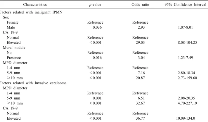
Table 2. Multivariate analysis of factors predictive of malignant or invasive IPMN
Characteristics p-value Odds ratio 95% Confidence Interval
Factors related with malignant IPMN Sex
Female Reference Reference
Male 0.036 2.93 1.07-8.01
CA 19-9
Normal Reference Reference
Elevated <0.001 29.03 8.08-104.25
Mural nodule
No Reference Reference
Presence 0.016 3.04 1.23-7.49
MPD diameter
1-4 mm Reference Reference
5-9 mm <0.001 7.16 2.80-18.34
≥10 mm <0.001 20.87 2.73-159.60
Factors related with Invasive carcinoma MPD diameter
1-4 mm Reference Reference
5-9 mm 0.001 6.51 2.08-20.35
≥10 mm <0.001 32.67 4.70-227.19
CA 19-9
Normal Reference Reference
Elevated <0.001 36.77 10.09-134.0
IPMN, intraductal papillary mucinous neoplasm; MPD, main pancreatic duct
Patient survival
Patient survival curves are shown in Fig. 1A and 1B.
The 5-year survival rates were 98.1% in LGD, 95.3% in IMGD, 100% in HGD, and 71.8% in invIPMC cases (Fig.
1A). Although HGD was included in malignant IPMN, the three survival curves for LGD, IMGD, and HGD were not significantly different. We did detect a significant differ-
ence between noninvasive IPMN and invasive IPMC (p
<0.001; Fig. 1B).
Multivariate analysis of factors associated with malignant or invasive IPMN
Among the factors associated with malignant IPMN de- scribed in Table 1, we identified four statistically sig-
Table 3. Characteristics of the patients who had recurrent disease following surgical resection No.Age/sexDFS (mos)Tumor locationTumor size (mm)
MPD diameter (mm)
Mural noduleCA19‐9 (U/ml)Name of operationDisease pathology Resection margin pathologyRecurrent siteDiagnosis of recurrence
Treatment for recurrent diseaseFinal status 171/M 2.9head7020presence314PPPDInvasiveLGDPeritoneal seedingCTObservationDead, 10.6 mos 265/F 3.5head30 5presence364PPPDInvasiveLGDLiver mets and peritoneal seedingCTChemotherapyDead, 10.1 mos 360/M 6.5head35 6no456PPPDInvasiveNegative Liver mets and peritoneal seedingCTObservationAlive, 15.8 mos 473/M 7.6Multiple (head and body)
10 2no326PPPDInvasiveNegativeLung metsCT and PETChemotherapyAlive, 19.7 mos 557/M12.7Multiple (head and tail) 40 3presence46.2TPInvasiveNegativeAround Celiac trunk and SMACT and PETCCRTAlive, 13.1 mos 679/M15.1Multiple (head and body)
38 3no12.7PPPDInvasiveNegativeHepatoduodenal ligamentCT and PETObservationDead, 16.2 mos 768/M16.0tail17 2no1.5Lap‐DPLGDNegativeRemnant pancreasCTObservationAlive, 99.9 mos 847/F22.3Multiple (head and tail)
20 6no1.5Lap‐DPIMGDIMGDProgression of remnant lesionCTObservationAlive, 27 mos 969/M22.5Head 42 5presence7.6Lap‐PPPDIMGDNegativePeritoneal seedingCTChemotherapyDead, 28.3 mos 1077/F24.6Head52 8No42.6PPPDInvasiveNegativeMesenteric rootCT and PETChemotherapyDead, 38.8 mos 1167/M24.9Head30 4NoNAPDIMGDNegativeLiver mets following recurrence at remnant pancreas
CT and liver biopsy
ChemotherapyDead, 42.1 mos 1270/M31.1Head25 5No1.5Lap‐PPPDLGDNegativeRemnant pancreasCTObservationAlive, 47.2 mos 1361/F32.0head3112Presence 2.0PDInvasiveLGDLung metsCT and PETChemotherapyDead, 32.9 mos 1461/M32.8Tail20 2No6.5DPLGDNegativeResected margin CTObservationAlive, 157.9 mos 1565/M71.6Head35 8No34.4PPPDInvasiveNegativeRemnant pancreasCTObservationAlive, 147.2 mos 1667/M84.3Tail55 1Presence1.7DPIMGDNegativeResected marginCTObservationAlive, 103.6 mos DFS, disease-free survival; MPD, main pancreatic duct; PPPD, pylorus-preserving pancreaticoduodenectomy; NA, not available; LGD, low grade dysplasia; TP, total pan- createctomy; SMA, superior mesenteric artery; CCRT, concurrent chemo-radiotherapy; Lap-DP, laparoscopic distal pancreatectomy; IMGD, intermediate grade dysplasia; Lap-PPPD, laparoscopic pylorus-preserving pancreaticoduodenectomy; PD, pancreaticoduodenectomy; DP, distal pancreatectomy; mets, metastasis; CT, computed tomography; PET, positron emission tomography
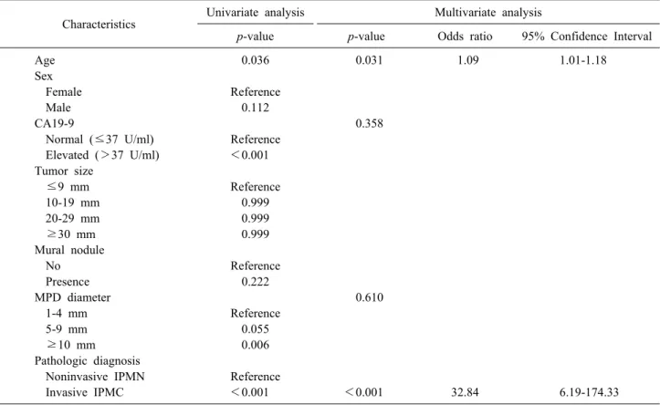
Table 4. Univariate and multivariate analyses for disease recurrence in branch duct IPMN following surgical resection Characteristics Univariate analysis Multivariate analysis
p-value p-value Odds ratio 95% Confidence Interval
Age 0.036 0.031 1.09 1.01-1.18
Sex
Female Reference
Male 0.112
CA19-9 0.358
Normal (≤37 U/ml) Reference Elevated (>37 U/ml) <0.001 Tumor size
≤9 mm Reference
10-19 mm 0.999
20-29 mm 0.999
≥30 mm 0.999
Mural nodule
No Reference
Presence 0.222
MPD diameter 0.610
1-4 mm Reference
5-9 mm 0.055
≥10 mm 0.006
Pathologic diagnosis
Noninvasive IPMN Reference
Invasive IPMC <0.001 <0.001 32.84 6.19-174.33
MPD, main pancreatic duct; IPMN, intraductal papillary mucinous neoplasm; IPMC, intraductal papillary mucinous carcinoma nificant factors, via a multivariate analysis (Table 2), that
included the following: male sex (odds ratio: OR 2.928), the presence of a mural nodule (OR 3.093), the dilated MPD diameter (OR 7.158 in 5-9 mm and 20.872 in more than 10 mm), and an elevated level of CA19-9 (OR 29.019). The Multivariate analysis identified two factors as being predictive of invasive carcinoma (Table 2) that included the following: a dilated MPD diameter (OR 6.512 in 5-9 mm and 32.674 in more than 10 mm) and an elevated level of CA19-9 (OR 36.774).
Disease recurrence following surgical resection There were 16 patients who experienced disease re- currence following surgical resection; their clinicopatho- logical characteristics are described in Table 3. The af- fected patients included 9 invasive carcinoma, 4 IMGD, and 3 LGD cases at the initial diagnosis. Of the 7 patients with initially noninvasive IPMN, 5 had recurrences or a progression of disease in the remnant pancreas (No. 7, 8, 12, 14, and 16; Table 3). 2 of the initially noninvasive IPMN patients experienced carcinomatous recurrences
(peritoneal seeding and liver metastasis) at 22.5 and 24.9 months following surgical resection, and died as a con- sequence of disease progression (No. 9 and 11; Table 3).
Old age, an elevated level of CA19-9 (>37 U/ml), a dilated MPD diameter (≥10 mm), and invasive pathology were associated with disease recurrence via a univariate analysis (p=0.036, p<0.001, p=0.006, and p<0.001, re- spectively; Table 4). According to the multivariate analy- sis, old age (OR 1.092) and invasive IPMC (OR 32.840) patients were independently significant, in regard to dis- ease recurrence following surgical resection.
DISCUSSION
IPMN is a disease entity that has a wide range of histo- logical subtypes, and a natural history that follows an ad- enoma-to-carcinoma sequence. Knowledge of the natural history and disease traits of each subtype of IPMN is im- portant for devising treatment strategies that avail patients of optimal treatment opportunities. International consensus guidelines for the management of IPMN were generated
to assist in devising treatment strategies, by the working group of the International Association of Pancreatology.
The 2012 guidelines adopted new concepts, ‘high-risk stigmata of malignancy’ and ‘worrisome features,’ so we stratified patients into these two categories.13 The recom- mendation of these guidelines was to let the stratified pa- tients follow different therapeutic flows. In this retro- spective single-institutional study, we investigated pre- operative clinical predictors of malignant and invasive dis- eases of BD-IPMN of the pancreas, and validated the flows recommended by the 2012 international consensus guidelines. To the best of our knowledge, this is the larg- est series- to date- of patients with BD-IPMNs that were resected and confirmed pathologically.
The prevalence of malignant IPMN was reported to range from 10% to 32% in patients with BD-IPMN.5,10,15,16 In the series presented here, malignant IPMN (HGD and invIPMC) was 13.0%, and invasive carcinoma (invIPMC) was 7.7% of the entire resected BD-IPMN cohort.
Therefore, the proportion of malignant or invasive dis- eases of BD-IPMN would be reduced if non-resected non- invasive BD-IPMNs under observation had been included.
Our survival analysis showed that the 5-year survival rate of non-invasive IPMN was 98.2%. Although HGD was included in the malignant category, its survival curve resembled that of benign IPMNs. The postoperative sur- vival rate of HGD of BD-IPMN was already verified by a previous study.2 However, Winner et al.17 showed that carcinoma in situ (HGD) of IPMN showed a higher re- currence rate after a postoperative period of 5 years.
Tamura et al also demonstrated that HGD of MD-IPMN showed a high recurrence rate and poor survival, when long-term surveillance of more than 5 years was carried out.11 Therefore, although HGD shows favorable survival outcomes for the initial 5 years after surgical resection, it should be followed as a malignant disease using long-term surveillance.
The strategic factors that were considered to be ‘high risk stigmata’ and/or ‘worrisome features’, from the 2012 international consensus guidelines, were validated in this study. Although we did not investigate the presence of a thickened cystic wall or cytological pathology, additional factors that were associated with malignant and/or in- vasive disease were identified. We also revealed other prognostic factors, including sex and CA19-9, which were
correlated with malignant and/or invasive IPMN in the multivariate analysis. Pedrazzoli et al.18 reported that posi- tron emission tomography was more accurate than the guidelines, in distinguishing benign from malignant IPMN. Additionally, more predictive factors will likely be identified in future studies. When more prognostic and di- agnostic data are accumulated, these guidelines should then be revised. Therefore, various trials, including those with retrospective and prospective designs, should be re- peated to identify more accurate factors. The present algo- rithm for management is quite complicated for application in an outpatient clinical setting, and it only focuses on the morphological features, without regard to patient factors or biomarkers that are capable of reflecting the propensity of the disease.
Previously, patients with invasive IPMC were reported to be more likely to recur with a probability of hazard ratio of 5.2, but the original pathology did not predict dis- ease severity on recurrence.17 Here, we observed recur- rences in 16 (4.9%) of the 324 patients with resected BD-IPMN, including 36% of the invasive IPMC and 2.5%
of the non-invasive IPMN cases. We determined that the initial disease pathology was the most powerful predictor of recurrence (OR 32.84: 95% CI 6.19-174.33), and that the initial pathology was related to the recurrence pattern (or severity) following resection. There were 8 of 9 pa- tients with initially invasive IPMN who had distant metastases. or peritoneal seeding, while 5 of 7 patients with originally non-invasive IPMN had recurrences on the remnant pancreas or resected margin (Table 3). The latter 5 patients have remained alive and are under periods of regular observation that have ranged from 27.0 to 157.9 months. We experienced 2 cases of malignant recurrences of IMGD in the two year follow-up period after surgical resection (No. 9 and 11 in Table 3). Those cases initially had a single lesion in the pancreas, and the resected mar- gins were clear from disease. Therefore, there must still be undisclosed biological characteristics of IPMN, and molecular or large scale clinical studies to prevent the deaths of patients with initially benign disease should be considered.
In conclusion, in a high-volume single-institutional study, we investigated the clinicopathological character- istics of BD-IPMN of the pancreas. Although the current treatment guidelines for BD-IPMN reflect malignant and
invasive features appropriately, they are confined to the morphological characteristics of the disease. Patient fac- tors and biological features should be considered when devising optimal therapeutic or surveillance strategies.
REFERENCES
1. Conlon KC. Intraductal papillary mucinous tumors of the pancreas. J Clin Oncol 2005;23:4518-4523.
2. Rodriguez JR, Salvia R, Crippa S, Warshaw AL, Bassi C, Falconi M, et al. Branch-duct intraductal papillary mucinous ne- oplasms: observations in 145 patients who underwent resection.
Gastroenterology 2007;133:72-79.
3. Sohn TA, Yeo CJ, Cameron JL, Iacobuzio-Donahue CA, Hruban RH, Lillemoe KD. Intraductal papillary mucinous neoplasms of the pancreas: an increasingly recognized clinicopathologic entity.
Ann Surg 2001;234:313-321.
4. Crippa S, Fernández-Del Castillo C, Salvia R, Finkelstein D, Bassi C, Domínguez I, et al. Mucin-producing neoplasms of the pancreas: an analysis of distinguishing clinical and epidemio- logic characteristics. Clin Gastroenterol Hepatol 2010;8:213-219.
5. Mimura T, Masuda A, Matsumoto I, Shiomi H, Yoshida S, Sugimoto M, et al. Predictors of malignant intraductal papillary mucinous neoplasm of the pancreas. J Clin Gastroenterol 2010;
44:e224-e229.
6. Salvia R, Fernández-del Castillo C, Bassi C, Thayer SP, Falconi M, Mantovani W, et al. Main-duct intraductal papillary mucinous neoplasms of the pancreas: clinical predictors of malignancy and long-term survival following resection. Ann Surg 2004;239:678-685.
7. Schmidt CM, White PB, Waters JA, Yiannoutsos CT, Cummings OW, Baker M, et al. Intraductal papillary mucinous neoplasms:
predictors of malignant and invasive pathology. Ann Surg 2007;
246:644-651.
8. Shin SH, Han DJ, Park KT, Kim YH, Park JB, Kim SC.
Validating a simple scoring system to predict malignancy and invasiveness of intraductal papillary mucinous neoplasms of the pancreas. World J Surg 2010;34:776-783.
9. Sohn TA, Yeo CJ, Cameron JL, Hruban RH, Fukushima N,
Campbell KA, et al. Intraductal papillary mucinous neoplasms of the pancreas: an updated experience. Ann Surg 2004;239:
788-797.
10. Sugiyama M, Izumisato Y, Abe N, Masaki T, Mori T, Atomi Y. Predictive factors for malignancy in intraductal papil- lary-mucinous tumours of the pancreas. Br J Surg 2003;90:
1244-1249.
11. Tamura K, Ohtsuka T, Ideno N, Aso T, Shindo K, Aishima S, et al. Treatment strategy for main duct intraductal papillary mu- cinous neoplasms of the pancreas based on the assessment of recurrence in the remnant pancreas after resection: a retro- spective review. Ann Surg 2014;259:360-368.
12. Tanaka M, Chari S, Adsay V, Fernandez-del Castillo C, Falconi M, Shimizu M, et al; International Association of Pancreatology.
International consensus guidelines for management of intraductal papillary mucinous neoplasms and mucinous cystic neoplasms of the pancreas. Pancreatology 2006;6:17-32.
13. Tanaka M, Fernández-del Castillo C, Adsay V, Chari S, Falconi M, Jang JY, et al; International Association of Pancreatology.
International consensus guidelines 2012 for the management of IPMN and MCN of the pancreas. Pancreatology 2012;12:183-197.
14. Bosman FT, Carneiro F, Hruban RH, Theise ND. WHO classi- fication of tumours of the digestive system. Lyon, France: IARC Press; 2010.
15. Lee SY, Lee KT, Lee JK, Jeon YH, Choi D, Lim JH, et al.
Long-term follow up results of intraductal papillary mucinous tu- mors of pancreas. J Gastroenterol Hepatol 2005;20:1379-1384.
16. Nagai K, Doi R, Kida A, Kami K, Kawaguchi Y, Ito T, et al.
Intraductal papillary mucinous neoplasms of the pancreas: clin- icopathologic characteristics and long-term follow-up after resection. World J Surg 2008;32:271-278.
17. Winner M, Epelboym I, Remotti H, Lee JL, Schrope BA, Chabot JA, et al. Predictors of recurrence in intraductal papillary muci- nous neoplasm: experience with 183 pancreatic resections. J Gastrointest Surg 2013;17:1618-1626.
18. Pedrazzoli S, Sperti C, Pasquali C, Bissoli S, Chierichetti F.
Comparison of International Consensus Guidelines versus 18-FDG PET in detecting malignancy of intraductal papillary mucinous neoplasms of the pancreas. Ann Surg 2011;254:
971-976.