Surgical outcomes of multifocal branch duct intraductal papillary mucinous neoplasms of pancreas
Jae Hyun Kwon, Song Cheol Kim, Ki-Byung Song, Jae Hoon Lee, Dae Wook Hwang, Kwang-Min Park, and Young-Joo Lee
Division of Hepatobiliary and Pancreatic Surgery, Department of Surgery, Asan Medical Center, University of Ulsan College of Medicine, Seoul, Korea
Backgrounds/Aims: Appropriate management for multifocal branch duct type intraductal papillary mucinous neoplasms (BD-IPMNs) of the pancreas is still controversial. This study was intended to reveal surgical outcomes of surgical re- section for multifocal BD-IPMNs, with BD-IPMNs in the remnant pancreas. Methods: Between January 1995 and December 2013, 699 patients underwent the pancreatic resection due to IPMN of pancreas in our institution. Among them, 37 patients showed multifocal BD-IPMNs. After excluding patients who had BD-IPMNs completely resected, medi- cal records of 22 patients with remained BD-IPMNs in the remnant pancreas were retrospectively reviewed. Results:
Mean patient age was 65±6.4 years. Types of surgery included central pancreatectomy (n=1), distal pancreatectomy (n=14), and standard pylorus-preserving pancreaticoduodenectomy (n=7). Specimen pathology showed that IPMN was either at low/intermediate-grade dysplasia (n=17) or at high-grade dysplasia (n=2). Three patients had IPMN associated with invasive carcinoma. Their mean follow-up period was 40.4 months. During follow-up, one mortality occurred 35.2 months after the operation which was not associated with IPMN. There was no clinically significant disease progression or recurrence of IPMN in the remnant pancreas during the follow-up period. Conclusions: Our results support that we can safely preserve the pancreas parenchyma with multifocal BD-IPMNs. Benign-looking multifocal BD-IPMNs in the remnant pancreas do not affect the survival of patients. (Korean J Hepatobiliary Pancreat Surg 2014;18:152-158) Key Words: Pancreatectomy; Intraductal papillary mucinous neoplasms; Branch duct; Multifocal; Follow-up
Received: August 23, 2014; Revised: October 14, 2014; Accepted: October 30, 2014 Corresponding author: Song Cheol Kim
Department of Surgery, Asan Medical Center, University of Ulsan College of Medicine, 388-1 Poongnap-dong, Songpa-gu, Seoul 138-736, Korea Tel: +82-2-3010-3936, Fax: +82-2-3010-8082, E-mail: [email protected]
Copyright Ⓒ 2014 by The Korean Association of Hepato-Biliary-Pancreatic Surgery
This is an Open Access article distributed under the terms of the Creative Commons Attribution Non-Commercial License (http://creativecommons.org/
licenses/by-nc/3.0) which permits unrestricted non-commercial use, distribution, and reproduction in any medium, provided the original work is properly cited.
Korean Journal of Hepato-Biliary-Pancreatic Surgery ∙ pISSN: 1738-6349ㆍeISSN: 2288-9213
INTRODUCTION
Intraductal papillary mucinous neoplasms (IPMNs) of the pancreas have been increasingly diagnosed due to the widespread use of advanced imaging modalities such as computed tomography (CT) and magnetic resonance imaging (MRI). IPMNs are typically classified into three groups: main duct type IPMN (MD-IPMN), branch duct type IPMN (BD-IPMN), and mixed type IPMN. In 2012, the international consensus guidelines for the management of IPMNs have been revised by the working group of the International Association of Pancreatology (IAP) follow- ing the 14th meeting of the IAP (Fukuoka, Japan in 20101). Despite some limitations in predicting malignant BD-IPMN,2 the utility of these guidelines for management of BD-IPMN has been validated by some studies.3,4
For MD-IPMNs, surgical resection is strongly recom- mended for all patients. However, management of BD-IPMNs is still controversial. Due to lower possibility of malignancy in BD-IPMNs (reported as about 25%1), surgical resection of BD-IPMNs is only considered for se- lected patients. In patients with multifocal BD-IPMNs (14.5-41%5,6 of BD-IPMNs are known to be multifocal), the extent of pancreatic resection needs to be determined carefully considering both the risk of tumor progression from BD-IPMNs in the remnant pancreas and the reduc- tion of quality of life from aggressive resection with cura- tive intent.
In this study, we evaluated the surgical outcomes of multifocal BD-IPMNs after resection. The long-term re- sult of the remnant pancreas in these patients were fol- lowed-up. We especially investigated whether we could
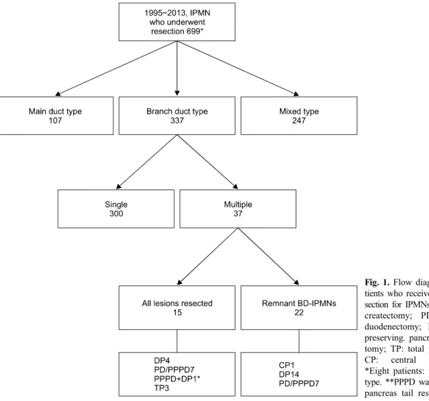
Fig. 1. Flow diagram of 699 pa- tients who received pancreatic re- section for IPMNs. DP: distal pan- createctomy; PD: pancreatico- duodenectomy; PPPD: pylorus- preserving. pancreaticoduodenec- tomy; TP: total pancreatectomy;
CP: central pancreatectomy.
*Eight patients: unknown ductal type. **PPPD was combined with pancreas tail resection.
safely preserve parenchyma of the pancreas that had mul- tifocal BD-IPMNs.
MATERIALS AND METHODS
Between January 1995 and December 2013, 699 pa- tients underwent pancreatic resection for IPMN of the pancreas at the Department of Surgery in Asan Medical Center. The diagnosis of IPMN was confirmed by patho- logic result for all cases. Among them, 337 patients had BD-IPMN. Of the 337 patients, 37 showed multifocality.
Because the purpose of this study was to review the prog- nosis and fate of remaining pancreas with BD-IPMNs, we excluded 15 patients whose multifocal BD-IPMNs were completely removed through pancreatic resection (which means no residual BD-IPMNs in the remnant pancreas).
Patient selection flow is illustrated at Fig. 1. Medical re-
cords of 22 patients who still had BD-IPMNs (with or without mixed type IPMNs) were retrospectively re- viewed, including past medical histories, radiologic find- ings, laboratory data, and pathologic results.
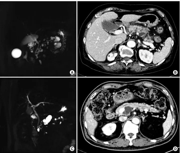
We defined multifocal BD-IPMNs as two or more cyst- ic lesions in any part of the pancreas that had communica- tion with the main pancreatic duct. Multifocal BD-IPMN was diagnosed using computed tomography (CT) scan or magnetic resonance cholangiopancratography (MRCP) (Fig. 2). MRCP was performed on a 1.5 Tesla MR scan- ner (Magnetom Avanto, Siemens Medical Solutions) with intravenous injection of Gd-BOPTA (gadobenate dimeglu- mine, MultihanceⓇ). Of the 22 patients, 20 were evaluated by both CT and MRCP. Malignancy of BD-IPMNs was suspected when the followings were observed in pre- operative imaging studies: diffused main pancreatic duct dilatation over 10 mm, tumor size over 3 cm, enhanced
Fig. 2. Radiologic findings of multifocal intraductal papillary mucinous neoplasms. (A and B) MRI and CT scan revealed pancreas swelling with peri-pancreatic infiltration, implying acute pancreatitis. There were variable sized multiple cystic lesions communi- cated with the main pancreatic duct in the entire pancreas, suggesting multiple BD-IPMNs. Main pancreatic duct size was meas- ured at 3 mm. (C and D) Multifocal BD-IPMNs in the pancreas with maximal diameter measured at 3.5 cm in the uncinate process without evidence of dilatation of main pancreatic duct or mural nodules.
mural nodules, and lymphadenopathy. Pancreatic resection was performed for all suspicious malignant lesions, whereas benign-looking BD-IPMNs were left intact within the remnant pancreas.
Ductal types of IPMNs of resected specimens were classified according to radiologic study and pathologic results. Pathologic classification of resected pancreas was in accordance with World Health Organization classi- fication of tumors.7 Staging system of IPMN carcinoma was based on the latest pancreatic cancer staging system by the American Joint Committee on Cancer (AJCC).8 After operation, patients were followed up by CT scans
immediately after the operation (within one month) and at three months, and six months post-operatively. If there was no change in lesions in the remnant pancreas, CT scans with chemical battery and CA 19-9 were performed annually.
RESULTS
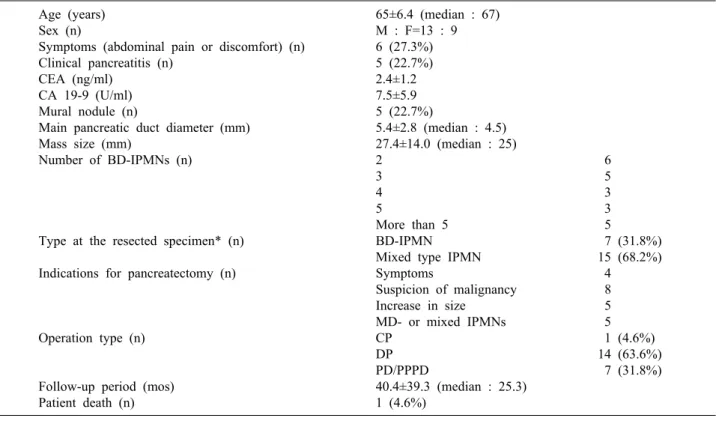
Clinical and pathological characteristics of the 22 pa- tients (13 males and 9 females, mean age 65 at the time of operation) analyzed in this study are summarized in Table 1. Of the 22 patients, six visited hospital due to ab-
Table 1. Clinical and pathological characteristics of patients with multifocal branch duct type IPMNs in the remnant pancreas (n=22)
Age (years) Sex (n)
Symptoms (abdominal pain or discomfort) (n) Clinical pancreatitis (n)
CEA (ng/ml) CA 19-9 (U/ml) Mural nodule (n)
Main pancreatic duct diameter (mm) Mass size (mm)
Number of BD-IPMNs (n)
Type at the resected specimen* (n) Indications for pancreatectomy (n)
Operation type (n)
Follow-up period (mos) Patient death (n)
65±6.4 (median : 67) M : F=13 : 9 6 (27.3%) 5 (22.7%) 2.4±1.2 7.5±5.9 5 (22.7%)
5.4±2.8 (median : 4.5) 27.4±14.0 (median : 25)
2 6
3 5
4 3
5 3
More than 5 5
BD-IPMN 7 (31.8%)
Mixed type IPMN 15 (68.2%)
Symptoms 4
Suspicion of malignancy 8
Increase in size 5
MD- or mixed IPMNs 5
CP 1 (4.6%)
DP 14 (63.6%)
PD/PPPD 7 (31.8%)
40.4±39.3 (median : 25.3) 1 (4.6%)
CP, central pancreatectomy; DP, distal pancreatectomy; PD, pancreaticoduodenectomy; PPPD, pylorus-preserving pancreati- coduodenectomy. *Ductal types of BD-IPMNs in remnant pancreas were all branch-duct types in all 22 patients
dominal pain or discomfort. Of the six patients, three had pancreatitis. A total of five patients were diagnosed with pancreatitis, of which three had symptom of abdominal pain. Both mean CEA (2.4 ng/ml) and CA 19-9 (7.5 U/ml) were within the normal range (0-6 ng/ml for CEA, and 0-37 U/ml for CA 19-9).
Mural nodules were found in five (22.7%) patients.
Mean diameter of the main pancreatic duct was 5.4 mm.
Mean diameter of the largest lesion (measured by patholo- gists) was 27.4 mm. The number of BD-IPMNs evaluated by pathologists or radiologists was: two in 6 patients, three in 5 patients, four in 3 patients, five in 3 patients, and more than five in 5 patients. In 15 patients, IPMNs in the resected pancreas initially assessed as BD-IPMNs by radiologic studies were diagnosed as mixed type pathologically. All 22 patients showed BD-IPMNs in their remnant pancreas revealed by CT or MRCP.
Pancreatic resection was proposed and performed in pa- tients with symptoms of abdominal pain or recurrent pan- creatitis (n=4), suspicion of malignancy in radiologic stud- ies (n=8), increase in size during follow-up (n=5), and
main duct-IPMN or mixed type IPMN suspected in pre- operative radiologic study (n=5). The operation types of pancreatectomy included distal pancreatectomy (n=14), pancreaticoduodenectomy or pylorus-preserving pan- creaticoduodenectomy (n=7), and central pancreatectomy (n=1).
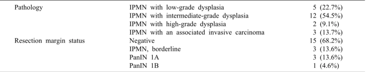
Pathologic results of resected pancreas are summarized in Table 2. A total of 17 patients showed low- or inter- mediate-grade dysplasia. Two patients were diagnosed as high-grade dysplasia. Three patients had IPMN with an associated invasive carcinoma. TNM stage of invasive IPMN carcinomas were IA in one patient and IB in two patients. None of them underwent postoperative adjuvant chemotherapy. Instead, they were observed regularly with- out evidence of recurrence during follow-up period. All pancreatic resection margins except three patients were negative for tumors. In these three patients, resection mar- gins were IPMN with intermediate-grade dysplasia.
Another three patients showed pancreatic intraepithelial lesions 1A (PanIN 1A) in resection margins. One patient had PanIN 1B in resection margin. No perioperative mor-
Table 2. Pathological characteristics of resected specimen in patients with multifocal branch duct type IPMNs in the remnant pancreas (n=22)
Pathology
Resection margin status
IPMN with low-grade dysplasia IPMN with intermediate-grade dysplasia IPMN with high-grade dysplasia
IPMN with an associated invasive carcinoma Negative
IPMN, borderline PanIN 1A PanIN 1B
5 (22.7%) 12 (54.5%)
2 (9.1%) 3 (13.7%) 15 (68.2%) 3 (13.6%) 3 (13.6%) 1 (4.6%) PanIN, pancreatic intraepithelial lesion
Fig. 3. Patient showing progressive dilatation of BD-IPMN. A 68 years-old male patient underwent pylorus-preserving pan- creaticoduodenctomy due to increase in size of multifocal BD-IPMNs with mural nodules in the head. At 17 months after pancre- atic resection, follow-up CT scan showed increase in diameter of two remnant BD-IPMNs in the pancreatic tail: (A) from 8.2 mm (B) to 11 mm, (C) from 10 mm (D) to 13.5 mm. Because both of them did not have any high-risk stigmata or worrisome features, we chose to cautiously observe these lesions.
tality occurred. The mean follow-up period was 40.4 months. During the follow-up, no disease-associated mor- tality occurred. One patient who had received renal trans- plantation six years before the pancreatic resection died 35.2 months after the operation. There was no disease
progression (no increase in diameter) or recurrence of IPMN in the remnant pancreas during follow-up except in one patient who showed increase in diameters of two remnant BD-IPMNs (from 8.2 mm to 11 mm and from 10 mm to 13.5 mm) 17 months after the pancreatic re-
section (Fig. 3). Because there was no evidence of malig- nant features in the remnant lesions, this patient was ob- served regularly without any intervention. There was no perioperative morbidity. However, one patient showed re- current pancreatitis after PPPD with pancreatic duct stone.
The patient underwent re-operation for pancreaticojejuno- stomy (PJ) revision 40 months after the first surgery PPPD. After PJ revision, the patient has been doing well without any complication. The remaining twenty patients are now currently alive without any change in follow-up CT scans. No patient had tumor recurrence.
Among the 37 patients with multifocal BD-IPMNs, 15 were able to remove all multifocal BD-IPMNs through pancreatectomy. One patient had IPMN with an associated invasive carcinoma. Another patient showed IPMN with high-grade dysplasia. A total of 13 patients had low- or intermediate-grade dysplasia. Indications for pancreatec- tomy were symptoms of abdominal pain or recurrent pan- creatitis (n=3), suspicion of malignancy in radiologic stud- ies (n=7), increase in size during follow-up (n=3), mixed type suspected in preoperative radiologic study (n=1), and patient’s desire (n=1). There was no recurrence or disease progression except in one patient who was diagnosed with IPMN associated with invasive carcinoma. Pathologic staging of invasive IPMN was stage IA. The patient didn’t receive any adjuvant chemotherapy. Six months after the surgery, the patient was found to have multiple lung meta- stases on follow-up CT scans which were treated by chemotherapy. The remaining 14 patients have been doing well without any recurrence or progression during the mean follow-up period of 40.4 months.
Total pancreatectomy was performed in three patients, including two patients who had pancreatitis with multi- focal IPMNs involving the entire pancreas and one patient who had increase in size and number of IPMNs affecting the entire pancreas. Pathologic results of these three pa- tients were BD-IPMNs with low- or intermediate-grade dysplasia.
DISCUSSION
According to the international consensus guidelines for the management of IPMNs,1 surgical resection is recom- mended for BD-IPMN with ‘high-risk stigmata of malig- nancy’ or ‘worrisome features’ that suggests malignant
potential of IPMNs. “High-risk stigmata” include ob- structive jaundice, enhanced solid component within cyst, and main pancreatic duct size over 10 mm. “Worrisome features” include pancreatitis, cyst size over 3 cm, thick- ened or enhanced cyst walls, non-enhanced mural nodules, main pancreatic duct size of 5-9 mm, abrupt change in the main pancreatic duct caliber with distal pancreatic atrophy, and lymphadenopathy. They also advocated sur- gical resection only for the highest oncological risk even in multifocal BD-IPMN cases using the same treatment strategy as single BD-IPMN. In our study, multifocal BD-IPMNs in remnant pancreas do not have any
‘high-risk stigmata’ nor ‘worrisome features’. They were followed up safely without significant changes in CT scans. Although BD-IPMN itself is bearing the potential to progress into malignancy, if BD-IPMNs do not have any features implying the malignancy, we do not have to remove all BD-IPMNs to avoid the risk of pancreatic insufficiency.
There are several reports on the follow-up of BD-IPMN patients. About 82-97% of BD-IPMNs without mural nod- ules showed no progression during 42-61 months.9-11 Therefore, resection is only indicated for lesions with malig- nant features. Considering the natural course and the clinical behavior of BD-IPMNs, cautious clinical and radiological follow-up is suggested for asymptomatic small (less than 3 cm in diameter) BD-IPMNs. Similar recommendation could be applied to multifocal BD-IPMNs. Although the natural history and precise prognosis of multifocal BD-IPMNs remain controversial, multifocal BD-IPMNs showed slow growth and non-significant evolution as time elapsed in radiologic follow-up.12 However, Tanno et al.11 suggested that patients with BD-IPMNs had a high risk for metachronous pancreatic ductal adenocarcinoma even though pancreatic cancer developed separately from BD- IPMN. Therefore, benign course of multifocal BD-IPMNs should not be overlooked. We need to pay attention to the importance of careful observation and regular follow-up in these patients. In our study, of 37 patients, recurrence developed in only one patient who had all IPMNs in the pancreas removed through PPPD, suggesting that no remnant IPMNs was in the pancreas tail. Recurrence appeared in the form of distant metastasis in the lung 6 months after the pancreatic resection. On the other hand, for all patients who had remnant BD-IPMNs in the remnant pancreas, no
recurrence or clinically significant progression of the disease occurred.
In 22 patients with remnant BD-IPMNs in the remain- ing pancreas, indications for the pancreatic resection were consistent with the ‘International Consensus Guidelines 2012’ mentioned above, suggesting that BD-IPMNs might have malignant features when CT or MRCP findings show mural nodules, enhancing solid portions, lymphadenop- athy, or tumor size over 3 cm.
In this study, we specified our subject to remnant pan- creas with multiple BD-IPMNs. Our conclusion is that we can safely preserve pancreas parenchyma with multifocal BD-IPMNs in the remnant pancreas by performing pan- creatic resection because they would remain silent without disease progression or recurrence. Regardless of patho- logic status of the resected specimen of IPMNs, BD-IPMNs in the remnant pancreas do not affect the sur- vival of patients.
REFERENCES
1. Tanaka M, Fernández-del Castillo C, Adsay V, Chari S, Falconi M, Jang JY, et al; International Association of Pancreatology.
International consensus guidelines 2012 for the management of IPMN and MCN of the pancreas. Pancreatology 2012;12:
183-197.
2. Goh BK, Tan DM, Ho MM, Lim TK, Chung AY, Ooi LL.
Utility of the sendai consensus guidelines for branch-duct intra- ductal papillary mucinous neoplasms: a systematic review. J Gastrointest Surg 2014;18:1350-1357.
3. Jang JY, Park T, Lee S, Kang MJ, Lee SY, Lee KB, et al.
Validation of international consensus guidelines for the resection of branch duct-type intraductal papillary mucinous neoplasms. Br J Surg 2014;101:686-692.
4. Aso T, Ohtsuka T, Matsunaga T, Kimura H, Watanabe Y, Tamura K, et al. "High-risk stigmata" of the 2012 international consensus guidelines correlate with the malignant grade of branch duct intraductal papillary mucinous neoplasms of the pancreas. Pancreas 2014;43:1239-1243.
5. Schmidt CM, White PB, Waters JA, Yiannoutsos CT, Cummings OW, Baker M, et al. Intraductal papillary mucinous neoplasms:
predictors of malignant and invasive pathology. Ann Surg 2007;246:644-651.
6. Rodriguez JR, Salvia R, Crippa S, Warshaw AL, Bassi C, Falconi M, et al. Branch-duct intraductal papillary mucinous ne- oplasms: observations in 145 patients who underwent resection.
Gastroenterology 2007;133:72-79.
7. Bosmanm FT, Carneiro F, Hruban RH, Theise ND. WHO classi- fication of tumours of the digestive system. 4th ed. Lyon, France: IARC Press, 2010.
8. Edge SB, Byrd DR, Compton CC, Fritz AG, Greene FL, Trotti A, et al. AJCC cancer staging manual. 7th ed. New York:
Springer-Verlag, 2010.
9. Maguchi H, Tanno S, Mizuno N, Hanada K, Kobayashi G, Hatori T, et al. Natural history of branch duct intraductal papil- lary mucinous neoplasms of the pancreas: a multicenter study in Japan. Pancreas 2011;40:364-370.
10. Kobayashi G, Fujita N, Maguchi H, Tanno S, Mizuno N, Hanada K, et al; Working Group for the Natural History of IPMN of the Japan Pancreas Society. Natural history of branch duct intra- ductal papillary mucinous neoplasm with mural nodules: a Japan pancreas society multicenter study. Pancreas 2014;43:532-538.
11. Tanno S, Nakano Y, Koizumi K, Sugiyama Y, Nakamura K, Sasajima J, et al. Pancreatic ductal adenocarcinomas in long-term follow-up patients with branch duct intraductal papillary muci- nous neoplasms. Pancreas 2010;39:36-40.
12. Castelli F, Bosetti D, Negrelli R, Di Paola V, Zantedeschi L, Ventriglia A, et al. Multifocal branch-duct intraductal papillary mucinous neoplasms (IPMNs) of the pancreas: magnetic reso- nance (MR) imaging pattern and evolution over time. Radiol Med 2013;118:917-929.