165
서 론
췌장의 관내 유두상 점액성 종양(Intraductal papillary mu- cinous tumor, IPMT)은 췌관에서 기원하는 임상적으로 증가 하고 있는 질병군이다. 1982년 Ohhashi 등(1)은 내시경적 역 행성 담췌관 조영술(ERCP)상 조영 결손을 동반한 주췌관 확장 소견을 보이고 Vater 팽대부를 통해 점액이 분비되는 질환을 처음으로 ‘mucin secreting pancreatic cancer’라는 용 어로 기술하였다. 이후에는 mucinous ductal ectasia, mucin producing tumor, mucin-hypersecreting tumor, ductectatic cy- stadenoma or cystadenocarcinoma, intraductal mucin-hyperse- creting neoplasm, mucinous villous adenomatosis, intraductal papillary neoplasm, intraductal papillary mucinous neoplasm, IPMT 등 여러 용어로 혼용해 왔으나 최근에는 WHO 정의 에 의해 IPMT로 표기하고 있다.(2) 이 질환의 진단은 종종 만성 췌장염이나 낭성 췌장종양 등과 감별이 어려워 진단 이 늦어지기도 한다. Obara 등(3)이 140예의 IPMT 증례를 1988년도에 보고한 이후 최근에는 미국, 유럽에서 60예를 보고하기도 하였다.(4-7) 최근 들어 이 질환의 보고가 증가 되고 있으며, 이는 컴퓨터 전산화 단층 촬영(CT)이나 ERCP 등의 영상의학의 발달이 중요한 역할을 하리라 생각된다.
병리학적으로 과증식, 선종, 악성 선종을 모두 포함하며 이 는 췌장주변 조직을 침윤하거나 간문맥을 침윤한 악성선종
췌장의 관내 유두상 점액종의 임상적, 병리학적 고찰
연세대학교 의과대학 외과학교실, 1내과학교실 및 2병리학교실
윤지섭․조신일․이효상․정준표1․박영년2․김경식․윤동섭․최진섭․이우정․지훈상․김병로
책임저자:윤동섭, 서울시 강남구 도곡동 146-92 ꂕ 135-720, 영동세브란스병원 외과 Tel: 02-3497-2444, Fax: 02-3462-5994 E-mail: [email protected]
접수일:2002년 10월 17일, 게재승인일:2002년 11월 5일
Clinical & Pathological Characteristics of Intra- ductal Papillary Mucinous Tumor of the Pancreas
Ji Sup Yun, M.D., Sin Il Cho, M.D., Hyo Sang Lee, M.D., Jun Pyo Chung, M.D.1, Young Nyon Park, M.D.2, Kyung Sik Kim, M.D., Dong Sup Yoon, M.D., Jin Sup Choi, M.D., Woo Jung Lee, M.D., Hoon Sang Chi, M.D. and Byong Ro Kim, M.D.
Purpose: Intraductal papillary mucinous tumors of the pancreas (IPMT) are becoming increasingly recognized. De- spite a better understanding of these conditions, IPMT still present difficulty relating to the predictive factors and the risk of relapse after surgery. The aim of this study was to inve- stigate the clinical, and pathological characteristics of IPMT.
Methods: Between October 1998 and July 2002, 22 patients with IPMT underwent surgery. We retrospectively examined the clinicopathological features and surgical outcomes of these patients.
Results: The types of IPMT were as follows: dysplasia (1);
adenoma (4); borderline malignancy (9); carcinoma in situ (3); and carcinoma, both non-invasive (3) and invasive (2).
Lymph node metastasis was not found, but stromal invasion was found in the 2 cases of invasive carcinoma. The locations of the IPMT were as follows: head (6); uncinate process (11); body (4); and tail (1). There were 11 main duct types, 10 branched duct types and 1 combined. All patients underwent surgical resection, including 3 pancrea- ticoduodenectomies, 12 pylorus-preserving pancreaticoduo- denectomies, 4 distal pancreatectomies with splenectomies, 2 near-total pancreatectomies with splenectomies, and 1 enucleation. There were no operative or hospital deaths. A recurrence of the IPMT following surgery occurred in 2 cases. Their pathological features were a carcinoma in situ and a borderline malignancy, but not the invasive type.
However, one case of recurrence expired 7 month after surgery. A combination of other malignancies in these
patients was found in 2 cases.
Conclusion: IPMT has a favorable prognosis, when compared with pancreatic duct carcinoma. However, long- term follow-up after surgery is necessary, even for a curative resection due to a recurrence or a combination of other malignancies. Because combination of other malignancies exist infrequently, surgeons should be aware of the possibility of co-existing other malignancies. (J Korean Surg Soc 2003;64:165-169)
Key Words: IPMT, Pancreas, Characteristics 중심 단어: 관내 유두상 점액종, 췌장, 특징 ꠏꠏꠏꠏꠏꠏꠏꠏꠏꠏꠏꠏꠏꠏꠏꠏꠏꠏꠏꠏꠏꠏꠏꠏꠏꠏꠏꠏꠏꠏꠏꠏꠏꠏꠏꠏꠏꠏꠏꠏꠏꠏꠏꠏꠏꠏꠏꠏꠏꠏ Departments of Surgery, 1Internal Medicine and 2Pathology, Yonsei University College of Medicine, Seoul, Korea
ꠏꠏꠏꠏꠏꠏꠏꠏꠏꠏꠏꠏꠏꠏꠏꠏꠏꠏꠏꠏꠏꠏꠏꠏꠏꠏꠏꠏꠏꠏꠏꠏꠏꠏꠏꠏꠏꠏꠏꠏꠏꠏꠏꠏꠏꠏꠏꠏꠏꠏꠏꠏꠏꠏꠏꠏꠏꠏꠏꠏꠏꠏꠏꠏꠏꠏꠏꠏꠏꠏꠏꠏꠏꠏꠏꠏꠏꠏꠏꠏꠏꠏꠏꠏꠏꠏꠏꠏꠏꠏꠏꠏꠏꠏꠏꠏꠏꠏꠏꠏꠏꠏꠏꠏꠏꠏꠏꠏꠏꠏꠏꠏꠏꠏꠏ 이 아니라면 수술 전 감별은 어렵다. 그러나 다른 관내 악성
선종과는 달리 악성인 경우에도 절제가 용이하거나 예후가 좋은 경우가 많으며, 이는 종양의 성장속도가 늦고 전이가 적은 분자 생물학적 특성과 관련이 있다고 생각되나 절제 후 예후나 재발에 관계되는 인자는 아직까지 정확히 밝혀지지 않고 있다. 본 연구에서는 연세의료원에서 1998년 10월부 터 2002년 6월까지 IPMT로 수술을 시행받은 22예의 환자에 대한 임상적, 진단병리학적 특징을 살펴보고자 한다.
방 법
1998년 10월부터 2002년 6월까지 연세의료원에서 IPMT로 수술을 시행받은 22명의 환자를 대상으로 임상적 특성 및 병리학적 소견은 의무기록 및 영상검사를 중심으로 후향적 으로 분석하였으며, 질병의 재발 유무와 생존 여부는 외래 의무기록의 분석 및 전화통화를 통하여 연구를 진행하였 다. 남여비는 17:5이었고 평균 연령은 64.8세(범위 44∼80 세)였다. 환자들은 수술 후 진단병리학적으로 확진이 되었 고 임상적으로도 반복적인 췌장염, 점액으로 가득 찬 주췌
관의 확장, 주췌관과 연결된 낭성병변, 점액에 의해 열린 십 이지장 팽대부 등 IPMT의 전형적 특징을 보이는 환자들이 었다. 각각의 환자들은 임상적 특징과 병변이 위치해 있는 췌장의 위치, 진단 병리학적인 소견과 악성의 유무, 수술 방 법, 영상 진단을 위해 사용한 방법과 동반된 다른 악성 종양 의 유무 등을 조사해 보았다. IPMT는 종양이 생긴 부위를 기준으로 Kuroda(9) 분류에 의거하여 세 가지 형태로 분류 하였다: 1) main duct type (n=6); 주로 종양이 주췌관에 위치 하며 주췌관의 확장을 동반한다. 2) branch duct type (n=3);
종양이 주췌관에 위치하지 않으며 가지췌관만 확장된 소견 을 보인다. 3) combined type (n=1); 위 두 가지 형태가 복합 적으로 존재하는 경우이다. 악성종양일 경우는 TNM stage 분류에 의해 암의 진행정도를 알아보았다(Table 1).
결 과
1) 임상적, 병리학적 특성
근치적 절제술을 시행받은 22명의 환자 중 양성은 4예, 경계부는 9예, 악성은 9예였다. 악성인 환자 2예에서는 양
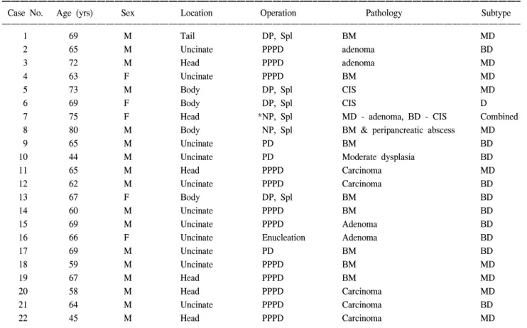
Table 1. Clinicopathologic features of 22 patients with IPMT treated by curative resection
ꠚꠚꠚꠚꠚꠚꠚꠚꠚꠚꠚꠚꠚꠚꠚꠚꠚꠚꠚꠚꠚꠚꠚꠚꠚꠚꠚꠚꠚꠚꠚꠚꠚꠚꠚꠚꠚꠚꠚꠚꠚꠚꠚꠚꠚꠚꠚꠚꠚꠚꠚꠚꠚꠚꠚꠚꠚꠚꠚꠚꠚꠚꠚꠚꠚꠚꠚꠚꠚꠚꠚꠚꠚꠚꠚꠚꠚꠚꠚꠚꠚꠚꠚꠚꠚꠚꠚꠚꠚꠚꠚꠚꠚꠚꠚꠚꠚꠚꠚꠚꠚꠚꠚꠚꠚꠚꠚꠚꠚꠚꠚꠚꠚꠚꠚ
Case No. Age (yrs) Sex Location Operation Pathology Subtype
ꠏꠏꠏꠏꠏꠏꠏꠏꠏꠏꠏꠏꠏꠏꠏꠏꠏꠏꠏꠏꠏꠏꠏꠏꠏꠏꠏꠏꠏꠏꠏꠏꠏꠏꠏꠏꠏꠏꠏꠏꠏꠏꠏꠏꠏꠏꠏꠏꠏꠏꠏꠏꠏꠏꠏꠏꠏꠏꠏꠏꠏꠏꠏꠏꠏꠏꠏꠏꠏꠏꠏꠏꠏꠏꠏꠏꠏꠏꠏꠏꠏꠏꠏꠏꠏꠏꠏꠏꠏꠏꠏꠏꠏꠏꠏꠏꠏꠏꠏꠏꠏꠏꠏꠏꠏꠏꠏꠏꠏꠏꠏꠏꠏꠏꠏ
1 69 M Tail DP, Spl BM MD
2 65 M Uncinate PPPD adenoma BD
3 72 M Head PPPD adenoma MD
4 63 F Uncinate PPPD BM MD
5 73 M Body DP, Spl CIS MD
6 69 F Body DP, Spl CIS D
7 75 F Head *NP, Spl MD - adenoma, BD - CIS Combined
8 80 M Body NP, Spl BM & peripancreatic abscess MD
9 65 M Uncinate PD BM BD
10 44 M Uncinate PD Moderate dysplasia BD
11 65 M Head PPPD Carcinoma MD
12 62 M Uncinate PPPD Carcinoma BD
13 67 F Body DP, Spl BM BD
14 60 M Uncinate PPPD BM BD
15 69 M Uncinate PPPD Adenoma BD
16 66 F Uncinate Enucleation Adenoma BD
17 69 M Uncinate PD BM BD
18 59 M Uncinate PPPD BM MD
19 67 M Head PPPD BM MD
20 58 M Head PPPD Carcinoma MD
21 64 M Uncinate PPPD Carcinoma BD
22 45 M Head PPPD Carcinoma MD
ꠏꠏꠏꠏꠏꠏꠏꠏꠏꠏꠏꠏꠏꠏꠏꠏꠏꠏꠏꠏꠏꠏꠏꠏꠏꠏꠏꠏꠏꠏꠏꠏꠏꠏꠏꠏꠏꠏꠏꠏꠏꠏꠏꠏꠏꠏꠏꠏꠏꠏꠏꠏꠏꠏꠏꠏꠏꠏꠏꠏꠏꠏꠏꠏꠏꠏꠏꠏꠏꠏꠏꠏꠏꠏꠏꠏꠏꠏꠏꠏꠏꠏꠏꠏꠏꠏꠏꠏꠏꠏꠏꠏꠏꠏꠏꠏꠏꠏꠏꠏꠏꠏꠏꠏꠏꠏꠏꠏꠏꠏꠏꠏꠏꠏꠏ DP, Spl: distal pancreatectomy with splenectomy; PPPD: pylorus-preserving pancreatoduodenectomy; NP, Spl: near-total pancreatectomy with splenectomy; BM: borderline malignancy; CIS: carcinoma in situ; MD: main duct; BD: branched duct. *concurrent radical total gastrectomy with Roux-en-Y esophagojejunostomy due to stomach cancer
ꠏꠏꠏꠏꠏꠏꠏꠏꠏꠏꠏꠏꠏꠏꠏꠏꠏꠏꠏꠏꠏꠏꠏꠏꠏꠏꠏꠏꠏꠏꠏꠏꠏꠏꠏꠏꠏꠏꠏꠏꠏꠏꠏꠏꠏꠏꠏꠏꠏꠏꠏꠏꠏꠏꠏꠏꠏꠏꠏꠏꠏꠏꠏꠏꠏꠏꠏꠏꠏꠏꠏꠏꠏꠏꠏꠏꠏꠏꠏꠏꠏꠏꠏꠏꠏꠏꠏꠏꠏꠏꠏꠏꠏꠏꠏꠏꠏꠏꠏꠏꠏꠏꠏꠏꠏꠏꠏꠏꠏꠏꠏꠏꠏꠏꠏ 성 선종과 악성 선암의 소견을 함께 동반하였으며 주변 췌
장 실질로의 미세한 침윤 소견이 있었다. 이러한 경우 IPMT에 합당한지에 대한 이견이 있을 수 있으나 본 연구에 서는 이 2예의 환자를 포함하였다. 전체 22예 중 주췌관을 침범한 경우가 11예, 부췌관을 침범한 경우가 10예, 그리고 주췌관과 부췌관을 동시에 침범한 경우가 1예였다. 병변의 위치는 췌장 두부에 위치한 경우가 6예, 미상엽에 위치한 경우는 11예, 체부는 4예, 미부는 1예였다. 두부에는 악성이 6예였으며 미상엽에는 양성 4예, 악성이 7예였으며 체부는 악성만 4예, 미부는 악성이 1예가 관찰되었다(Table 2). 주 췌관의 확장 소견은 17예에서 보였으며 부췌관에서도 17예 에서 낭성 확장 소견을 관찰하였다. 점액 분비가 확인된 경 우는 19예였고 mural nodule이 병리학적 혹은 방사선학적으 로 확인된 경우는 5예였다. 이 5예 모두 악성인 것으로 확인 되었다(Table 3). 주변 림프절을 침윤한 예는 없었다.
2) 영상진단 검사
내시경적 역행성 담췌관 조영술은 16명에서 시행하였는 데 주췌관의 이상소견(부분적, 전반적 확장 및 협착)은 14 명에서, 부췌관의 낭성 확장도 14명에서, mural nodule은 2 명에서만이 관찰이 가능했다. 유두의 확대소견은 1명의 환 자에서 관찰할 수 있었고 15명에서 점액의 과다 분비를 확 인할 수 있었으며 모두가 주유두에서 분비됨을 관찰할 수 있었다.
복부 초음파 검사를 시행한 21명의 환자 중 주췌관의 확 장은 17명에서, 부췌관의 낭성 확장도 17명에서, mural nodule 은 5명에서만 관찰할 수 있었다.
22명의 환자 모두에서 복부 전산화 단층촬영을 시행하였 다. 이 중 17명에서 주췌관의 확장 소견을 관찰할 수 있었 고, 이 중 1명에서 부분적 주췌관의 협착을 관찰할 수 있었 다. 17명의 환자에서 부췌관의 낭성 확장소견이 관찰되었 고, mural nodule은 5명에서만 관찰할 수 있었다.
내시경적 초음파 검사는 15명에서 시행하였는데 주췌관 의 확장은 11명, 부췌관 낭성 확장은 13명, mural nodule은 4명에서 관찰이 가능했다.
MRCP는 6명만이 시행하였는데 이 중 주췌관 이상, 부췌 관의 낭성 확장은 각각 4명에서, mural nodule은 2명에서만 관찰되었다(Table 4).
3) 외과적 치료
시행된 수술의 방법은 췌두부십이지장 절제술 3명, 유문 보존 췌두부십이지장 절제술 12명, 원위부 췌장 절제술 및 비장 절제술이 4명에서 시행되었으며, 2예에서는 아전 췌 장 절제술 및 비장 절제술이 시행되었다. 아전 췌장 절제술 및 비장 절제술을 시행받은 2명 중 1명은 위암을 동반하여 근치적 위전절제술 및 Roux-en-Y 식도 공장 문합술을 동시 에 시행받았다. 1명에서는 종괴 적출술만을 시행하였다.
절제연은 동결절편검사를 통하여 병변의 잔존 여부를 확 인하였는데 수술을 시행한 22예에서 모두 음성으로 보고되 었다. 수술 후 합병증은 없었다.
4) 장기 추적관찰 결과
수술 후 3개월에서 50개월 동안 추적관찰이 가능했다(평 Table 3. Relationship between mural nodule, mucus secretion and
pathology in IPMT
ꠚꠚꠚꠚꠚꠚꠚꠚꠚꠚꠚꠚꠚꠚꠚꠚꠚꠚꠚꠚꠚꠚꠚꠚꠚꠚꠚꠚꠚꠚꠚꠚꠚꠚꠚꠚꠚꠚꠚꠚꠚꠚꠚꠚꠚꠚꠚꠚꠚꠚꠚꠚꠚꠚꠚ Mural nodule Mucin secretion ꠏꠏꠏꠏꠏꠏꠏꠏꠏꠏꠏꠏ ꠏꠏꠏꠏꠏꠏꠏꠏꠏꠏꠏꠏꠏꠏ
Y N Y N
ꠏꠏꠏꠏꠏꠏꠏꠏꠏꠏꠏꠏꠏꠏꠏꠏꠏꠏꠏꠏꠏꠏꠏꠏꠏꠏꠏꠏꠏꠏꠏꠏꠏꠏꠏꠏꠏꠏꠏꠏꠏꠏꠏꠏꠏꠏꠏꠏꠏꠏꠏꠏꠏꠏꠏ
Operated Benign (4) 1 3 3 1
(N=22) Borderline (9) 2 7 7 2
*Malignancy (9) 4 5 8 1
ꠏꠏꠏꠏꠏꠏꠏꠏꠏꠏꠏꠏꠏꠏꠏꠏꠏꠏꠏꠏꠏꠏꠏꠏꠏꠏꠏꠏꠏꠏꠏꠏꠏꠏꠏꠏꠏꠏꠏꠏꠏꠏꠏꠏꠏꠏꠏꠏꠏꠏꠏꠏꠏꠏꠏ
*In one case, CIS is combined in branching duct and adenoma is combined in main duet.
Table. 4 Detection of ductal lesions and mural nodules by imaging modalities
ꠚꠚꠚꠚꠚꠚꠚꠚꠚꠚꠚꠚꠚꠚꠚꠚꠚꠚꠚꠚꠚꠚꠚꠚꠚꠚꠚꠚꠚꠚꠚꠚꠚꠚꠚꠚꠚꠚꠚꠚꠚꠚꠚꠚꠚꠚꠚꠚꠚꠚꠚꠚꠚꠚꠚꠚꠚꠚꠚꠚꠚꠚꠚꠚꠚꠚꠚꠚꠚꠚꠚꠚꠚꠚꠚꠚꠚꠚꠚꠚꠚꠚꠚꠚꠚꠚꠚꠚꠚꠚꠚꠚꠚꠚꠚꠚꠚꠚꠚꠚꠚꠚꠚꠚꠚꠚꠚꠚꠚꠚꠚꠚꠚꠚꠚ Abdominal U/S (n=21) Abdominal CT (n=15) EUS (n=22) ERCP (n=16) MRCP (n=6) ꠏꠏꠏꠏꠏꠏꠏꠏꠏꠏꠏꠏꠏꠏꠏꠏꠏꠏꠏꠏꠏꠏꠏꠏꠏꠏꠏꠏꠏꠏꠏꠏꠏꠏꠏꠏꠏꠏꠏꠏꠏꠏꠏꠏꠏꠏꠏꠏꠏꠏꠏꠏꠏꠏꠏꠏꠏꠏꠏꠏꠏꠏꠏꠏꠏꠏꠏꠏꠏꠏꠏꠏꠏꠏꠏꠏꠏꠏꠏꠏꠏꠏꠏꠏꠏꠏꠏꠏꠏꠏꠏꠏꠏꠏꠏꠏꠏꠏꠏꠏꠏꠏꠏꠏꠏꠏꠏꠏꠏꠏꠏꠏꠏꠏꠏ
MD dilatation 17 *17 11 *14 4
BD cystic dilatation 17 17 13 14 4
Mural nodules 5 5 4 2 2
ꠏꠏꠏꠏꠏꠏꠏꠏꠏꠏꠏꠏꠏꠏꠏꠏꠏꠏꠏꠏꠏꠏꠏꠏꠏꠏꠏꠏꠏꠏꠏꠏꠏꠏꠏꠏꠏꠏꠏꠏꠏꠏꠏꠏꠏꠏꠏꠏꠏꠏꠏꠏꠏꠏꠏꠏꠏꠏꠏꠏꠏꠏꠏꠏꠏꠏꠏꠏꠏꠏꠏꠏꠏꠏꠏꠏꠏꠏꠏꠏꠏꠏꠏꠏꠏꠏꠏꠏꠏꠏꠏꠏꠏꠏꠏꠏꠏꠏꠏꠏꠏꠏꠏꠏꠏꠏꠏꠏꠏꠏꠏꠏꠏꠏꠏ
*1 case is combined with main duct stenosis.
Table 2. Tumor Location in pancreas
ꠚꠚꠚꠚꠚꠚꠚꠚꠚꠚꠚꠚꠚꠚꠚꠚꠚꠚꠚꠚꠚꠚꠚꠚꠚꠚꠚꠚꠚꠚꠚꠚꠚꠚꠚꠚꠚꠚꠚꠚꠚꠚꠚꠚꠚꠚꠚꠚꠚꠚꠚꠚꠚꠚꠚ Benign (n=4) Borderline (9) Malignant (n=9) ꠏꠏꠏꠏꠏꠏꠏꠏꠏꠏꠏꠏꠏꠏꠏꠏꠏꠏꠏꠏꠏꠏꠏꠏꠏꠏꠏꠏꠏꠏꠏꠏꠏꠏꠏꠏꠏꠏꠏꠏꠏꠏꠏꠏꠏꠏꠏꠏꠏꠏꠏꠏꠏꠏꠏ
*Head 1 5
Uncinate 4 5 2
Body 2 2
Tail 1
ꠏꠏꠏꠏꠏꠏꠏꠏꠏꠏꠏꠏꠏꠏꠏꠏꠏꠏꠏꠏꠏꠏꠏꠏꠏꠏꠏꠏꠏꠏꠏꠏꠏꠏꠏꠏꠏꠏꠏꠏꠏꠏꠏꠏꠏꠏꠏꠏꠏꠏꠏꠏꠏꠏꠏ
*Adenoma is combined in two cases.
ꠏꠏꠏꠏꠏꠏꠏꠏꠏꠏꠏꠏꠏꠏꠏꠏꠏꠏꠏꠏꠏꠏꠏꠏꠏꠏꠏꠏꠏꠏꠏꠏꠏꠏꠏꠏꠏꠏꠏꠏꠏꠏꠏꠏꠏꠏꠏꠏꠏꠏꠏꠏꠏꠏꠏꠏꠏꠏꠏꠏꠏꠏꠏꠏꠏꠏꠏꠏꠏꠏꠏꠏꠏꠏꠏꠏꠏꠏꠏꠏꠏꠏꠏꠏꠏꠏꠏꠏꠏꠏꠏꠏꠏꠏꠏꠏꠏꠏꠏꠏꠏꠏꠏꠏꠏꠏꠏꠏꠏꠏꠏꠏꠏꠏꠏ
균, 22.6개월). 이 기간 동안 IPMT로 인한 암종증으로 1명이 사망하였고 1명은 수술 후 48개월만에 재발이 확인되었으 며 2명은 추적관찰이 불가능하였다. 수술 후 추적관찰기간 이 짧아서 생존율에 대한 통계화는 어렵지만 병리학적으로 경계부의 악성도, 관상피내암으로 관찰된 2명이 총수담관 과 폐, 직장 등에서 재발하였고 이 중 1명이 사망하였다(Table 5). 수술 전 또는 수술 후에 타장기에서의 원발성 암이 발생 한 경우를 22예 중 2예에서 관찰할 수 있었다.
고 찰
IPMT의 개념은 일본에서 처음으로 발표를 하였지만 아 직까지도 질병의 개념이 명확히 확립되지는 못한 실정이 다.(1,8) 이 질환은 과거 매우 다양한 이름으로 명명되어 왔 으며 그 발생 빈도도 낮아 정확한 진단 기준, 발생 원인 및 기전, 병태생리학적 특성 및 치료 후의 성적 등에 대한 이해 가 매우 부족하였으나, 최근 영상진단 기술의 발전으로 진 단 예가 증가하고 비교적 양호한 예후를 갖는다는 보고가 많다. 전 세계적으로 이 질환에 대한 개념 정립의 필요성의
공감대가 형성되었다.(4-7,10) IPMT는 췌장에 발생하는 pancreatic adenocarcinoma, cystadenocarcinoma 등과 비교 해 볼 때 성장속도가 느리고 타 장기로의 전이율이 낮아 좋은 예후를 보이는 치유 가능한 종양으로 알려져 있 다.(1,4,5,8,11-19) 그러므로 정확한 진단과 적절한 치료방침 이 IPMT 환자에서 정립되어야 하겠다.
IPMT는 주로 60세 이상의 남자에게서 호발하는 것으로 보고되고 있으며 본 연구에서도 82.8%의 환자가 60세 이상 이었다.(20) IPMT는 종양의 성장 속도가 느리고 절반정도 에서는 증상이 없기도 하나 급성 췌장염이 일반적인 임상 적 양상으로 나타나는데 쉽게 반복되지만 증상은 심하지 않다. 일부에서는 폐쇄성 췌장염으로 인한 exocrine pancreatic insufficiency를 보이는 경우도 있다.(11,21,22) 5∼25% 정도 에서 폐쇄성 황달을 동반하기도 하는데 이는 직접적으로 Vater 팽대부에 종양이 퍼져서 발생하거나 배출되는 mucin plug에 의해서 발생할 수 있다.(19) 그러므로 이 폐쇄성 황 달은 양성과 악성 IPMT 모두에서 보일 수 있는 소견이다.
IPMT를 진단하기 위해 여러 가지 영상진단법이 사용될 수 있다. 현재까지 IPMT의 진단을 위한 가장 유용한 검사 는 ERCP이다. Barbe 등(23)은 CT, ERCP, EUS의 정확도를 각각 72%, 83%, 94%로 보고하기도 하였다. 이 수치는 후향 적 연구로 인한 bias가 일부 정확도를 높였다고 생각되나 각각의 방법이 진단에 중요한 역할을 하고 있다는 사실은 분명하겠다. 최근 들어 MRCP가 IPMT를 진단하기 위해 많 이 사용되고 있다. Sugiyama 등(15)은 MRCP가 병변의 위치 를 정확하게 파악하는 데 도움을 줄 수는 있지만 낭성 병변 과 췌관과의 연결을 확인할 수는 없다는 점과 유두부위에 서 점액을 배출하는 모습을 보여줄 수 없다는 한계를 제기 하기도 했다.(24) 여러 영상진단 방법이 사용되고는 있지만 양성 선종과 악성 선암을 감별진단하기는 어렵다. 그러므 로 병변의 완전절제가 IPMT의 치료에 있어서 중요하다.
관내 유두선종이나 상피내 선암은 수술 후 매우 좋은 예후 를 보이며 재발도 거의 하지 않는 것으로 알려져 있다.(19) 본 연구에서는 상피 내 선암 1예와 경계부 선암 1예에서 각각 수술 후 48개월, 7개월만에 재발했음을 알 수 있었다. 림프절 전이는 IPMT에서 침윤성 선암이 아닌 경우라면 매우 드물다.
본 연구에서도 림프절 전이는 발견되지 않았다.
IPMT는 독립적인 질병군으로 인식되어 왔으나 정확한 생물학적 특징은 명확히 밝혀지지 않았다. 분자생물학과 유전학의 발전은 IPMT의 발병과 병태생리학적 과정을 이 해하는 데 많은 도움을 주고 있다. oncogene (K-ras, c- erbB2), tumor suppressor gene (p53)의 이상발현이 IPMT에서 보고되고 있다.(25-29) 그러나 아직까지는 일부에서만 보 고되고 있는 경우이므로 더 많은 연구가 필요하리라 생각 된다.
본 연구에서는 IPMT뿐 아니라 동반된 다른 종류의 암을 가진 경우가 2명에서 관찰되었으며 이 보고에는 포함시키 Table 5. Pattern of recurrence & survival after operation
ꠚꠚꠚꠚꠚꠚꠚꠚꠚꠚꠚꠚꠚꠚꠚꠚꠚꠚꠚꠚꠚꠚꠚꠚꠚꠚꠚꠚꠚꠚꠚꠚꠚꠚꠚꠚꠚꠚꠚꠚꠚꠚꠚꠚꠚꠚꠚꠚꠚꠚꠚꠚꠚꠚꠚ F/U Initial detection Recurrence
Case No. Survival
duration of recurrence site
ꠏꠏꠏꠏꠏꠏꠏꠏꠏꠏꠏꠏꠏꠏꠏꠏꠏꠏꠏꠏꠏꠏꠏꠏꠏꠏꠏꠏꠏꠏꠏꠏꠏꠏꠏꠏꠏꠏꠏꠏꠏꠏꠏꠏꠏꠏꠏꠏꠏꠏꠏꠏꠏꠏꠏ
1 1 month Y
2 45 month Y
3 45 month Y
4 8 month ?
5 18 month Y
6 50 month Y 48 month CBD & lung
7 12 month ?
8 9 month Expired 7 month Liver,
rectal shelf, peritoneum
9 38 month Y
10 6 month Y
11 10 month Y
12 50 month Y
13 12 month Y
14 17 month Y
15 23 minth Y
16 27 month Y
17 30 month Y
18 29 month Y
19 32 month Y
20 7 month Y
21 22 month Y
22 6 month Y
ꠏꠏꠏꠏꠏꠏꠏꠏꠏꠏꠏꠏꠏꠏꠏꠏꠏꠏꠏꠏꠏꠏꠏꠏꠏꠏꠏꠏꠏꠏꠏꠏꠏꠏꠏꠏꠏꠏꠏꠏꠏꠏꠏꠏꠏꠏꠏꠏꠏꠏꠏꠏꠏꠏꠏ
ꠏꠏꠏꠏꠏꠏꠏꠏꠏꠏꠏꠏꠏꠏꠏꠏꠏꠏꠏꠏꠏꠏꠏꠏꠏꠏꠏꠏꠏꠏꠏꠏꠏꠏꠏꠏꠏꠏꠏꠏꠏꠏꠏꠏꠏꠏꠏꠏꠏꠏꠏꠏꠏꠏꠏꠏꠏꠏꠏꠏꠏꠏꠏꠏꠏꠏꠏꠏꠏꠏꠏꠏꠏꠏꠏꠏꠏꠏꠏꠏꠏꠏꠏꠏꠏꠏꠏꠏꠏꠏꠏꠏꠏꠏꠏꠏꠏꠏꠏꠏꠏꠏꠏꠏꠏꠏꠏꠏꠏꠏꠏꠏꠏꠏꠏ 지 않았지만 현재 영동세브란스 병원에서 수술을 시행받지
않고 추적 관찰 중인 8명의 환자 중 2예의 환자에서도 타장 기 암의 동반을 관찰할 수 있었다. 따라서 재발이나 전이가 아니고 원발성 암을 동반한 경우이므로 IPMT에서 다른 원 발성 암을 동반하는 것이 통계학적 의미를 가지는 지와 그 이유에 대한 연구가 필요할 것으로 생각한다.
REFERENCES
1) Ohashi K, Murakami Y, Murayama M, Takekoshi T, Ohta H, Ohhashi H, et al. Four cases of “mucin-producing” cancer of the pancreas on specific findings of the papilla Vater. Progress of Digestive Endoscopy [Japanese] 1982;20:348-51.
2) Kloppel G, Solcia E, Longnecker DS, Capella C, Sobin LH.
Histological typing of tumors of the exocrine pancreas. Second edition. WHO International histological classification of tumors.
Berlin: Springer, 1996.
3) Obara T, Saitoh Y, Maguchi H, Ura H, Yokota K, Okamura K, et al. Papillary adenoma of the pancreas with excessive mu- cin secretion. Pancreas 1992;7:114-7.
4) Rickaert F, Cremer M, Deviere J, Tavares L, Lambilliotte JP, Schroder S, et al. Intraductal mucin-hypersecreting neoplasm of the pancreas. Gastroenterology 1991;101:512-9.
5) Shyr YM, Su CH, Tsay SH, Lui WY. Mucin-producing neo- plasm of the pancreas. Ann Surg 1996;223:141-6.
6) Procacci C, Graziani R, Bicego E, Mainardi P, Zamboni G, Pederzoli P, et al. Intraductal mucin-producing tumors of the pancreas: imaging findings. Radiology 1996;198:249.
7) Tian F, Myles J, Howard JM. Mucinous pancreatic ductal ec- tasia of latent malignancy: an emerging clinicopathologic en- tity. Surg 1992;111:109.
8) Pour PM, Konishi Y, Kloppel G, Longnecker DS. Atlas of exocrine pancreatic tumors. Budapest, Springer 1994:43-66.
9) Kuroda A. Recent progress in clinicopathology of pancreatic tumors. Tan To Sui 1988;9:1459-72(in Japan).
10) Milchgrub S, Campuzano M, Casillas J, Albores-Saaverdra J.
Intraductal carcinoma of the pancreas. Cancer 1992;69:651.
11) Loftus EV, Olivares-Pakzad BA, Batts KP, Adkins MC, Stephens DH, Sarr MG, et al. Intraductal papillary mucinous tumors of the pancreas: clinicopathologic features, outcome, and nomenclature. Gastroenterology 1996;110:1909-18.
12) Rivera JA, Catillo CF, Pins M, Compton CC, Lewandrowski KB, Rattner DW, et al. Pancreatic mucinous ductal ectasia and intraductal papillary neoplasm. A single malignant clinicopathologic entity. Ann Surg 1997;225:637-46.
13) Itali Y, Kokubo T, Atomi Y, Kuroda A, Haraguchi Y, Terano A. Mucin-hypersecreting carcinoma of the pancreas. Radiology 1987;165:51-5.
14) Morohoshi T, Kanda M, Asanuma K, Kloppel G. Intraductal papillary neoplasm of the pancreas. A clinicopathologic study of six patients. Cancer 1989;64:1329-35.
15) Sugiyama M, Atomi Y, Kuroda A. Two types of mucin-pro- ducing cystic tumors of the pancreas: diagnosis and treatment.
Surgery 1997;122:617-25.
16) Itali Y, Ohashi K, Nagai H, Murakami Y, Kokubo T, Makita K, et al. “Ductectatic” mucinous cystadenoma and cystadeno- carcinoma of the pancreas. Radiology 1986;161:697-700.
17) Bastid C, Bernard JP, Sarles H, Payan MJ, Sahel J. Mucinous ductal ectasia of the pancreas: a premalignant disease and a cause of obstructive pancreatitis. Pancreas 1991;6:15-22.
18) Yamaguchi K, Ogawa Y, Chijiiwa K, Tanaka M. Mucin- hypersecreting tumors of the pancreas: assessing the grade of malignancy preoperatively. Am J Surg 1996;171:427-31.
19) Kimura W, Sasahira N, Yoshikawa T, Muto T, Makuuchi M.
Duct-ectatic type of mucin producing tumor of the pancreas- new concept of pancreatic neoplasia. Hepatogastroenterology 1996;43:692-7.
20) Partensky C, Berger F, Ponchon T, Valette PJ. Pancreatectomy for intraductal papillary mucinous tumor of pancreas. Gastro- enterol Clin Biol 1996;20:938-45.
21) Payan MJ, Xerri L, Moncada K, Bastid C, Argostini S, Sastre B, et al. Villous adenoma of the main pancreatic duct: A potentially malignant tumor? Am J Gastroenterol 1990;85:
459-63.
22) Raijman I, Kortan P, Walden D, Kandel G, Marcon NE, Haber GB. Mucinous ductal ectasia: Cholangio-pancreatographic and endoscopic findings. Endoscopy 1994;26:303-7.
23) Barbe L, Ponsot P, Vilgrain V, Terris B, Flejou JF, Sauvanet A, et al. Tumeurs intracanalaires papillaires mucineuses pancreatiques. Aspects cliniques et morphologique chez 30 patients. Gastroenterol Clin Biol 1997; 21:278-86(in French).
24) Sugiyama M, Atomi Y, Saito M. Intraductal papillary tumors of the pancreas: Evaluation with magnetic resornance chol- angiopancreatography. Am J Gastroenterol 1998;93:156-9.
25) Yanagisawa A, Kato Y, Ohtake K, Kitagawa T, Ohashi K, Hori M, et al. c-Ki-ras point mutations in ductectatic type mucinous cystic neoplasm of the pancreas. Jpn J Cancer Res 1991;82:1057.
26) Sessa F, Solcia E, Capella C, Bonato M, Scarpa A, Pellegata NS, et al. Intraductal papillary-mucinous tumors represent a distinct group of pancreatic neoplasm: an investigation of tumor cell differentiation and K-ras, p53 and c-erbB-2 abnor- malities in 26 patients. Virchows Arch 1994;425:327.
27) Satoh K, Sasano H, Shinosegawa T, Koizumi M, Yamazaki T, Mochizuki F, et al. An immunohistochemical study of the c-erbB-2 oncogene product in intraductal mucin-hypersecreting neoplasms and in ductal cell carcinomas of the pancreas. Can- cer 1993;72:51.
28) Tada M, Omata M, Ohto M. Ras genemutation in intraductal papillary neoplasms of the pancreas. Cancer 1991;67:634.
29) Satoh K, Shimosegawa T, Moriizumi S, Koizumi M, Toyota T. K-ras mutation and p53 protein accumulation in intraductal mucin hypersecreting neoplasms of the pancreas. Pancreas 1996;12:362.