Differences in the mandibular premolar positions in Angle Class I subjects with different vertical facial types: A cone-beam computed tomography study
Objective: To compare the positions of the mandibular premolars in Angle Class I subjects according to vertical facial type. The results will provide a theoretical basis for predicting effective tooth movement in orthodontic treatment.
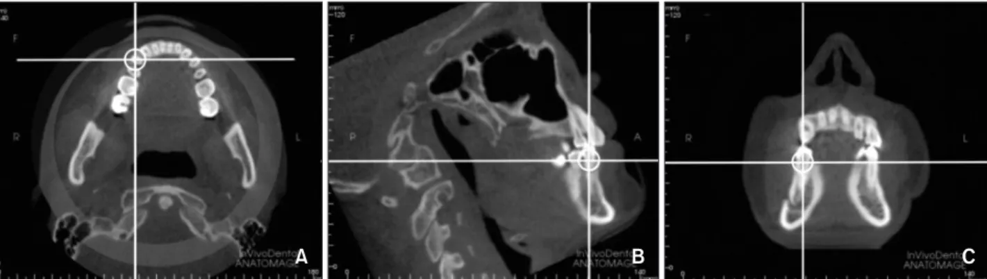
Methods: Cephalometric parameters were determined using cone-beam com- puted tomography in 120 Angle Class I subjects. Subjects were categorized as short, normal, and long face types according to the Frankfort mandibular angle. Parameters indicating the position of the mandibular right premolars and the mandible were also measured. Results: The angle between the mandibular first premolar axis and buccal cortex, the distance between the root apex and buccal cortex, angle of vestibularization, arc of vestibularization, and root apex maximum movable distance were significantly greater in the short face type than in the long and norm face types. The angle between the mandibular second premolar axis and buccal cortex, the distance from root apex to buccal cortex, and the arc of vestibularization were significantly greater in the short face type than in the normal face type. Conclusions: There are significant differences in the mandibular premolar positions in Class I subjects according to vertical facial type.
[Korean J Orthod 2015;45(4):180-189]
Key words: Cone-beam computed tomography, Mandibular premolar, Tooth position, Vertical facial type
Jun Duan
aFeng Deng
b,cWan-Shan Li
aXue-Lei Li
dLei-Lei Zheng
b,cGui-Yuan Li
aYan-Jie Bai
b,ca
Department of Stomatology, Ministry of Education Key Laboratory of Child Development and Disorders, Children's Hospital of Chongqing Medical University, Chongqing, China
b
Department of Orthodontics, College of Stomatology, Chongqing Medical University, Chongqing, China
c
Chongqing Key Laboratory for Oral Diseases and Biomedical Sciences, Chongqing Medical University, Chongqing, China
d
Department of Otorhinolaryngology, Children’s Hospital of Chongqing Medical University, Chongqing, China
Received August 17, 2014; Revised October 29, 2014; Accepted November 24, 2014.
Corresponding author: Yan-Jie Bai.
Assistant Professor, Department of Orthodontics, College of Stomatology, Chongqing Medical University, 426 Songshibei Road, Chongqing 401147, China.
Tel +86-23-88860107 e-mail [email protected]
*The study was supported by program for the natural science foundation of China (No.81470772 & No.31301021), the health bureau of Chongqing(No.2012-2-132 &
2012-2-218).
© 2015 The Korean Association of Orthodontists.
The authors report no commercial, proprietary, or financial interest in the products or companies described in this article.
This is an Open Access article distributed under the terms of the Creative Commons Attribution Non-Commercial License (http://creativecommons.org/licenses/by-nc/4.0) which permits unrestricted non-commercial use, distribution, and reproduction in any medium, provided the original work is properly cited.