이용한하악제3대구치부의부하악관평가
3
부산대학교 치의학전문대학원 구강악안면방사선학교실 조 봉 혜, 정 연 화
콘빔 CT를 이용한 하악 제3대구치부의 부하악관 평가
Assessment of accessory mandibular canal in mandibular third molars using cone- beam computed tomography
Department of Oral and Maxillofacial Radiology, School of Dentistry, Pusan National University Bong-Hae Cho, Yun-Hoa Jung
Purpose: This study was performed to examine distribution of accessory mandibular canal and its characteristics in mandibular third molars.
Materials and methods: A total of 251 subjects (166 males and 85 females) having mandibular third molars bilaterally were included in the study. Cone-beam computed tomographic images were reviewed for bifid or trifid accessory mandibular canal. The prevalence of accessory mandibular canal was evaluated according to gender, side and its branching type. Proximity and cross- sectional position of accessory mandibular canal to mandibular third molar was analyzed.
Results: Accessory mandibular canals were found in 66 (26.3%) of 251 patients and 86 (17.1%) of 502 hemi-mandibles. Gender and sides showed no statistically significant differences in prevalence. Retromolar canal (46.1%) was the most common branching type. Proximity of accessory canal to mandibular third molars showed mean distance of 2.8 mm from third molar and a statistically significant difference was found among types of accessory canal. Dental canal was the closest to tooth among branching types and closer to tooth than main canal. On cross-sectional view, accessory canal was generally located on buccal side of mandibular third molar.
Conclusion: Accessory mandibular canal was common and well detected with cone-beam computed tomography. Their localization is significant in all anesthetic and surgical procedures involving mandibular third molars.
Key words : bifid mandibular canal; cone-beam computed tomography; molar, third ABSTRACT
Corresponding Author Dr. Yun-Hoa Jung
(Department of Oral and Maxillofacial Radiology, School of Dentistry, Pusan National University, E-mail : [email protected])
* 이 논문은 부산대학교 자유과제 학술연구비(2년)에 의하여 연구되었음
Ⅰ. 서론
하악관은 하악공에서 이부로 하치조 신경혈관다발 이 주행하는 구조물로 일반적으로 단일구조물로 알려 져 있으나 이열 혹은 삼열하악관 등 부하악관이 꾸준 히 보고되어 왔다1~10). 부하악관은 하악 제3대구치 발 치, 이식 골 채취, 시상골절단술 등 하악골을 포함하 는 외과적 술식과 관련하여 과다 출혈이나 감각이상 등의 합병증을 야기할 수 있으며10, 11) 하치조 신경마취 에 어려움을 줄 수 있으므로12) 존재여부와 경로에 대 한 정보를 파악하여야 한다.
과거 파노라마 방사선사진을 이용한 연구에서는 이 열하악관의 발생빈도가 1% 미만으로 보고되었으나13,
14) 최근 콘빔 CT를 이용한 연구에서는 환자의 10.2~65%에서 이열 혹은 삼열하악관이 관찰된다는 연구결과가 보고되어1, 2, 4, 6, 8, 15~18) 부하악관이 상당히 흔히 나타나는 해부학적 변이임을 말해준다. 콘빔 CT 는 고해상도의 3차원 및 2차원 다면상이 가능하고 이 미지 뷰어상에서 판독자가 원하는 단면상을 실시간으 로 재구성할 수 있으므로 하악관의 분지를 쉽게 인식 할 수 있다. 대부분의 부하악관은 하악 제3대구치 후 방에서 분지되며 한국인의 평균 길이가 14.97㎜로 보 고2)된 바와 같이 주로 제3대구치부에서 끝나므로 제3 대구치의 발치나 이식골 채취 등 제3대구치 포함된 술 식을 시행할 때 특히 주의가 필요하다.
본 연구의 목적은 콘빔 CT를 이용하여 한국인에 있 어서 하악 제3대구치부에서의 부하악관의 발생빈도와 특성을 파악하고자 함에 있다.
Ⅱ. 재료 및 방법
1) 연구재료
연구재료는 부산대학교치과병원에서 2013년 1월부 터 2014년 5월 사이에 촬영한 환자의 콘빔 CT 영상
중 양측 하악 제3대구치가 모두 존재하는 20세에서 55세 사이(평균 31.2 세)의 총 251명(남 166명, 여 85명)의 환자 영상을 대상으로 하였다. 콘빔영상은 PaX-Zenith3D(Vatech, Kihung, Korea)을 이 용하여 촬영범위(field of view) 12x9cm, 관전압 120 kVp, 관전류 5-6mA, 24초의 노출시간으로 촬 영되었다. 영상은 촬영 즉시 재구성알고리즘을 이용 하여 14비트, 0.3㎣ 복셀의 해상도로 재구성되었다.
2) 부하악관 판독
Ez3D 2009 3D image viewer(Vatech, Kihung, Korea) 상에서 실시간으로 3차원 영상과 2차원 다면영상을 재구성하여 판독하였다. 판독은 10 년 이상의 경험을 가진 구강악안면방사선 전공자 1명 이 Coronis 5MP monitors(Barco, Brussels, Belgium 2048 × 2560 image matrices, 10- bit viewable gray scale, and 145.9-ft- lambert luminescence) 상에서 시행하였으며 영 상 외에 환자에 대한 정보는 제공되지 않았다.
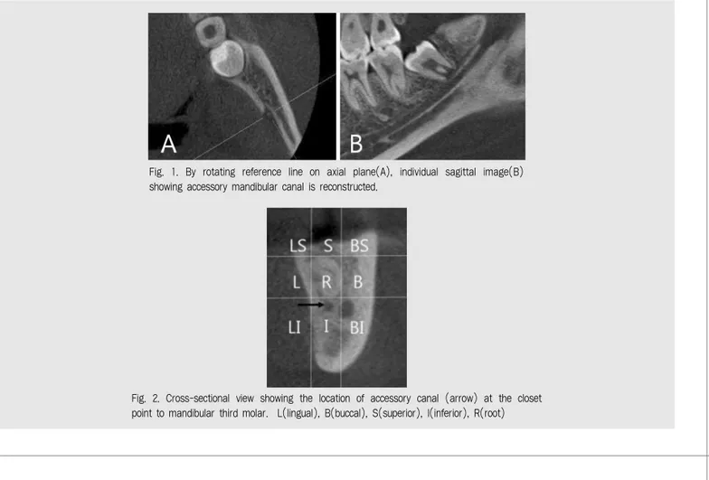
부하악관은 하악관에서 분지되는 피질골 경계를 보 이는 소관이 관찰될 경우‘있음’으로 판정하였다. 부 하악관을 확인하기 위하여 먼저 축상면의 하악공에서 기준축을 회전하여 시상단면에서 하악관이 분명하게 관찰되는 개별 시상단면을 얻었다. 그 후 시상단면상 을 협설측으로 연속적으로 이동시키면서 하악관에서 분지되는 소관이 있는지 관찰하였고 횡단면상에서도 전후방으로 축을 이동시켜 하악관의 분지를 확인하였 다(Fig. 1). 판독은 축상면, 시상면, 횡단면의 다면 2 차원 영상을 사용하였으며 원하는 단면상을 얻기 위한 기준축의 자유로운 설정이 허용되었으며 확대, 대조 도, 밝기의 조정도 허용되는 등 이미지 뷰어 프로그램 상의 기능에 제한을 두지 않았다.
3) 부하악관 분류
부하악관은 주관(main canal)에서 분지되는 양상
이용한하악제3대구치부의부하악관평가 에 따라 Naitoh 등8)의 분류기준에 예비연구에서 관
찰된 주하악관 하방으로 분지되는 inferior canal을 추가하여 5가지 유형으로 분류하였다.
분류기준은 다음과 같다.
1) Retromolar canal ; 부하악관이 후삼각 치조 정부위로 개구
2) Forward canal ; 부하악관이 상전방으로 주행 하다가 악골 내에서 정지
3) Dental canal ; 제2, 제3 대구치 치근부로 주행 4) Buccolingual canal ; 부하악관이 주하악관
의 협측 혹은 설측으로 주행
5) Inferior canal ; 부하악관이 주하악관 하방으 로 주행
4) 부하악관의 하악 제3대구치 근접도 평가 다면 재구성 영상에서 부하악관이 하악 제3대구치 에 가장 근접하는 지점까지의 거리를 계측하여 치아
근접도를 평가하였다. 계측은 두 번 시행하여 평균치 를 사용하였으며 두 번의 계측치 차가 0.2㎜ 보다 클 경우에는 재계측을 시행하였다. 또한 주하악관에서 하악 제3대구치까지의 거리를 계측하여 주-부하악관 의 치아 근접도 차를 정량적으로 평가하고 이를 사용 하여 부하악관이 주하악관보다 제3대구치에 더 근접 한지 더 떨어져 있는지의 정성적 평가도 시행하였다.
5) 절단면에서의 부하악관의 위치 평가
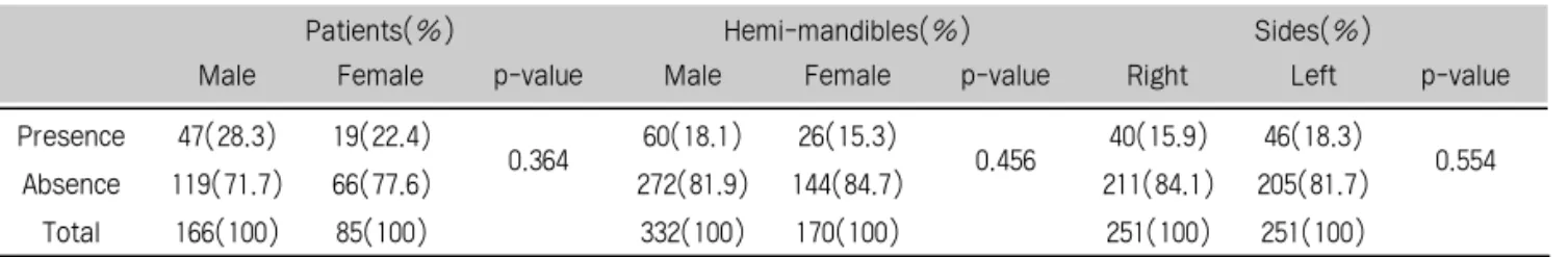
축상면상에서는 하악체에 평행하게, 시상면과 횡단 면에서는 각각 하악 제2대구치의 협설 및 근원심 장축 에 평행하게 기준축을 설정한 뒤 부하악관이 제3대구 치부위에 가장 근접한 횡단면을 기준 절단면으로 설정 하였다. 기준 절단면상에서 치아의 협설상하의 수직 및 수평 접선을 설정하여 치아에 대한 부하악관의 위치를 그림 2에서와 같이 9부위로 분류하였다(Fig. 2).
Fig. 1. By rotating reference line on axial plane(A), individual sagittal image(B) showing accessory mandibular canal is reconstructed.
Fig. 2. Cross-sectional view showing the location of accessory canal (arrow) at the closet point to mandibular third molar. L(lingual), B(buccal), S(superior), I(inferior), R(root)
6) 하악 제3대구치 평가
하악 제3대구치는 맹출 여부에 따라 맹출, 부분맹 출, 매복으로 분류하였고, 악골내 위치에 따라 수직, 수평, 근심경사로 분류하였다. ‘수직’은 하악 제2대구 치 장축에 근원심 10도이내의 장축각도를 가질 때,
‘수평’은 하악 제2대구치 장축의 수직선에 상하 10도 이내의 장축각도를 가질 때였으며, 그 이외는‘근심경 사’로 분류하였다. 예비연구에서 원심경사나 역위 매 복된 치아는 없었으므로 제외하였다.
7) 통계분석
부하악관의 발생빈도의 성별, 좌우 차이는 χ2 test 로 분석하였다. 부하악관의 유형, 치아위치, 맹출 여 부에 따른 부하악관의 치아 근접도는 analysis of variance(ANOVA)로 분석하였고, 주-부하악관의 치아 근접도 차는 analysis of variance(ANOVA) 를 이용한 정량적 분석과 χ2 test를 이용한 정성적 분 석을 시행하였다. 모든 통계는 PASW Statistics
version 18 software (SPSS Inc, Chicago, Ill, USA)를 이용하였으며 통계학적 유의성은 .05로 고 정하였다.
Ⅲ. 결과
1) 부하악관 발생빈도
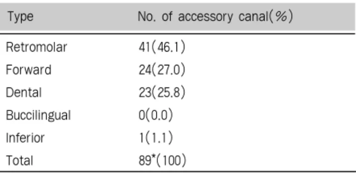
부하악관은 총 251명의 환자 중 66명의 환자에서 관찰되어 26.3%의 발생빈도를 나타내었으며, 20명 의 환자가 양측성을 보여 부하악관을 가진 편측 하악 은 모두 86개로 평균 17.1%의 발생빈도를 보였다. 대 부분 이열하악관이었으나 삼열하악관이 3례 관찰되었 다. 남자가 여자보다 높은 발생빈도를 보였으나 통계 학적으로 남녀간 유의성있는 차이는 없었으며 좌우의 발생빈도도 유의성 있는 차이가 나타나지 않았다 (Table 1&2).
Table 1. Prevalence of accessory mandibular canal according to gender and sides
Presence 47(28.3) 19(22.4)
0.364 60(18.1) 26(15.3)
0.456 40(15.9) 46(18.3)
0.554
Absence 119(71.7) 66(77.6) 272(81.9) 144(84.7) 211(84.1) 205(81.7)
Total 166(100) 85(100) 332(100) 170(100) 251(100) 251(100)
Patients(%) Hemi-mandibles(%) Sides(%)
Male Female p-value Male Female p-value Right Left p-value
Table 2. Distribution of bifid and trifid canal
Bifid
Unilateral 34(72.3) 11(57.9) 45(68.2)
Bilateral 12(25.3) 7(36.8) 19(28.8)
Trifid
Unilateral 0(0.0) 1(5.3) 1(1.5)
Bilateral 1(2.1) 0(0.0) 1(1.5)
Total 47(100) 19(100) 66(100)
No. of accessory canal(%)
Male (n=47) Female(n=19) Total(n=66)
이용한하악제3대구치부의부하악관평가
2) 부하악관 분지 유형
삼열하악관은 각각 2개의 부하악관으로 분류하여 총 89개의 부하악관이 연구에 사용되었다. 부하악관 의 분지 유형은 retromolar canal이 41례(46.1%) 로 가장 많았고, 하방으로 분지하는 inferior canal 이 1례 관찰되었으나 buccolingual canal은 관찰 되지 않았다(Table 3). 부하악관 분지의 4가지 유형 은 그림 3에서 보는 바와 같다(Fig. 3). 대부분의 dental canal은 후방에서 분지하여 제3대구치 치근 으로 수렴하였지만 특이하게 제3대구치 전방에서 분
지하여 제2대구치 치근으로 주행하는 경우도 1례 관 찰되었다(Fig. 4).
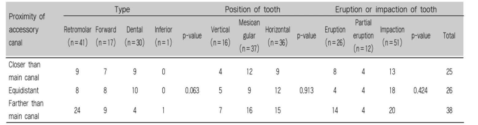
3) 부하악관의 치아근접도
부하악관에서 하악 제3대구치에서까지의 최단거리 는 평균 2.8㎜였으며, dental canal 유형이 평균 0.8㎜로 치아와 가장 가까웠다. 주하악관과 부하악관 에서 제3대구치까지의 거리 비교에서는 부하악관이 주하악관보다 평균 1.1㎜ 더 멀었으나 dental canal 은 주하악관 보다 평균 0.4 ㎜ 치아에 더 가까운 것으 Table 3. Rate of types in 89 accessory mandibular canals
Retromolar 41(46.1)
Forward 24(27.0)
Dental 23(25.8)
Buccilingual 0(0.0)
Inferior 1(1.1)
Total 89*(100)
Type No. of accessory canal(%)
* Each of 3 trifid canals has 2 accessory canals and the number of the accessory canals totals 89 from 86 hemi-mandibles.
Fig.3. Four types of accessory canal(arrow); A. retromolar, B. forward, C. dental, D. inferior
Fig. 4. Accessory canal(arrow) branching anteriorly from the mandibular third molar and reaching the root of mandibular second molar
로 나타났다. 부하악관의 치아 근접도와 주-부하악관 의 근접도 차의 정량적 분석은 부하악관의 유형에 따 라 통계적으로 유의성 있는 차이를 보였으나 치아의 위치, 맹출여부는 유의성있는 차이를 보이지 않았다 (p<.05, Table 4). 주-부하악관의 근접도 차의 정성 적 분석에서는 총 25개(28.1%)의 부하악관이 주하악 관보다 치아에 더 근접한 결과를 보였지만 통계적 유 의성은 나타나지 않았다(Table 5).
4) 절단면에서의 부하악관 위치
89개의 부하악관 중 1례는 제3대구치 전방에서 분 지를 보였고 17례는 종말부가 제3대구치 후방에서 끝 나 제 3대구치가 관찰되는 절단면상에서 관찰할 수 없 었다. 제3대구치 절단면에서 관찰된 71례중 49례가
제3대구치의 협측에서 관찰되었고 단 4례만이 설측에 위치한 것으로 나타났으며 7례가 치아의 상방에서, 11 례가 치아의 하방에서 관찰되었다(Table 6).
Ⅳ. 고찰
이열 혹은 삼열하악관으로 불리는 부하악관은 발치, 임플란트 식립, 이식골 채취 등의 외과적 술식과 관련 하여 불완전한 마취를 야기하거나 시술과정 중의 과다 출혈, 시술 후 감각이상 등의 합병증을 야기할 수 있으 므로 술전에 주하악관의 주행경로와 함께 그 존재여 부나 위치를 파악하여야한다8, 10, 11). 악골의 해부학적 변이를 정확히 관찰하기 위하여 주로 사용되는 촬영법
Table 4. Proximity of accessory canal to mandibular third molar.
Proximity to
3.7±3.5 3.3±2.5 0.8±2.2 4.8±0.0 0.003* 3.7±2.6 2.7±2.7 2.6±3.7 0.446 3.8±2.7 2.6±1.6 2.4±3.5 0.185 2.8±3.1 tooth† (mm)
Difference of
-2.1±3.8 -0.6±2.6 0.4±2.5 -4.8±0.0 0.019* -1.1±2.6 -0.8±2.8 -1.4±4.1 0.765 -1.6±2.7 0.4±2.9 -1.1±3.6 0.219 -1.1±3.3) proximity‡ (mm)
Type Position of tooth Eruption or impaction of tooth
Retromolar Forward Dental Inferior Vertical Mesioan
Horizontal Eruption Partial Impaction (n=41) (n=17) (n=30) (n=1) p-value
(n=16) gular
(n=36) p-value
(n=26) eruption
(n=51) p-value Mean
(n=37) (n=12)
† Proximity to tooth was the closest distance of accessory canal to the mandibular third molar.
‡ Difference of proximity was the value gained by subtracting the proximity of accessory canal to tooth from that of main canal. Minus value means accessory canal is farther to tooth than main canal.
* Statistically significant (P<.05)
Table 5. Comparison of proximity of main and accessory canal to mandibular third molar.
Closer than
9 7 9 0 4 12 9 8 4 13 25
main canal
Equidistant 8 8 10 0 0.063 5 9 12 0.913 4 4 18 0.424 26
Farther than
24 9 4 1 7 16 15 14 4 20 38
main canal
Proximity of Type Position of tooth Eruption or impaction of tooth
accessory Retromolar Forward Dental Inferior Vertical Mesioan
Horizontal Eruption Partial Impaction canal (n=41) (n=17) (n=30) (n=1) p-value
(n=16) gular
(n=36) p-value
(n=26) eruption
(n=51) p-value Total
(n=37) (n=12)
이용한하악제3대구치부의부하악관평가
은 멀티 슬라이스 CT와 콘빔 CT이다. 콘빔 CT는 멀 티 슬라이스 CT에 비하여 경제적이고 비교적 저선량 의 방사선을 사용하여 고해상도의 다면영상을 얻을 수 있는 장점이 있으며 하악관의 경로나 해부학적 변이를 평가하는데 있어 멀티 슬라이스 CT보다 우수함이 보 고되었다7).
콘빔 CT를 이용한 연구1, 2, 4, 6, 8, 15~18)에서 이열하악관 의 발생빈도는 10~65%까지 상당한 편차를 보이는데 이러한 발생빈도의 차이는 유전적, 환경적 요소 뿐 아 니라 표본 선택, 직경 및 피질골 유무 등 판정기준이 상이하기 때문이다. 본 연구에서는 환자의 26.3%에 서 부하악관이 관찰되었는데 이는 한국인을 대상으로 한 강 등2)의 연구(10.2%)와 Rashsuren 등1)의 연구 (22.6%)결과 보다 높았다. 편측 하악을 기준으로 한 발생빈도는 17.1%로 Rashsuren 등2)의 연구 (16.2%)와 유사한 결과를 보였다. Fu 등5)은 여자보 다 남자의 발생빈도가 더 높았다고 보고하였지만 본 연구에서는 다른 대부분의 연구1~3, 8, 18)에서와 마찬가 지로 성별이나 좌우의 발생빈도는 통계학적으로 유의 성있는 차이를 보이지 않았다. 본 연구에서 편측성의
이열하악관이 가장 흔하게 나타났지만 502개의 편측 하악 중에서 3례의 삼열하악관이 관찰되었고, 755개 의 편측 하악을 대상으로 한 Rashsuren 등2)의 연구 에서도 7례가 보고되어 삼열하악관이 드물기는 하지 만 가끔 관찰되는 소견으로 생각된다.
부하악관의 분지유형에 있어서 Orhan 등17) 및 Naitoh 등8)은 forward canal이 가장 높은 빈도로 나타났다고 보고하였지만, 본 연구를 포함한 한국인 대상의 연구1, 2)에서는 retromolar canal이 가장 흔 히 관찰되었다. 본 연구나 Rashsuren 등1)의 연구에 서는 buccolingual canal이 관찰되지 않았으나 강 등2)의 연구에서는 빈도는 아주 낮지만 4례가 보고된 것으로 보아 표본 수가 증가한다면 buccolingual canal도 관찰 가능할 것으로 판단된다. 하악 제3대구 치에 대한 근접도 평가에서 dental canal이 다른 유 형에 비하여 가장 근접한 결과를 보였다. 특히 주-부 하악관의 근접도를 비교했을 때 총 23례의 dental canal 중 19례가 주하악관과 같거나 주하악관보다 제3대구치에 더 근접한 결과를 보여 부하악관 유형 중 에서 dental canal의 위치평가가 특히 중요한 의미 Table 6. Location of accessory canal at the closest point to
mandibular third molar on cross-sectional view
Bucco-superior 11(12.4)
Buccal 28(31.5)
Bucco-inferior 10(11.2) Linguo-superior 1(1.1)
Lingual 3(3.4)
Linguo-inferior 0(0.0)
Superiorl 7(7.9)
Inferior 11(12.4)
Anterior* 1(1.1)
Posterior* 17(19.1)
Total 89(100)
Location Number(%)
* Accessory canals positioned anterior or posterior to mandibular third molar were not presented on cross-sectional view.
를 가질 것으로 생각된다. dental canal의 종말부는 대구치의 치근과 완전히 접촉하는 경우도 있었지만 대 부분은 치근과의 사이에 피질골이 개재되어 있어 치근 첨과 약간 떨어져 있는 경우가 많았다. 본 연구에서는 다른 연구에서 보고된 적이 없었던 inferior canal 도 관찰되었는데 이 유형은 주하악관 하방으로 주행하 므로 술식과 관련한 부작용은 드물 것으로 생각된다.
절단면 상에서 제3대구치와 부하악관의 위치를 평가 한 결과, 주로 치아의 협측에 위치하고 있었으므로 협 측골을 채취하는 이식골 채취술을 시행할 때 부하악관 의 위치에 대한 고려가 필요하다. 수평매복이나 근심
경사 치아의 경우에는 부하악관이 치근의 위 혹은 아 래에서 관찰되었다.
본 연구는 한국인에 있어 부하악관의 발생빈도 및 하악 제3대구치와의 위치관계에 대한 정보를 얻고자 이루어졌다. 본 연구 결과는 부하악관이 비교적 흔하 게 나타나며 일부는 하악 제3대구치에 상당히 근접해 있음을 보여준다. 하악 제3대구치는 종종 발치나 다 른 외과적 술식의 대상부위가 되므로 임상의들은 외과 적 술식을 시행하기 전에 콘빔 CT 등 고해상도의 영 상을 이용하여 부하악관의 존재 경로를 사전에 인지하 는 것이 필요하다.
이용한하악제3대구치부의부하악관평가
1. Rashsuren O, Choi JW, Han WJ, Kim EK.
Assessment of bifid and trifid mandibular canals using cone-beam computed tomography. Imaging Sci Dent. 2014;44(3):229-36.
2. Kang JH, Lee KS, Oh MG, Choi HY, Lee SR, Oh SH, Choi YJ, Kim GT, Choi YS, Hwang EH. The incidence and configuration of the bifid mandibular canal in Koreans by using cone-beam computed tomography. Imaging Sci Dent. 2014;44(1):53-60.
3. Kuczynski A, Kucharski W, Franco A, Westphalen FH, de Lima AA, Fernandes A. Prevalence of bifid mandibular canals in panoramic radiographs: a maxillofacial surgical scope. Surg Radiol Anat. 2014
;36(9):847-50.
4. Lizio G, Pelliccioni GA, Ghigi G, Fanelli A, Marchetti C.
Radiographic assessment of the mandibular retromolar canal using cone-beam computed tomography. Acta Odontol Scand. 2013;71(3-4):650-5.
5. Earl Fu, Michelle Peng, Cheng-Yang Chiang, Hsiao- Pei Tu, Yaoh-Shiang Lin, E-Chin Shen. Bifid mandibular canals and the factors associated with their presence: a medical computed tomography evaluation in a Taiwanese population. Clin. Oral Impl.
Res. 2014;25(2):e64-7.
6. Kuribayashi A, Watanabe H, Imaizumi A, Tantanapornkul W, Katakami K, Kurabayashi T. Bifid mandibular canals: cone beam computed tomography evaluation. Dentomaxillofac Radiol.
2010;39(4):235-9.
7. Naitoh M, Nakahara K, Suenaga Y, Gotoh K, Kondo S, Ariji E. Comparison between cone-beam and multislice computed tomography depicting mandibular neurovascular canal structures. Oral Surg Oral Med Oral Pathol Oral Radiol Endod. 2010;109(1):e25-31.
8. Naitoh M, Hiraiwa Y, Aimiya H, Ariji E. Observation of bifid mandibular canal using cone-beam computerized tomography. Int J Oral Maxillofac Implants. 2009;24(1):155-9.
9. 이현우, 김여갑, 이백수,권용대, 최병준,김영란. 이 열하악관(Bifid mandibular canal): 방사선 소견과 임상적 의의 -증례보고- 대한치과마취과학회지.
2009;9(1):24-9.
10. Kaufman E, Serman NJ, Wang PD. Bilateral mandibular accessory foramina and canals: a case report and review of the literature. Dentomaxillofac Radiol. 2000;29(3):170-5.
11. Bilecenoglu B, Tuncer N. Clinical and anatomical study of retromolar foramen and canal. J Oral Maxillofac Surg. 2006;64(10):1493-1497.
12. Grover PS, Lorton L. Bifid mandibular nerve as a possible cause of inadequate anesthesia in the mandible. J Oral Maxillofac Surg. 1983;41(3):177-9.
13. Nortjé CJ, Farman AG, Grotepass FW. Variations in the normal anatomy of the inferior dental (mandibular) canal: a retrospective study of panoramic radiographs from 3612 routine dental patients. Br J Oral Surg. 1977;15:55-63.
14. Langlais RP, Broadus R, Glass BJ. Bifid mandibular canals in panoramic radiographs. J Am Dent Assoc. 1985;110(6):923-6.
15. Patil S, Matsuda Y, Nakajima K, Araki K, Okano T.
Retromolar canals as observed on cone-beam computed tomography: their incidence, course, and characteristics. Oral Surg Oral Med Oral Pathol Oral Radiol. 2013;115(5):692-9.
16. de Oliveira-Santos C, Souza PH, de Azambuja Berti-Couto S, Stinkens L, Moyaert K, Rubira- Bullen IR, Jacobs R. Assessment of variations of the mandibular canal through cone beam computed tomography. Clin Oral Investig.
2012;16(2):387-93.
17. Orhan K, Aksoy S, Bilecenoglu B, Sakul BU, Paksoy CS. Evaluation of bifid mandibular canals with cone-beam computed tomography in a Turkish adult population a retrospective study. Surg Radiol Anat. 2011;33(6):501-7.
18. von Arx T, Hänni A, Sendi P, Buser D, Bornstein MM. Radiographic Study of the Mandibular Retromolar Canal An Anatomic Structure with Clinical Importance. J Endod. 2011;37(12):1630-5.
참 고 문 헌