한국인의하악절치관평가
5
부산대학교 치의학전문대학원 영상치의학교실 조 봉 혜, 정 연 화
콘빔 CT를 이용한 한국인의 하악 절치관 평가
Assessment of mandibular incisive canal using cone-beam computed tomography in Korean population
Department of Imaging dentistry, School of Dentistry, Pusan National University Bong-Hae Cho, Yun-Hoa Jung
Purpose: This study was performed to investigate the characteristics of mandibular incisive canal (MIC) in Korean population.
Materials and methods: A total of 97 subjects (60 males and 37 females) who underwent cone-beam computed tomography were included in the study. The anatomic features of MIC was assessed according to gender. Length, diameter and distance to inferior, lingual and buccal border were measured at the origin and the terminal. Also the distribution of MIC at each tooth position was evaluated.
Results: Of 97 patients included, 75(77.3%) presented bilateral MIC and 13(13.4%) presented unilateral MIC. Of 194 hamimandibles, MIC was detected in 102(85%) sites in male and 61(82.4%) sites in female. Gender and side showed no statistically significant differences in detectability. The length, diameter and distance to adjacent structures were bigger in male than in female except the distance to lingual border. MIC travelled anteriorly in a slightly downward and lingual direction and usually terminated between the first premolar and the canine. On cross-sectional view, MIC showed individually scattered distribution both buccolingually and superoinferiorly.
Conclusion: MIC is well detected with cone-beam computed tomography. Considered that the length and the location of MIC has large variations between individuals, its localization using cone-beam CT is highly recommended before performing surgical procedures such as implant placement and bone harvesting.
Key words : Mandible; mandibular nerve; cone-beam computed tomography ABSTRACT
Corresponding Author Dr. Yun-Hoa Jung
(Department of imaging dentistry, School of Dentistry, Pusan National University, E-mail : [email protected])
* 이 논문은 2015년 부산대학교치과병원 임상연구비 지원으로 이루어졌음
Ⅰ. 서론
하악 절치관은 이공 전방으로 연장되는 하악관의 종말부로 제1소구치, 견치 및 하악 절치부에 신경과 혈관을 분포시킨다1). 따라서 하악 소구치 사이 하악 전방부에 임플란트 식립이나 이식골 채취 등의 외과 적 술식을 시행하기 전에 하악 절치관의 위치와 경로 를 평가하는 것이 필요하다. 사체 하악을 이용한 해부 학적 연구2, 3)에서 하악 절치 신경혈관다발이 모든 하 악에서 존재한다고 보고되었고, 건조 하악을 이용한
연구4, 5)에서 하악 절치관이 대부분의 하악에서 관찰
되었다고 보고되었다. 방사선 영상을 이용한 연구에 서 파노라마방사선촬영술6~10)은 하악 절치관의 탐지 율이 매우 낮았다는 결과를 보여 최근의 연구는 대부 분 콘빔 CT영상이 이용되고 있다. 콘빔 CT는 골해상 도가 높고, 비교적 적은 선량으로 3차원 및 2차원 다 면상이 가능하며 계측정확도도 높아서 하악 절치관의 임상적 평가에 매우 유용하다. 콘빔 CT를 이용한 연 구에서 하악 절치관의 관찰빈도는 80%이상으로 보 고되었다6, 7, 11~16).
해부학적 구조는 인종적 차이를 보이므로 한국인을 대상으로 한 연구가 필요하다. 한국인 대상의 하악 절 치관 연구는 CT를 이용한 김 등17)의 연구가 있으나 활 발한 연구는 이루어지지 않고 있다. 본 연구의 목적은 콘빔 CT를 이용하여 한국인에 있어서 절치관의 관찰 빈도와 해부학적 특성을 파악하고자 함에 있다.
Ⅱ. 재료 및 방법
연구재료는 부산대학교 치과병원에 내원하여 진단 적 목적으로 콘빔 CT를 촬영한 환자의 영상으로 골 병소나 발육성 이상이 있는 경우와 제3대구치 외에 치 아 상실이 있는 영상은 제외하였다. 연구 대상 환자는 총 97명(남 60, 여 37)으로 20세에서 54세 사이(평
균 29.4 세)였다.
콘 빔 CT영 상 은 PaX-Zenith3D(Vatech, Kihung, Korea)을 이용하여 촬영범위(field of view) 16x14cm, 관전압 120kVp, 관전류 5-6 mA, 24초의 노출시간으로 촬영되었다. 영상은 촬영 즉시 재구성알고리즘을 이용하여 14비트, 0.3㎣ 복셀 의 해상도로 재구성되었으며 판독 시에는 Ez3D 2009 3D image viewer(Vatech, Kihung, Korea) 상에서 실시간으로 3차원영상과 2차원 다면 영상을 재구성하였다.
판 독 은 Coronis 5MP monitors(Barco, Brussels, Belgium 2048 × 2560 image matrices, 10-bit viewable gray scale, and 145.9-ft-lambert luminescence) 상에서 시행 하였으며 영상 외에 환자에 대한 정보는 제공되지 않 았다. 판독은 다면 2차원영상을 자유로이 사용하였으 며 확대, 대조도, 밝기의 조정도 허용되었다. 하악 절 치관의 유무 판독과 계측은 10년 이상의 경험을 가진 구강악안면방사선 전공자 2명의 동의로 이루어졌다.
1. 하악 절치관의 탐지 ; 하악관이 전방으로 주행하 다가 후상방의 이관으로 이행하는 부위에서 분지 되어 전방으로 계속 주행하는 신경관이 관찰될 때 하악 절치관이 있다고 판단하였다. 일차적으로 시상면에서 관찰한 후 횡단면상에서 하악관과의 연속성을 확인하였으며 절치관의 피질골이 더 이 상 관찰되지 않는 지점을 종말부로 판단하였다.
2. 계측 ; 관상면에서 하악제1소구치 치근부의 협설 피질골에 수직이며 시상면과 횡단면에서 하악 제 1소구치 장축을 지나는 선을 설정한 후 관상면에 서 하악궁을 따라 panoramic curve를 그려 시상면과 횡단면을 얻었다. 시상면에서 절치관 의 길이를 계측하였으며 횡단면에서 기시부와 종 말부에서의 절치관 내경, 하악 하연까지의 거리, 설측 및 협측 피질골까지의 거리를 계측하였다.
치아의 치근단 하방으로 절치관이 지나가면 그
한국인의하악절치관평가 치아의 횡단면에서도 계측을 시행하였다.
각 계측항목은 다음과 같다(Fig 1).
1) 길이 ; 하악관에서 이관과 절치관이 분지되는 지 점(기시부)에서 전방의 끝나는 지점(종말부)까지 의 직선거리
2) 폭경 ; 절치관의 협설 내경
3) 하악 하연까지의 거리 ; 절치관의 최하방 지점에 서 하악 하연까지의 수직거리
4) 설측 피질골까지의 거리 ; 절치관의 최설측 지점 에서 설측 피질골까지의 수평거리
5) 협측 피질골까지의 거리 ; 절치관의 최협측 지점 에서 협측 피질골까지의 수평거리
6) 치근첨까지의 거리 ; 절치관의 최상방 지점에서 치근첨까지의 수직거리
3. 치근단면에서의 절치관의 분포
치근단과 절치관의 위치관계를 보기 위하여 치근단 면에서의 절치관의 위치를 산포도로 나타내었다. 하 악 하연까지의 거리는 실계측치이며 협설분포는 설측 및 협측 피질골까지의 실계측치를 10mm의 협설 폭 경으로 환산한 상대적 위치이다.
통계학적 분석은 PASW Statistics version 18
software(SPSS Inc, Chicago, Ill, USA)를 이용 하여 χ2 test로 남녀간, 좌우간 차이를, paired t- test로 기시부와 종말부의 차이를 분석하였다. 통계 학적 유의성은 .05로 고정하였다.
Ⅲ. 결과
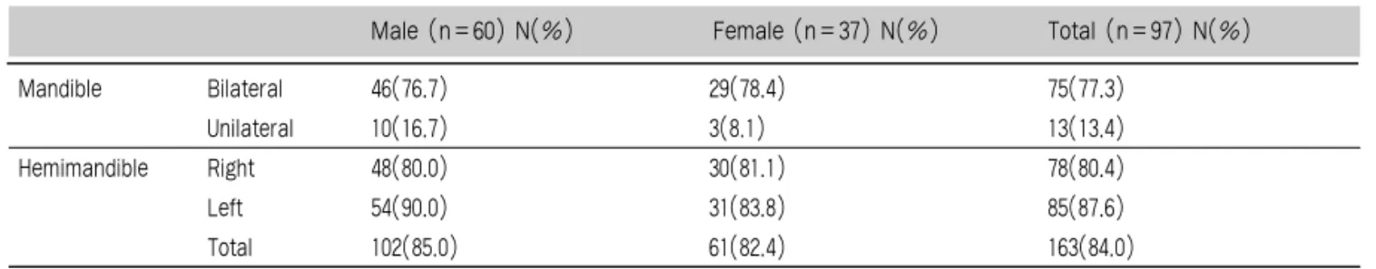
하악 절치관이 양측 모두에서 관찰된 환자는 75명 (77.3%)이었으며 총 194 편악 중 163 편악(84.0%) 에서 하악 절치관이 확인되었다. 남녀나 좌우에 따른 관찰빈도의 통계학적인 차이는 없었다(Table 1).
절치관의 길이는 최소 2.0mm에서 최대 20.5mm 까지의 범위를 보였으며 평균길이는 남자 6.71mm, 여자 5.80mm로 남자에서 더 길었다(p<.05). 대부 분의 계측항목에서 남자가 여자보다 크게 나타났으며 기시부에서는 절치관의 폭경, 하악 하연까지의 거리, 설측 피질골까지의 거리가, 종말부에서는 하악 하연 까지의 거리와 협측 피질골까지의 거리가 통계학적 유 의성을 보였다(Table 2, p<.05). 기시부와 종말부의 비교에서 남자의 설측 피질골까지의 거리 항목을 제외
Fig. 1. a. Length(L) of MIC, b. Diameter(D) and distance from MIC to root tip(R), inferior border(I), lingual(L) and buccal(B) plate.
하고는 모두 유의성있는 차이를 보였는데 기시부에 비 하여 종말부에서 폭경이 줄었고 하악 하연까지의 거리 가 짧아졌으며 협측 피질골까지의 거리가 더 커져 기 시부에서 종말부로 갈수록 약간 하방, 설측으로 주행
하였다(Table 2, p<.05).
치아와의 관계 평가에서 하악 제1소구치 하방에서 관찰된 절치관은 194 편악 중 145(74.7%)개로 13부 위는 절치관의 길이가 너무 짧아서 관찰되지 않았고, Table 1. Detectability of mandibular incisive canal(MIC) on CBCT
There were no statistically significant differences in incidence by gender and sides.
Mandible Bilateral 46(76.7) 29(78.4) 75(77.3)
Unilateral 10(16.7) 3(8.1) 13(13.4)
Hemimandible Right 48(80.0) 30(81.1) 78(80.4)
Left 54(90.0) 31(83.8) 85(87.6)
Total 102(85.0) 61(82.4) 163(84.0)
Male (n=60) N(%) Female (n=37) N(%) Total (n=97) N(%)
Table 2. Length, diameter and distance of MIC to inferior, lingual and buccal border at the origin and the terminal
* statistically significant differences between male and female (p <.05).
† statistically significant differences between the origin and the terminal (p <.05).
Canal Length 6.71(3.05) 5.80(2.48)*
Canal diameter 2.99(0.46) 2.64(0.33)*
Origin Distance to Inferior border 10.22(1.42) 9.20(1.13)*
Distance to lingual border 4.99(0.96) 5.34(1.08)*
Distance to buccal border 2.61(0.66) 2.51(0.67)
Canal diameter 1.77(0.32)† 1.71(0.28)†
Terminal Distance to Inferior border 9 (1.45)† 8.78(1.47)*,†
Distance to lingual border 4.84(0.84) 5.11(1.12)†
Distance to buccal border 3.72(1.07)† 3.08(0.72)*,†
Measurement(mm) Male Mean(SD) Female Mean(SD)
Table 3. Distance of MIC to root apex, inferior, lingual and buccal border at each tooth
Distance to 12.6(1.4) 12.4(1.5) 8.3(2.2) 7.0(2.8) 8.2(2.4) 7.1(2.0)
root apex 10.3-14.2 10.9-13.8 4.0-11.7 3.7-11.8 4.7-14.9 3.0-11.3
Distance to 9.1(2.5) 8.3(2.0) 9.2(1.2) 8.7(2.0) 10.1(1.4) 9.2(1.4)
Inferior border 6.5-12.6 6.3-10.2 6.5-10.8 6.1-12.3 7.5-13.1 6.5-15.5
Distance to 6.0(1.1) 6.2(1.4) 5.1(1.0) 5.1(0.9) 4.9(1.0) 5.3(1.1)
lingual border 4.6-7.0 4.8-7.5 2.8-7.0 3.8-7.0 3.2-8.4 3.2-8.4
Distance to 5.5(1.7) 4.1(1.1) 4.0(1.3) 3.0(0.7) 2.8(0.7) 2.7(0.6)
buccal border 2.7-7.2 3.0-5.2 1.8-6.7 1.7-4.1 1.4-4.8 1.6-4.2
Lateral incisor Canine First premolar
Measurement Male (n=5) Female (n=2) Male (n=23) Female (n=10) Male (n=91) Female (n=54)
(mm) Mean(SD) Mean(SD) Mean(SD) Mean(SD) Mean(SD) Mean(SD)
Range Range Range Range Range Range
한국인의하악절치관평가 5부위는 절치관이 하악 제1소구치 전방에서 분지하고
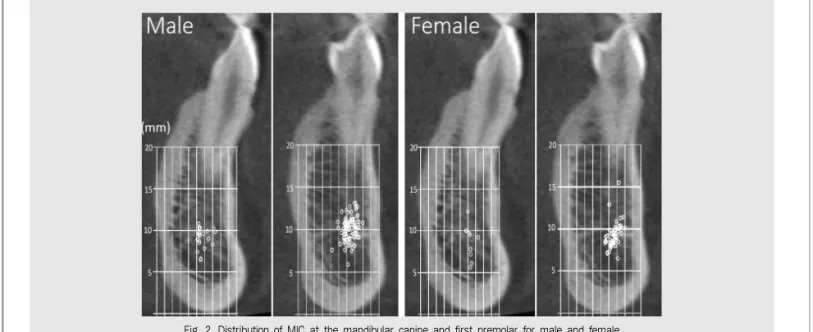
있었다. 견치는 33부위(17.0%)에서, 측절치는 7부위 (3.6%)에서 절치관이 관찰되었다. 하악 절치관과 해 부학적 구조물까지의 거리는 개인마다 상당한 차이를 보였다(Table 3). Fig. 2는 제1소구치와 견치부에서 의 절치관의 분포를 보여준다. 전반적으로 제1소구치 부에 비하여 견치부에서 절치관이 더 하방, 더 설측에 위치하고 있으나 분포가 매우 산재된 양상을 보였다.
Ⅳ. 고찰
하악 정중부는 종종 골 이식 공여부로 이용되는 부 위이다18). 이공 사이 하악 전방부에 임플란트를 식립 하였을 때 감각이상이나 불편감을 호소하는 증례가 보
고되면서19, 20)하악 절치관의 해부학적 특징에 대한 관
심이 높아졌다. 골 채취술이나 임플란트 시술과 관련 하여 하악 절치관이 손상되면 출혈이나 감각이상 등의 합병증이 발생할 수 있으므로 외과적 술식을 시행하기 전에 하악 절치관에 대한 평가가 필요하다. 임상적 평 가는 주로 방사선사진을 이용하여 이루어지며 현재로
서는 콘빔 CT가 선량, 편의성, 화질의 측면에서 가장 유용한 것으로 생각된다. 이런 이유로 본 연구는 콘빔 CT를 이용하였으며 해면골 내의 경로는 골소주와 감 별이 어려우므로 이관에서 전방으로 연장되는 피질골 이 관찰될 때 절치관으로 간주하였고 피질골이 소실되 는 부위를 종말부의 기준으로 삼았다.
건조 하악을 이용한 연구에서 Obradovic 등5)은 유 치악에서는 92%에서 절치관이 분명하게 관찰되었으 나 무치악에서는 31%에서만 분명히 관찰되었다고 보 고한 반면 Mraiwa 등4)은 무치악 40개, 부분 유치악 10개, 총 50개의 건조 하악의 96%에서 절치관이 관 찰되었다고 보고하였다. 치아의 유무가 절치관의 탐 지율에 어떤 영향을 미치는지는 분명하지 않으나 본 연구에서는 치아와의 위치관계를 파악하기 위하여 유 치악 만을 대상으로 하였다. Xu 등2)과 Mardinger 등3)은 사체 하악 연구에서 모든 하악에서 절치 신경속 이 존재하고 있음을 보고하였고, Xu 등2)은 20%의 하 악 절치관이 피질골 없이 해면골로만 이루어져 있었다 고 보고하면서 피질골이 없는 경우 방사선사진에서 절 치관의 인지가 어려울 수 있다고 하였다. 콘빔 CT를 이용한 연구에서 절치관의 관찰빈도는 79%-100%로
Fig. 2. Distribution of MIC at the mandibular canine and first premolar for male and female
건조 하악의 결과와 유사한 정도의 높은 관찰빈도를
보여6, 7, 11~13,15~17, 21~23) 콘빔 CT가 절치관의 탐지에 유
용한 수단이라는 것을 알 수 있다. 본 연구의 관찰빈도 는 84%로 한국인을 대상으로 한 김 등17)의 연구 결과 (88%)와 유사하였다.
하악 절치신경의 종말부는 사체2)나 건조 하악 연구4) 에서는 대부분 측절치 하방이나 측절치와 중절치사이 인 것으로 보고되었다. 그러나 콘빔 CT 연구12)는 제1 소구치와 견치사이로 보고되었으며 본 연구에서도 대 부분이 견치와 제1소구치 사이에 위치하여 사체 연구 에 비하여 콘빔 CT 상에서 절치관의 길이가 더 짧게 나타났다. 이것은 절치관이 종말부에 근접하면서 직 경이 작아지고 피질골이 소실되어 콘빔 CT 상에서 절 치관이 명백히 관찰되지 않았기 때문인 것으로 해석되 며 임상 술식을 시행할 때 절치신경이 해면골 내로 더 연장되어 있을 가능성을 염두에 두어야한다. CT를 이 용한 연구에서 김 등17)은 견치부에서 50%의 절치관 이, 측절치부에서 20%의 절치관이 관찰되었다고 보 고하였으나 본 연구에서는 견치부에서 17.0%, 측절 치부에서 3.6%만이 관찰되었다. 이 결과는 본 연구에 서 절치관이 더 짧게 나타난 것으로 생각할 수 있으나 김 등17)의 연구는 절치관의 경로를 추적하지 않고 각 치아부위에서의 기시율을 평가한 것으로 본 연구에서 74.7%가 관찰된 제1소구치부에서 18%의 절치관이 관찰되었다고 보고한 바 연구방법의 차이로 인하여 두 연구 결과의 직접적인 비교는 어렵다. 본 연구에서 절 치관의 평균 길이는 남자 6.71mm, 여자 5.80mm로 7-9 mm를 보인 다른 콘빔 CT 연구7, 14, 15)에서보다 약간 짧았다. 길이의 차이는 인종적 차이, 촬영법 차 이, 피질골 유무 기준의 차이에 기인한 것으로 생각되 는데 본 연구의 길이 범위(2.0mm~20.5mm)를 감 안할 때 본 연구와 다른 연구사이의 평균값 차이는 임 상적 의미가 거의 없는 것으로 판단된다.
절치관은 기시부에서 종말부로 갈수록 약간 하방으 로 주행하였으나 종말부에서 하악 하연까지의 거리는
최소 6.1mm에서 최대 15.5mm까지 큰 차이를 보였 다. 절치관의 손상을 막기 위하여 임플란트 시술시에 는 하악 하연에서 가장 높은 지점을 기준으로 삼고 골 채취시에는 하연에서 가장 낮은 지점을 기준으로 삼는 것이 권장되지만 절치관의 위치가 상당한 개인 차를 보이므로 술식 전에 개별적인 위치 평가가 추천된다.
절치관의 협설 위치는 기시부에서는 이관에서 연장되 므로 협측 편위를 보이며 전방으로 갈수록 약간 설측 방향으로 이동하나 여전히 설측보다 협측 피질골에 더 근접한 양상을 보였다. 이 같은 결과는 Fig. 2의 절치 관 분포에서 잘 드러나는데 절치관이 제1소구치에 비 하여 견치에서 약간 설측 하방 위치를 보이지만 상하, 협설 모두 상당히 산재된 분포를 보여 개인 차가 크다 는 것을 알 수 있다. 절치관에서 협측 피질골까지의 거 리는 안전한 골 이식편 채취를 위하여 반드시 평가되 어야한다. Pommer 등24)은 CT 연구에서 협측 피질 골까지의 평균 거리가 제1소구치부 3.4mm, 견치 4.2mm, 측절치 4.2mm, 중절치 4.4mm임을 보고 하면서 최대 골 채취 두께를 4mm로 보고하였고, 김 등17)은 제 1소 구 치 부 는 남 자 4.19mm, 여 자 3.48mm, 견치부는 남자 4.32mm, 여자 3.48mm 로, 남자는 4.1mm, 여자는 3.4mm 이내에서 골 채 취를 하면 절치관의 손상을 최소로 할 수 있다고 하였 다. 그러나 본 연구 결과는 제1소구치부는 남자 2.8mm, 여자 2.7mm, 견치부는 남자 4.0mm, 여 자 3.0mm로 작았으며 특히 제1소구치와 견치부 모 두 협측 피질골까지의 거리의 최소값은 2.0mm 미만 으로 두 연구에서 제시한 두께에 훨씬 미치지 못했다.
그러므로 평균값을 이용하여 술식의 안전거리를 설정 하는 것은 위험할 수 있다.
본 연구는 절치관의 위치가 개인차가 크므로 이관 사이 하악 전방부의 외과적 술식을 시행하기 전에 반 드시 개별 위치를 파악할 필요가 있으며 절치관의 평 가에 콘빔 CT가 매우 유용한 수단이라는 것을 보여 준다.
한국인의하악절치관평가 1. De Andrade E, Otomo-Corgel J, Pucher J,
Ranganath KA, St George N Jr. The intraosseous course of the mandibular incisive nerve in the mandibular symphysis. Int J Periodontics Restorative Dent. 2001;21(6):591-597.
2. Xu Y, Suo N, Tian X, Li F, Zhong G, Liu X, Bao Y, Song T, Tian H. Anatomic study on mental canal and incisive nerve canal in interforaminal region in Chinese population. Surg Radiol Anat.
2015;37(6):585-589
3. Mardinger O, Chaushu G, Arensburg B, Taicher S, Kaffe I. Anatomic and radiologic course of the mandibular incisive canal. Surg Radiol Anat.
2000;22(3-4):157-161.
4. Mraiwa N, Jacobs R, Moerman P, Lambrichts I, van Steenberghe D, Quirynen M. Presence and course of the incisive canal in the human mandibular interforaminal region, two-dimensional imaging versus anatomical observations. Surg Radiol Anat.
2003;25(5-6):416-423
5. Obradovic O, Todorovic L, Pesic V, Pejkovic B, Vitanovic V. Morphometric analysis of mandibular canal: clinical aspects. Bull Group Int Rech Sci Stomatol Odontol. 1993;36(3-4):109-113.
6. Raitz R, Shimura E, Chilvarquer I, Fenyo-Pereira M.
Assessment of the mandibular incisive canal by panoramic radiograph and cone-beam computed tomography. Int.J Dent.2014;2014:187085.(EPub) 7. Pires CA, Bissada NF, Becker JJ, Kanawati A,
Landers MA. Mandibular incisive canal, cone beam computed tomography. Clin Implant Dent Relat Res.
2012 ;14(1):67-73.
8. Jalili MR, Esmaeelinejad M, Bayat M, Aghdasi MM.
Appearance of anatomical structures of mandible on panoramic radiographs in Iranian population. Acta Odontologica Scandinavica. 2012;70(5):384-389 9. Romanos GE, Papadimitriou DE, Royer K,
Stefanova-Stephens N, Salwan R, Malmstr?m H, Caton JG. The presence of the mandibular incisive canal: a panoramic radiographic examination.
Implant Dent. 2012;21(3):202-206.
10. Jacobs R, Mraiwa N, Van Steenberghe D, Sanderink G, Quirynen M. Appearance of the mandibular incisive canal on panoramic radiographs. Surg Radiol Anat. 2004;26(4):329-333 11. Leite GM, Lana JP, de Carvalho Machado V, Manzi
FR, Souza PE, Horta MC. Anatomic variations and lesions of the mandibular canal detected by cone beam computed tomography. Surg Radiol Anat.
2014;36(8):795-804
12. Sahman H, Sekerci AE, Sisman Y, Payveren M.
Assessment of the visibility and characteristics of the mandibular incisive canal : conebeam computed tomography versus panoramic radiography. Int J Oral Maxillofac Implants. 2014
;29(1):71-78.
13. Huang H, Liu P, Li X, Pei Z, Yang X, Bai S, Huang Y. Mandibular incisive canal by cone beam CT. Hua Xi Kou Qiang Yi Xue Za Zhi. 2013
;31(5):479-482.
14. Rosa MB, Sotto-Maior BS, Machado Vde C, Francischone CE. Retrospective study of the anterior loop of the inferior alveolar nerve and the incisive canal using cone beam computed tomography. Int J Oral Maxillofac Implants. 2013
;28(2):388-392
15. Apostolakis D, Brown JE. The dimensions of the mandibular incisive canal and its spatial relationship to various anatomical landmarks of the mandible: a study using cone beam computed tomography. Int J Oral Maxillofac Implants. 2013
;28(1):117-124
16. Makris N, Stamatakis H, Syriopoulos K, Tsiklakis K, van der Stelt PF. Evaluation of the visibility and the course of the mandibular incisive canal and the lingual foramen using cone-beam computed tomography. Clin Oral Implants Res.
2010;21(7):766-771
17. 김지혁, 김세호, 권광준, 김성민, 박영욱. 하악정중 부에서 자가골 채취시 절치관의 해부학적인 변이 에 대한 평가. 대한구강악안면외과학회지 2006;32(3):226-229
18. Raghoebar GM, Louwerse C, Kalk WW, Vissink A.
참 고 문 헌
Morbidity of chin bone harvesting. Clin Oral Implants Res. 2001;12(5):503-507.
19. Wismeijer D,van Waas MA, Vermeeren JI, Kalk W.
Patients’perception of sensory disturbances of the mental nerve before and after implant surgery: a prospective study of 110 patients. Br J Oral Maxillofac Surg. 1997;35(4):254-259.
20. Kohavi D, Bar-Ziv J. Atypical incisive nerve:
clinical report. Implant Dent. 1996 ;5(4):281-283.
21. Parnia F, Moslehifard E, Hafezeqoran A, Mahboub F, Mojaver-Kahnamoui H. Characteristics of anatomical landmarks in the mandibular interforaminal region: a cone-beam computed tomography study. Med Oral Patol Oral Cir Bucal.
2012;17(3):e420-425.
22. Al-Ani O, Nambiar P, Ha KO, Ngeow WC. Safe zone for bone harvesting from the interforaminal region of the mandible. Clin. Oral Impl. Res.
2013;24 Suppl A100:115-121
23. Sokhn S, Nasseh I, Noujeim M. Using cone beam computed tomography to determine safe regions for implant placement. Gen Dent. 2011;59(2):e72- 77.
24. Pommer B, Tepper G, Gahleitner A, Zechner W, Watzek G. New safety margins for chin bone harvesting based on the course of the mandibular incisive canal in CT. Clin Oral Implants Res.
2008;19(12):1312-1316.
참 고 문 헌